Vol 11, No
l,
January-March2002The
cB of ebctrostatic fiel"dThe effect of continuous
exposure
to electromagnetic fïeld
on
four
successiYe
generations of mice
Abstrak
Tujuan peneLitian ini adalah untuk mengetahui efek biologik akibat dart pemajarwn medan elektrostatik terhndap 4 generasi mencit. Empat puluh delapan ekor mencit Strain Swiss Webster, masing-masing 24 ekar jantan dan betina, wnur 3 bulan, dengan berat badan antara 35 - 40 g, ditempatkan pada suatu ruan.Ban yang terkontrol, dan diberi diet makanan standar serta minum secukupnya. Mencit dibagi menjadi 6 kelompok, masing-masing terdiri
dari
4 pasang mencit. Kelompok pertama dipajan medan elektromagnit pada tegangan 1 kV/10 cm, kelompok kedua pada tegangan 2 kV/10 cm, dan kelompok ketiga pada medan elektromagnit dari tegangan 3kV/I0
cm. Ketiga kelompok tersebut disebut kelompok eksperimen, Tiga kelompok Ininnya digunal<an sebagai kelompok kontrotr,masing-masing untuk kelompok eksperimen perîama, kedua, dan ketiga. Setiap pasang mencit dimasukkan ke dalam l<andang plastik (26x20x1Icm) dengan penutup terbuat dari kawat knsa. Selanjutnya, kelompok mencit el<sperimen ditaruh di atas lempeng elektroda negatif, yang letaknya .rejajar dengan elektode positif, berjarak
l0
cm di atas elektrodanegatif. Kedua lempeng elektroda tersebutdibuat dari aluminium. Kedua elektroda tadi dihubungkan dengan stepup transformer, kemudian dengan sumber listrik PLN. Semua
pasangan mencit, baik
dari
kelompok eksperimen maupun keLompokkontol,
dibiarkan kawin, bunting, dan melahirkan generasi pertama sampai generasi keempat. Keempat generasi dari kelompok eksperimen terus mengalami pemajanan medan elektromagnit, sedangkan keempat generasi kelompok kontrol tidak dipajan medan elektromagnit. Selama penelitian berlangsung, semua mencit beradadi
ruanBan bersuhu 260C
dengan waktu pencahayaan gelap-terang 12:12jam. Hasil penelitian
menunjukkan, terjadi penurunan fertilitas pada keempat generasi kelompok eksperimen, tetapi tidak diikuti dengan perubahan rasio seks. Sebalilmya,pemajanan tersebut cukup efektif untuk menimbulkan beberapa macam kelainan kongenital,
seperti
mikroftalmi, mata berwama putih, kaki belakang pendek, kerdil, dan tumor pada generasi jantan maupun betina yang menyebabknn laju kematian setelah berumur 3 - 4 bulan cukup tinggi, terutamapala
generasi ke-3 dan ke-4. Padn kelompok kontrol tidak ditemul<an kelainan kongenital maupuntumor. Dari
penelitianini
at disimpulkan, bahwa beberapa kejadian yang diperoleh pada penelitianini
diduga karena terjadi pentbahan-perubahan materi genetikpada sel
sperrna atautelur
selama spermatogenesis atau oogenesis,yaitu
berupa efekmutagenik. Interaksi antara medan elektromagnit dengan sel hidup dapat menimbulkan efek biologik pada sel, jaringan, maupun
organ, sehingga merupakan konsekuensi bagi seluruh kehidupan organisme. (Med J Ind.ones 2002; 11: j-10)
Abstract
The objective
of
thi.rst
is to know the biobgical effects of electromagnetic f.eld treatment on four successiye generations of mice"Fnurty eight mnle and female mice
of
Swiss Webster Strain, 3 months of old, and 3 5 - 40g
body weight, were kept ina
controlledenvironment and
fed
a
standard diet. Mice were divided into 6 groupsof
four
couples each. Thefirst
group was exposed toelectromagnetic field of I kV/10 cm, the second group to 2 kV/10 cm, and the third group to 3 kV/10 cm. The remaining 3 groups were served as untreated controls
of
thefirst,
second, and third group, respectively. Each couple of mice was placed in a cage (26x20x1I
cm) with wire metal cage tops. The cages of experimental groups with mice inside, were then put on the negative terminal plate of a
pair
of parallel aLuminiwm plate electrodes. These cages were perpendicular to the positive electrode plate at a distance ofI0
cm.Subsequently, the electrodes were connected to stepup transformer as an alternating current power supply. AII mice belonging to
experimental and untreated control groups were allowed to mate, gastate, and
deliver
thefirst
up tofourth
generations, During investigation,all
generationsof
experimental groups were continuously treatedto
electromagneticfield,
while
generationsof
d control Êroups received no treatment to electromagnetic field, During the study,
all
m
n a room having a ture of 260 C and a light - dark cycle of 12: 12 hours. The resultsof this
study showedthnt
to electromagniticin inducing
congenital anomalies, such as micropthalmy, white eyes, short hind legs, dwarf mice, and tumorsin both
sexes of theoffspring
which
causedof
death after3
- 4 months of old.A
large mortality rate were found, especially in the third and fourth Senerations. No congenital anomalies and tumors were noted in untreated controls. ln conclusion, we suggest that severalfacts whichfound
in
this study were the resuLtof
changesin
genetic materialof
the sperrnor
eggs during spermatogenesisor
oogenesis,respectirely, i.e.
a
mutagenic effect. This interaction benveen electromagneticfield
and the livingcell,
may then cause biologicaleffects on celLs, tissues, and organs, so thntfinally there are consequencesfor the whole organisms. (Med J Inilones 2002;
I1:
3-10) Keywords: electromagneticfeld,
fertility, mortality rate, congenital anomalies, tumor, miceDepartment of BioLogy, Faculty
of
Medicine, University of Indonesia,Soeradi et aL
The increasing
demandfor
electrical
energyresulting
from population growth
andindustrial
expansionwill
require thetransmitting
of
electricity
at higher voltagesover
existing
and
new right
of
way.
Associated
with
thesehigher
voltages,
will
higher electric
strengths,to which
people and other
life form
may be
exposed.Knowledge
of
the
efTectsof
electric fields on living
organismsis
still
scanty asis
ourknowledge
about thetolerance
of
human being, animals and plants
tovarious
frequencies
of
these
fields. Hitherto,
mainly
phenomenological
observation
have been
made,which clearly
indicate that
the
processesof
live
may beinfluenced
by electric fields.
Biological effects of
low
level
electromagnetic radiation
have become the focusof
a numberof
studies.There are
some reports
on the biological effècts of
electrictields.
It
has been reported that the reproductiverisks
of
low-frequecy
electromagnetic
ftelds
(EMF)
causedreduced
fertility
and
congenital
anomalies in
the ofïspring.r'2'3
An
epidemiological study
tiom
the
greater Denver
areain
Colorado
showed
that
an
excessof
electrical
wiring
configuration
suggestiveof
high current
flow
(60Hz)
near the homesof
chrldrenwho
had develope cancer as comparedto
the homesof
control chtldren
Recentepidemiological findings
indicate
an increaserisk of
breast cancerin
workersoccupationally
exposedto
electromagnetic
field.t
Moreover,
chromosomal abenationin
lymphocytes
of
employeesin
transformerand
generator production exposed
to
EMF
andmineral
oil
also have been reported.6'7The
following
study was designedto examine
the possibleimpact
in successivegenerations
of
mice
from
the
continuous exposure toEMF.
METHODS
Fourty
eight adult
male
and
female
mice
(initial
parents)
of
Swiss Webster
Strain, 3
months
of
old, and 35- 40
g
body weight, were kept
in
acontrolled
environment
and
fed
a
standard
diet. Mice
weredivided randomly
into
6 groupsof
4 coupleseach
and treated asfollows.
The
first
group was
exposedto EMF
of
I
kV/10
cm,second group to
2kV/I0
cm, and thethird
groupto 3
kV/l0
cm.The remaining
3 groups served as untreatedcontrols
of
the first,
second,
and the
third
group, respectively.Twenty
four
plastic cages
(26x
20x
11cm)
with
wire
metal
cagetops were employed,
andMed J Indones
contain
acouple of mice
each,respectively.
The cagesof
experimental groups
with
mice
in
side, were
thenput
on negativeterminal
plate
(120x
34cm)
of
apair
of
parallel aluminium plate
electrodes.The
cageswith
mice inside
were
pelpendicular to positive
electrodeplate
(120
x
34 cm)
at a
distance
of
10
cm.Subsequently,
the
electrodes
were
connected
to an
alternating
current
(AC)
power
supply.
All
mice
belonging
toexperimental
and groups wereallowed
tomate,
gestate,
and
deliver
the
I't
generation.
The untreatedcontrols received no
treatmentto
EMF
andallowed
to
produce
offspring.
During the study,
all
mice were
housedin
aroom
having
a
temperatureof
260C
and
a light-dark
cycle
of
12: 12
hours.At
maturity, randomly
selected
individuals
of
micefiom
the
l't
generation were
similarly
allowed
tomate,
gestate,deliver
and
rear their offspring
of
thewas followed
for
the
control
groups, wherein
4generations
were produced
in
the
ambient
electric
fieid.
Pregnant fèmalesmice
were placedin
individual
cages and remained
with their otfspring until
weaning at about4
weeksafter birth.
The number
of offspring
andthe
sexratio
were
noted24
hours after birth.
The
number
and
type
of
anomalies
were also noted 60 days after birth.
The datawere
analyzedby 2
statistical
tests. Analysis of
variance was usedto
testfor
significant
dift'erences in the numberof offspring
betweenexperimental
groups compared to itscontrol
groups, whereas the contingencytest was
usedto
evaluate differences
in
the
sex
ratio
of
the
litters.
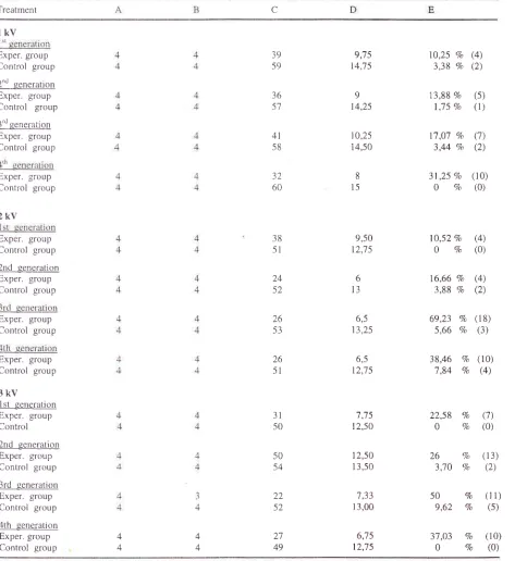
RESULTS
Number
ofoffspring
VoL I l, No
l,
January-
March 2002number
of offspring
between theexperimental
groups(Table
1).Mortality rate were noted
during
1
-
35
days
afterbirth.
A
large mortahty
rate
were fbund
in
all
The effects of electrosbtic
fieA
generations, even
in
males
or
females
offspring,
particularly in
the
third
andfourth
generationswhich
were rreared ro
EMF of
2kV and
3kV.
Table
I.
The eflects o1- electromagnetic field onfour
successive generationsof
miceTreatment
c
lkv
Exper. group
Control group 2t'd generatiofl Exper. group
Control
group 3'd qeneration Exper. groupControl group
Exper. group
Control group
2kv
Exper. group
Control group
2nd generation Exper. group
Control group
Exper. group
Control group
Exper. group
Control group
3kv
1st generation Exper. group Control
2nd generation
Exper. group
Control group
3rd
qenerationExper
groupControl group
4th
generation Exper. groupControl group
32 60 38 51 24 52 26 53 26 51 39 59 36 57 41 s8 22 52 27 49 9;15 t4,7 5
9 t4,25 I0,25 14,50 8 15 9,50 12,75 6 13 6,5 13,25 6,5 t2,75
7,75
22,58
Va
(7)12,50
0
Vo
(0) 10.25 Vo (4)3,38 Vo (2)
13,88
Vo
(5) 1,75Vo
(1)17,07
Vo
(7)3,44
7o
(2)31 ,25 Vo (10)
0
Vo
(0)10,s2
%
(4)0
Va
(0)16,66
Vo
(4)3,88
7o
(2)6e,23
%
(t8)
5,66 Vo
(3)38,46
%
(10)7,84
Vo
(4)26 Vo
(13)3.70
Vo
(2)s0 %
(11)9,62
Vo
(5)37
,03
Vo
(10)0
Vo
(0) 31 50 50 54 12,50 13,50 '7,33 13,00 6,75 12,75 4 4 4 4A=numberof
femalesmated;B-numberof
pregnancies;C=
numberof
pupsdelivered; D=averagelitter size;
[image:3.612.51.524.179.695.2]Soeradi et al
The sex
ratio
of offspring
The
sexratios
of offspring in
theexperimental
groupswere unaffected
by the different
treatments
of
EMF.
With
a
6x
2
contingency table
it
was clear that therewas
no
significant
difïerence
(P
>
0,2)
in
the
sexratio
between
the
various experimental groups
orcontrol
groups as determinedby
thetest (Table
2).Number
ofcongenital
anomaliesCongenital
anomalieswere
also notedin both
sexesof
the offspring
sired by
parents
of
each
generationMed J Indones
exposed to
EMF of
1kV,
2kV,
and 3kV,
respectively.However,
no
congenital anomalies were noted
in
offspring
of
untreated
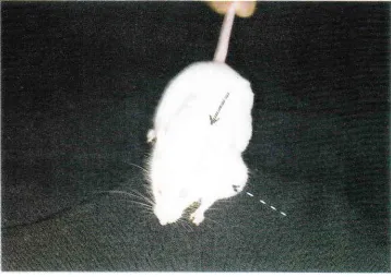
controls. Several
types
of
anomalies were obtained, such as
micropthalmy, white
eyes (Figure
1)
and
dwarf
mice (Figure
2).
Other interestingresult
of
our investigation
is
the
occunenceof
tumorsin
both
sexesof the
offsprrng
and causedof
deathafter
3 to 4 monthsof
old
(Figure
3).Table
2.
The effèct of electromagnetic freld on the sex ratioof the
offspring of four successive generationsof
micetkv
2kv
3kv
Treatment
M M
M
i st generation
Exper.
groupControl group 2nd qeneration
Exper.
group Control group3rd generation
Exper.
groupControl group
4th
generationExper. group
Control group
39 59 36 5'7 41 58 32 60 18 27 t1 30 2l 26
l6
3l
21 32 20 32l6
29 38 51 26 53 26 5l t6 1) 14 1^ 22 28 t4 24 12 29 l5 3r 3t 50 t4 25 t'7 25 27 2950
2350
2119
24
l0
2'7 52
2822 52 2'7 49
ll
20 r0 30 t5 22l2
22 t2 2'7T=
numberofottspring;M=
male;
F=tèma1eTable
3.
Numberof
tumors and anomalies found electromagnetic fieldin the
offipring
of
four successive generationsof
mice exposed tolkv
2kv
3kv
Treatment An Tm An Tm An Tm l.st eeneration
Exper.
groupControl group
2nd qeneratio!
Exper.
groupControl group
3rd generation
Exper. group
Control group
ath-sclcral-an
Exper. group
Control gror,rp
0 0 0 0 2 0 I 0 2 0 2 0 4 0 3 0 5 0 0 0 4 0 2 0 2 0 2 0 8 0 8 0 J 0 2 0 4 0 2 0 0 0 2 0 2 0 0 0
[image:4.612.53.480.277.466.2] [image:4.612.54.478.523.713.2]Vol I
l,
NoI,
January-
March 2002The
cts of electrostatic fieldSoeradi et al
't
[image:6.612.116.473.87.347.2] [image:6.612.116.474.433.687.2]Med J Indones
Figure 2. Anomalous mouse offspring showing a dwarf mouse (A) and a normal mouse offspring (B) of the same age
VoL I
l, No l, Januart'
-
March 2002DISCUSSION
The present results show that
although all
female micebecame pregnant
atter
berng mated
to
males which
also
exposed
to
EMF, the
litter
sizes
were
reducedsrgnificantly
as comparedto untreated controls,
while
litter
size
did not
vary
signrficantly
betweenexperimental groups.
The same
result have
also beenshown
earlier
for otfspring
in
rats after treatmentwith
electrostatic
field.2'r
It
seemslhe effect
of
EMF
onI'ertility was
not
immediately apparent;
probably
because
the
postgonial
cells (primary
and
secondaryspermatocytes, spermatids,
and
spermatozoa) are
relatively
resistant
to
the
lethal
etïects
of
EMF.
However. afier
exposure
to
moderate
or
even
high
doses,
there
is
an
initial fèrtile
period
foliowed
by
clecreaseci
fèrtility.s
Therefore,
the reduction
in
litter
size
(Table
l),
is probably
associatedwith
a decreasein
thenumber
of
spennatogenic celis. Electromagneticfields
can
affects
in
several biological
functions.
includrng hormone levels,
alterationsin
thebinding
of
ions
to
cell
membrane.
and
the
modification of
biochemical
processesinsid
the cell,
such as
RNA
tranccription
ancl
protein
ynthesis
of
enzymes'which
lt'e
needetl rn spermatogenesisor
oogenesis.It
has been
reported
previously, that one etruct ot
electric
fields
is to
increase
the trequency
of
sex-linked
recessivelethal mutation in
femaleDrosophila
melanogaster. Electrostatic
fleld
also has aneffect
on the fiequency of non-disjunction of the X-chromosome.l0This
will
cause the maleto
f-emaleratio
to be less thanunity. The
dataof
our
presentinvestigation
show. thatthe
sex
ratio
of
otïspring in
the experimental
groups werenot
significantly different,
which implies
that thenumber
of
male
andfèmale
otïspring
was unaffectedby
thedilferent
treatments
(Table
2).Micropthalmy
and
dwarf mice were
also
found
in
our
present
study
as an efïect
of
EMF,
while
theother types
of
anomalies werenot similar. We
suggestthat the
anomalies were
the result
of
changesin
the geneticmaterial
of
the spermor
ovaduring
spermato-genesis
or
oogenesis i.e.
amutagenic eifèct.
An
interesting
evidence
of
this investigalion is the
occurrence
of tumors
found
in
both
sexes
of
theoffspring and
causedof death
(Table 3). The
sameresult was also
reported
in
rats after exposure
to
anelectrostatic field.2
Researchers
at
Bettelle
Pasific
Dorthwest
Laboratory
in
Richland, Washington,
haveThe effects of electrostatic fieLd
come close
to
showing
a
direct
EMF-cancer
link
in
rats.
They
have
found
that
EMF
suppresslevels
of
the hormone melatonin, something that other researchershave shown makes female rats suseptible
to
chemicallyinduced
mammary
tumors.5'eIn
connection
with the
hormone
melatonin
shownin
rats whosepineal
glandshave been surgically removed
are
more
likely
todevelop tumors than rats
with
intact pineal glands.
However, after being given melatonin injections the
rats were
no
more
likely
to
develop tumors than
thecontrols.e
At
the level
of
human epidemiology,
50studies have examined
the
possible correlation of
electromagnetic
fields
exposures
with
adult
andchildhood
cancers.
The
studies suggest
that
electro-magneticfields might
be cancer promoters butunlikely
to be cancer initiators.r2
In
conclusion,
the resultsof
thrs investigation
suggestthat continuous exposure
to
electromagnetic
field
in
four successive
generationsof mice,
resultsin
reducedf'ertility with no
changein
the
sexratio, but
effective
in
inducin-qcongenital anomalies and tumors
in
bothsexes
of
the
ofïspring.
In
connection
with
these f'enomena,we
also suggest that theprimary
interactionbetween electromagnetic
field
and
the
living
cells is
more takes place
at
the
molecullar
level
than the
celullar level.
This
interaction
may
then
causebiological
effects
oncells,
tissues, and organs, so thatfinally
there are consequencesfor
thewhole
organism.REFERENCES
l.
KruegerMF,
GiarolaAJ
and BradleyJW.
E11ècts ofelectromagnetic flelds on fècundity in the chicken. Ann
NY Acad
Sci
1975.247:391-3.2.
Soeradi O, TadjudinMK.
Congenital anomaliesin
the offspringof
rats
attar exposureof
the
testesto
anelectrostaticfield
IntJAndrol 1986,9: I
-9.
3
Yurnadi. Pengaruh pemejanan medan elektrostatikter-hadap tèkunditas dan rasio seks mencit (Mus musculus) strain Swiss Webster BPMSOH pada dua generasi. Tesis
Magister Program Studi Biomedik Program pascasarjana
ul,
1999.4.
Tomenius L. 50Hz
electromagnetic environment and theincidence
of
childhood tumorsin
Stockholm county. Bioelectromagnetics 1986, '7 : l9'7 - 207 .5.
StevensRG,
Davis
C,
ThomasDB,
Anderson LE,Wilson
BW.
Electric power, pineal function, and the riskof
breast cancer. FASEBI
1992,6: 853-60.6.
Sari P. Pemajanan medan elektrostatik pada mencit (Mzs musculus) strain Swiss Webster dan pengaruhnya terhadap kromosomserta proliferasi
limfosit.
Tesis
Magister Program Studi Biomedik Program Pascasarjana UI, 1998.7.
Skyberg K, Lise HensteenI
and Vistnes AI. Chromosomall0
Soeradi et aI Med J Indonesand
generator production exposedto
electromagnetic
11. Griem ML.
The effectsof
radiationon
the fetus, ln: lrelds and mineraloil.
Bioelectromagnetics 2001,22:
Dalrymple DV, Gaulden ME, Koll-morgen GM, Vogel HH150-60.
(eds). Medical Radiation Biology. Philadelphia, Saunders8.
Dalrymple GV, Gaulden ME. Medical RadiationBiology.
WB, p. 97-9.Philadelphia, SaunderswB 1973,
p
12.
Hendee WR, Boteler JC. The questionof
health effects9.
Pool R. Electromagnetic Fields: The BiologicalEvidence.
from exposureto
electromagneticfields. Health
PhysScience
1990,249:1378-81
1994,66:127-36.10.
Portnov
FG,
ShakarinsVF,
Maiore
DJ.
Study
of mutageniceffect
of
static electric
fields
in
female Drosophila melanigaste. Genetika 1975, 1l:
177-81.I
I