DOI: 10.12928/TELKOMNIKA.v11i4.1583 791
Localizing Region-Based Level-set Contouring for
Common Carotid Artery in Ultrasonography
Yong Chen*, Xiao Ming Zhou, Dong C Liu
School of Computer Science, Sichuan University, ChengDu, P.R. China *Corresponding author, e-mail: [email protected]
Abstrak
Penelitian ini mengembangkan metode otomatis dan efisien secara penuh untuk mendeteksi kontur arteri karotid di bagian penampang sonografi B-mode dua-dimensi. Pertama, tapis pra-olah diterapkan pada citra ultrasonik demi mengurangi bintik. Sebuah metode contouring awal adaptif kemudian diterapkan untuk mendapatkan kontur awal untuk segmentasi set level. Terakhir, segmentasi set level berbasis wilayah lokal untuk pengekstraksian kontur secara presisi dari arteri karotid secara otomatis. Metode yang diusulkan mengevaluasi 130 citra ultrasonik dari tiga sukarelawan dan hasil segmentasi dibandingkan dengan batas-batas yang digariskan oleh seorang ahli. Hasil awal menunjukkan bahwa metode ini bisa mengidentifikasi kontur arteri karotid dengan akurasi yang memuaskan dalam dataset ini.
Kata kunci: kontur aktif, metoda set level, segmentasi citra, arteri karotid, ultrasonik
Abstract
This work developed a fully-automated and efficient method for detecting contour of common carotid artery in the cross section view of two-dimensional B-mode sonography. First, we applied a preprocessing filter to the ultrasound image for the sake of reducing speckle. An adaptive initial contouring method was then performed to obtain the initial contour for level set segmentation. Finally, the localizing region-based level set segmentation automatically extracted the precise contours of common carotid artery. The proposed method evaluated 130 ultrasound images from three healthy volunteers and the segmentation results were compared to the boundaries outlined by an expert. Preliminary results showed that the method described here could identify the contour of common carotid artery with satisfactory accuracy in this dataset.
Keywords: active contours, level set method, image segmentation, common carotid artery, ultrasound
1. Introduction
Compared to other imaging techniques, ultrasonography is inexpensive, noninvasive and more accessible. Therefore it has become a widely used diagnostic method in various medical disciplines, such as echocardiography, breast ultrasound, intravascular ultrasound (IVUS), etc.
Kalman filter [7], were also reported.
Traditional edge-based segmentation methods depend on the gradient of an image to locate the contour. But ultrasound image suffers from weak edges and speckle, so edge-based methods’ performance isn’t satisfactory on outlining ultrasound images. For the same reason, region-growing and morphological watershed transformation based methods perform poorly too when they were applied to sonography.
In this work, we proposed to use local region-based active contours introduced by [8] to extract the lumen-intima-boundary (LIB) from Ultrasound images. The localized active contour method using local image statistics, rather than global image statistics, to evolve the contour, which is capable of segmenting object with heterogeneous feature profiles that would be hard to extract using a traditional global method. The proposed method can be roughly divided into two steps. The first step is to adaptively generate the initial contour of LIB. The second is to apply the local region-based active contour method to the initial contour and evolve it to get the final contour.
The rest of this paper is organized as follows. We begin in section 2 by describing the proposed method in detail. Then section 3 presents the experiment result and discussion. Finally, we conclude our work in section 4.
2. Research Method 2.1. Data Acquisition
An ultrasound image database included 130 ultrasound images of three healthy volunteers aged 27 to 40 years. The common carotid arteries of the right sides of their necks, 2-3 cm proximal to the bifurcation, were captured by an expert. All digital ultrasound images were obtained using Saset iMago C21 system (SASET Healthcare, San Francisco, CA) with a 10 MHz linear transducer. The capturing resolution of the images was 0.07×0.07 mm2. Except for a few cases, the sonographic setting remained the same during the whole course of data acquisition.
2.2. Outline of the Proposed Method
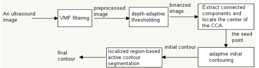
An outline of the proposed CCA segmentation algorithm is described in Figure 1, where the major components and data flow is illustrated. The algorithm includes the following computational steps: image preprocessing, automated region of interest (ROI) identification, adaptive initial contouring, and localized region-based active contour segmentation. These computational steps are detailed in the remainder of section 2.
2.3. Preprocessing
Figure 1. Flow Chart of the Proposed Method
2.4. Active Initial Contouring
After the image preprocessing, a depth-adaptive thresholding method was conducted to transform the gray-scale image into a binary image to locate the center of the CCA. After considering the characteristics of the intensity of blood: (a)it maybe different as different depths; (b)it is always the lower as compared to that of the non-blood pixels, we experimentally choose 20th percentile intensity of all pixels at the same depth as the blood threshold for that particular depth. Figure 2.(a) shows an example of the resulting binarized image with the blood area labeled as white and non-blood area as black.
Then the connected component areas in the binary image are labeled and the horizontal diameter (hd) and vertical diameter (vd) of each connected component area are calculated. After applying the knowledge that the CCA is always of circular appearance and the average diameter for adult males and females is 6.52 mm and 5.11 mm respectively [10], we claim that the connected component area whose hd is between 4mm and 10mm, vd is between 4mm and 10mm, and abs(hd – vd) <= 1mm is the CCA we persued. The initial contour is defined as a rectangle centered at the center point of the CCA. Its diameter is the minimum of hd and vd. The corresponding center point of the CCA and the initial contour in Figure 2.(a), being found through this method, are shown as red cross and green rectangle respectively in Figure 2.(b).
(a) (b)
Figure 2. The Process of Adaptive Initial Contouring (a)the binary image resulted from the depth-adaptive thresholing method, blood areas are shown as white and non-blood areas as
black. (b)the identified center point of the CCA (red cross) and the initial contour (green rectangle)
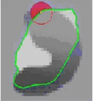
2.5. Localizing Region-based Level Set Contouring
[image:3.595.101.501.474.628.2]Figure 3. Illustratration of the Basic Idea of the Localizing Region-based Active Contour Algorithm
The authors have introduced local region-based framework and three specific energies: the uniform modeling energy, the means separation energy, and the histogram separation energy. As for our case, we chose the means separation energy. The local region-based flow is defined as:
2 2
( ( )
)
( ( )
)
( )
( )
( , )
( )
[image:4.595.88.504.569.724.2]( )
( )
| ( ) |
x x
y
I y
u
I y
v
x
x
B x y
y
dy
t
Au
Av
x
x div
x
(1)
Where
( )
x
is the Heaviside function and
is a smoothed version of Dirac function; B(x,y) isa local region mask function;
u
x andv
x represent the intensity means in the interior and exterior of the contour localized by B(x,y) at a point x respectively; Au and Av are the areas of the local interior and local exterior regions respectively;
is the gradient operator; parameter
controls the smoothness of zero level set. The evolving contour is hoped to settle whenu
xand
v
x are the most different at every x along the contour. Figure 4. presents an example of the segmentation result of the proposed method.(a) (b)
2.6. Contour Evaluation Metric
The quality of our segmentation method was validated using two measures: 1) Dice Metric (DM) and 2) Hausdorff Distance (HD) [11]. DM evaluates the similarity between two areas. It is defined as
2 * (
1
2)
*100
1
2
area
area
DM
area
area
(2)
So we know that the more similar two areas are, the higher value DM becomes, and vice verse. And a perfect match would yield a value of 100%.
The HD aims to evaluate the distance between two contours. If two contours are represented by a set of points A = {a1, a2, …, am} and B = {b1, b2, …, bm}, where each ai and bi is
an ordered point on the curve. The Distance to the closest point (DCP) for ai to B is defined as
( , )
imin ||
j i||
j
d a B
b
a
(3) and the HD is define as( , )
max(max{
( , )}, max{
i( , )})
ji j
HD A B
DCP a B
DCP b A
(4)3. Results and Discussion
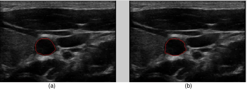
This section presents the evaluation of segmentation result to analyze the validation of the proposed method. This work experimented on a total of 130 ultrasound images, and then compared the segmentation results with the expert-drawn contours. In this dataset, it yielded a DM of 91.1% ± 4.2% for the LIB and a HD of 0.59 ± 0.35 mm. Figure 5 illustrates an example of segmentation result. Figure 5 (a) shows the contour manually drawn by the expert; Figure 5 (b) plots the contour determined by the proposed method and the result of measures (DM, HD) is (0.9475, 0.63 mm).
[image:5.595.88.514.449.604.2](a) (b)
Figure 5. Comparison of the segmentation result: (a) contour drawn by the expert (b) the contour determined by the proposed method
Localizing region-based level set utilize the statistical information inside and outside the contour to control the evolution, which are less sensitive to noise and are suitable for segmenting images with weak edges, such as the ultrasound images. But it also should be noted that it is sensitive to initialization, just as pointed out by the authors in [8]. Fortunetely, we have developed an adaptive initial contouring method for it and the experiment results showed that they worked together pretty well in this work.
the LIB. An image database with 130 cases was employed for evaluation in this work. Preliminary results showed that the method described here is able to locate the LIB contours that are very similar to expert-drawn contours and can be used as an alternative to manual contouring of the LIB from ultrasound images.
Future work would be focused on (a) optimizes the proposed method and improves the computing speed; (b) collects more unhealthy objects, such as those suffered from atherosclerosis plaque, and test our method on them.
Acknowledgement
The authors would like to thank the members of the Medical Imaging Laboratory at SiChuan University for volunteering for ultrasound scans; Dr. Zuo at SASET Healthcare for kindly providing the ultrasound data.
References
[1] N.Denarie, J.Gariepy, G.Chironi, M.Massonneau, F.Laskri, J.Salomon, J.Levenson, and A.Simon, Distribution of ultrasonographically-assessed dimensions of common carotid arteries in healthy adults of both sexes. Atherosclerosis. 2000; 148: 297-302.
[2] David C.Wang, Robert Klatzky, Bing Wu, Gregory Weller, Allan R.Sampson, and George D.Stetten, Fully Automated Common Carotid Artery and Internal Jugular Vein Identification and Tracking Using B-mode Ultrasound. IEEE Transactions on Biomedical Engineering. 2009; 56: 6.
[3] Xin Yang, Jiaoying Jin, Mengling Xu, Huihui Wu, Wanji He, Ming Yuchi, and Mingyue Ding, Ultrasound Common Carotid Artery Segmentation Based on Active Shape Model, Computational and Mathematical Methods in Medicine. 2013; 2013, article ID 345968.
[4] E.Ukwatta, J.Awad, A.D.Ward et al., Three-dimensional ultrasound of carotid atherosclerosis: semi-automated segmentation using a level set-based method. Medical Physics. 2011; 38(5): 2479-2493 [5] A. Abdel-Dayem and M.El-Sakka, Carotid ultrasound image segmentation using Fuzzy region
growing, Image Analysis and recognition. Springer, Berlin, Germany. 2005; 3656: 869-878.
[6] Mao F, Gill J, Downey D, and Fenster A. Segmentation of carotid Artery in Ultrasound images, Engineering in Medicine and Biology Society 2000 Proceedings of the 22nd Annual International Conference of the IEEE. 2000; 3: 1734-1737
[7] P.Abolmaesumi, M.Sirouspour, and S.Salcudean, real-time extraction of carotid artery contours from ultrasound images, proceedings of the 13th IEEE symposium on computer-based medical systems(CBMS’00), 2000; pp.181-186.
[8] Shawn Lankton, Allen Tannebaum. Localizaing Region-Based Active Contours. IEEE Transactions on Image Processing. 2008; 17(11): 2029-2039.
[9] Jaakko Astola, Petri Haavisto, and Yrjo Neuvo. Vector median filters. Proceedings of the IEEE. 1990; 79(4): 678-689.
[10] Jaroslaw Krejza, Michal Arkuszewski, et al. Carotid Artery Diameter in Men and Women and the Relation to Body and Nect Size, Stroke. 2006; 37:1103-1105