The Clinical Features of Ostiomeatal Complex in Chronic Maxillary
Sinusitis
by Nasoendoscopic Examination
De lfitri Munir
ENT De p a rtm e nt Me d ic a l Fa c ulty No rth Sum a tra Unive rsity
Abstrak: Lebih dari 90% sinusitis maksila kronis disebabkan oleh variasi anatomi ostiomeatal
kompleks. Variasi anatomi ostiomeatal kompleks dapat diketahui dengan pemeriksaan nasoendoskopi. Dengan pemeriksaan ini dapat diketahui kelainan di meatus media dan di bagian anterior hidung. Tujuan penelitian ini adalah untuk mengetahui kelainan di kompleks ostiomeatal pada sinusitis maksila kronis. Empat puluh pasien dengan 67 kelainan sinus maksila diperiksa dengan nasoendoskopi. Penelitian ini dilakukan di Departemen THT Fakultas Kedokteran Universitas Sumatra Utara/RS H. Adam Malik, Medan, Indonesia.
Dari 67 kasus sinusitis maksila, dijumpai 58 sinus (86,6%) kelainan pada kompleks ostiomeatal di antaranya 21 sinus (36,2%) adalah pembesaran bula etmoid, 16 sinus (37,6%) polip pada konka bulosa dan konka paradoksal, 10 sinus (17,3%) kelainan prosesus unsinatus, 7 sinus (12%) polip pada meatus media dan hiatus semilunaris, serta 4 sinus (6,9%) dijumpai deviasi septum.
Kata kunci: sinusitis maksila kronis, kompleks ostiomeatal, pemeriksaan nasoendoskopi
Abstract: More than 90% of chronic maxillary sinusitis are caused by anatomic variation of
ostiomeatal complex. The anatomic variation of ostiomeatal complex can be found by using nasoendoscopic examination. Nasoendoscopy reveals the feature of middle meatus and identifies the abnormality anteriorly. The aim of this study was to investigate the abnormality of ostiomeatal complex in patients with chronic maxillary sinusitis. Fourty patients with 67 abnormal maxillary sinus were examined by using nasoendoscope. The study took place in the center of ENT Department, Medicine Faculty, North Sumatera University/H. Adam Malik Hospital, Medan, Indonesia.
Out of 67 cases, 58 sinuses (86.6%) were found with abnormal ostiomeatal complex. Of the 58 sinuses, it was found that 21 sinuses (36.2%) were the enlargement of ethmoid bulla, and 16 (27.6%) were polyps in concha bullosa and concha paradoxal, 10 sinuses (17.3%) were abnormal uncinate process, 7 sinuses (12%) were polyps in middle meatus and hiatus semilunaris and 4 (6.9%) cases were septal deviation.
Keywords: chronic maxillary sinusitis, ostiomeatal complex, nasoendoscopic examination
INTRODUCTION
Chronic maxillary sinusitis is defined as an inflammation of the maxillary sinus mucosa which is more than 3 months.1 Loury describes symptom of chronic maxillary sinusitis as “triad”; nasal obstruction, nasal discharge and facial fullness.2 Acute maxillary sinusitis can usually be cured by a prompt medical treatment, but chronic maxillary sinusitis is often difficult to be cured by conservative treatment because of the ostiomeatal complex abnormality.3 The changes in maxillary sinus usually complication
of ostiomeatal complex abnormality.4
Messerklinger reported that infundibulum and middle meatus were the most common sites influenced by anatomic variation of ostiomeatal complex. Stammberger also describes that more than 90% of this disease are caused by anatomic
variation of ostiomeatal complex.5,6,7 The anatomic variation of ostiomeatal complex can be found by using nasoendoscopic examination. Nasoendoscopy reveals the feature of middle meatus and identifies the abnormality anteriorly. Nasoendoscopy is valuable to evaluate concha bullosa, concha paradoxal, nasal mucosal thickening, purulent drainage and small polyp which make chronic obstruction, disruption of ventilation and mucociliary system, subsequently predispose an inflammation of maxillary sinus mucosa.8,9
We performed this study as a cross-sectional study for patients who fulfil the inclusion and exclusion criteria. We wanted to know the anatomic variation of ostiomeatal complex by nasoendoscopic examination which
Delfitri Munir The Clinical Features of Ostiomeatal Complex…
is the most common cause for chronic maxillary sinusitis.
PATIENTS AND METHODS
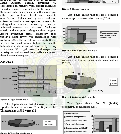
This cross-sectional study was performed from February to October 2000 in H. Adam Malik Hospital Medan, involving 40 consecutive out patients with chronic maxillary sinusitis. Sinusitis was judged to be present if the radiographic showed mucosal thickening and either an air-fluid level or complete opacification of the maxillary sinus. Inclusion criteria included minimal age was 15 years old, radiographic showed maxillary sinusitis, informed consent was obtained. Exclusion criteria included prior undergone sinus surgery. Before attempting nasal endoscopy each patient`s nasal cavity was anasthetized with pantocain 2% with epinephrine in a swab. It was inserted to nasal cavity below the middle turbinate and lateral wall of nasal cavity. Using
a 2.7-mm 30o rigid nasal endoscope, we
investigated and assesed the middle meatus and the ostiomeatal complex.
RESULTS
This figure shows that the most common age distribution is between 35 – 44 years old. The mean age is 38.3 years old.
52%
48% man
women
Fig ure 2. G e nd e r d istrib utio n
This figure shows that the most common gender is man.
This figure shows that the most common main symptom is nasal obstruction (60%)
0
This figure shows that the most common radiographic finding is complete opacification (57.5%). ostiomeatal complex are close.
0 Polyp in hiatus semilunaris Polyp in middle meatus Enlargement/polypoid in UP Enlargement ethmoid bulla
Fig ure 6. The a b no rm a lity o f o stio m e a ta l c o m p le x
This figure shows that enlargement of ethmoid bulla is the most common abnormality (36.2%).
Karangan Asli
DISCUSSION
The youngest patient participated in this study was 15 years of age. The reason is because the development of the maxillary sinus structure is completed at the age of 14. 10 Different research showed a different mean age. Nuty in Jakarta reported the mean age was 26 years old.12 Nadel reported the mean age was 47.3 years.13 In this study the mean age is 38.3 years. Massudi in Semarang 1991 reported that the most common age distribution was 21-25 years while in this study between 35-44 years.
The common symptoms of chronic maxillary sinusitis are nasal obstruction, nasal discharge and facial fullness. Benninger reported that nasal obstruction was the main
symptom.9 Massudi reported that the main
symptoms were nasal obstruction(42.4%), nasal discharge (33.4%), and headache (15.1%).11 In this study we found that the most common main symptom is nasal obstruction (60%). The rest were nasal discharge (25%) and headache (5%). Kennedy reported that nasal obstruction is related to abnormal nasal meatal and sinus that disrupt the ventilation and nasal drainage.2
Sinusitis is judged to be present if
radiographic finding shows that there is mucosal thickening, air-fluid level or complete opacification of the maxillary sinus.14 Nuty WN reported that the most common radiographic finding was complete opacification (87%).12 In this study it was found that the most common radiographic finding is complete opacification (57.5%). Cody reported that mucosal thickening is usually present in allergy and vasomotor reaction. Pathological change will enhance the mucosal thickening with sinus secretion if there is bacterial infection. Air-fluid level emerges because of transudation in submucosa fills the sinus cavity as an inflammation response .15 Rachelevsky reported that 37 % of chronic maxillary sinusitis patients are positive in allergy skin test.16
The etiology of chronic maxillary sinusitis are multifactorial. Ostiomeatal complex plays an important role in pathophysiology of chronic maxillary sinusitis. Stammberger reported 90 % of chronic maxillary sinusitis are commonly due to the abnormality of ostiomeatal complex. This study showed that 86.6 % abnormality is in ostiomeatal complex. Enlargement of ethmoid bulla (36%) was the most of ostiomeatal complex abnormality, and 16 (27.6%) were polyps in concha bullosa and concha paradoxal, 10 sinuses (17.3%) were abnormal uncinate process, 7 sinuses (12%) were polyps in middle
meatus and hiatus semilunaris,and 4 (6.9%) cases were septal deviation. Penttila reported that polyp was 54 %.17 In this study it was not clear why the enlargement of bulla ethmoid as the most common cause. According to the literature, anterior ethmoid bulla adjascent to middle turbinate which has a direct and strong contact with inspiration. The air-flow changes in this place, then settles the particles which size is more than 6 μm are trapped. These particles precipitate mucosal inflammation process and make the ostium narrow.2
CONCLUSION
Most chronic maxillary sinusitis (86.6%) were caused by ostiomeatal complex abnormality and the most common cause was the enlargement of ethmoid bulla (36.2%).
REFERENCES
1. Facer GW, Kern EB. Sinusitis: current
concepts and management. In: Bailey BJ, Editors. Head and neck surgery-otolaryngology. Philadelphia: J.B. Lippincott Company, 1993. p.366-8.
2. Loury MC, Kennedy DW. Chronic sinusitis
and nasal polyposis. In: TV Getchell et.al. Editors. Smell and taste in health and disease. New york: Raven Press, 1991. p. 517-27.
3. Nuty WN, Wardani RS. Anatomi
endoskopik hidung-sinus paranasal dan patofisiologi sinusitis. Disampaikan pada Kursus dan Pelatihan Bedah Sinus Endosopik Fungsional, Semarang, 29 September-1 Oktober,2000.
4. Kennedy DW, Loury MC. Nasal and sinus
pain. Current diagnosis and treatment. Seminars in Neurology, December 1998;3:303-13.
5. Stammberger H. Endoscopic endonasal
surgery. Concepts in treatment of recurring rhinosinusitis. Otolaryngol Head and Neck surg. 1986: p. 143-6.
6. Zinreich SJ, Kennedy DW. Paranasal
sinuses. CT imaging requirements for endoscopic surgery. Radiology 1987;163: p. 769-74.
7. Lee KJ. Text book of otolaryngology and
head and neck surgery. New York:Elsevier, 1989. p.222-3.
Delfitri Munir The Clinical Features of Ostiomeatal Complex…
Ma ja la h Ke d o kte ra n Nusa nta ra Vo lum e 39 y No . 1 y Ma re t 2006 15 8. Lang J. Clinical anatomy of the nose, nasal
cavity and paranasal sinuses. New York: Thieme. Medical publisher, 1989. p 56-125.
9. Benninger MS. Nasal endoscopy, it`s role in office diagnosis. Am J Rhinol 1997;11: p. 177-80.
10. Hollinshead WH. Anatomy for surgeons. A
Hoeber-Harper International Edition, 1996. p. 270-6.
11. Massudi RH. Pola dan kerentanan kuman
aerob secara invitro pada sinusitis maksilaris kronis pada rumah sakit Dr Kariadi Semarang. Disampaikan pada PIT Perhati, Batu Malang,1996.
12. Nuty WN, Damayanti S. Gambaran
sinuskopi pada sinusitis maksilaris kronis. Disampaikan pada Konas XI Perhati, Yogya. October 1995.
13. Nadel DM, Kennedy DW, Lanza DC.
Endoscopically guided cultures in chronic sinusitis. Am J Rhinol 1998;12: p. 240.
14. Itzhak B, David HT, Edith HF. Microbiology and management of chronic maxillary sinusitis. Arch Otolaryngol Head and Neck Surg 1994;120: p. 1317-9.
15. Cody DT, Kern EB. ENT disease. Jakarta:
EGC, 1993. p. 232-6.
16. Roland NJ. Key topics in otolaryngology. London: Toppan. Bios Scientific Publishers, 1995. p . 48.
17. Penttila MA, Pukander JS.. Clinical and