CLONING AND STEM CELLS Volume 8, Number 1, 2006 © Mary Ann Liebert, Inc.
Transfer of Inner Cell Mass Cells Derived from Bovine
Nuclear Transfer Embryos into the Trophoblast of
Bovine
In Vitro
–Produced Embryos
M. MURAKAMI,1C.E. FERGUSON,1 O. PEREZ,1 A. BOEDIONO,1 D. PACCAMONTI,2 K.R. BONDIOLI,1 and R.A. GODKE1,2
ABSTRACT
Presence of placental tissues from more normal noncloned embryos could reduce the preg-nancy failure of somatic cloning in cattle. In this study, inner cell mass (ICM) cells of in vitro–produced (IVP) embryos was replaced with those of nuclear transfer (NT) embryos to reconstruct bovine blastocysts with ICM and trophoblast cells from NT and IVP embryos, re-spectively. A total of 65 of these reconstructed embryos were nonsurgically transferred to 20 recipient beef females. Of those, two females were diagnosed pregnant by ultrasonography on day 30 of gestation. One pregnancy was lost at 60–90 days of gestation, and the other re-cipient cow remained pregnant at day 240 of gestation; however, this female died on day 252 of gestation. Gross pathology of the internal organs of the recipient female, a large fetus, and a large placental tissue mass suggested the massive size of the fetus and placental tissue were likely involved in terminating the life of the recipient female. Biopsy samples were harvested from the skin of the dead recipient cow, the fetus and from cotyledonary tissue. Microsatel-lite DNA analysis of these samples revealed that the genotype of the fetus was the same as that of the NT donor cells and different from that of the recipient cow. Correspondingly, nei-ther the fetus nor recipient cow had the same genotype with that of the fetal cotyledonary tis-sue. These results present the first known documented case of a bovine somatic NT preg-nancy with nonclone placental tissues after transfer of a blastocyst reconstructed by a microsurgical method to exchange of ICM cells and trophoblast tissue between NT and IVP blastocysts.
51 INTRODUCTION
A
N UNUSUALLY HIGH proportion of fetal orneonatal losses has been consistently docu-mented in bovine cloning by somatic cell nuclear transfer (NT), which is the major impediment to-wards widespread application of this methodol-ogy. Among the contributing factors, placental deficiency of the NT conceptus has been
associ-ated with lowered cloning efficiency (Stice et al., 1996; Hill et al., 2000; De Sousa et al., 2001, Bertolini and Anderson, 2002).
In cattle, both structural and epigenetic anom-alies have been detected in later stage somatic NT embryos, such as decreased trophectoderm cell to total embryonic cell ratio and trophectoderm-lo-calized methylation aberrancy (Kang et al., 2002; Koo et al., 2002; Han et al., 2003). Research
indi-1Embryo Biotechnology Laboratory, Department of Animal Sciences, 2Department of Clinical Sciences, Louisiana
cates a higher frequency of these and other alter-nations in developing NT embryos compared with those of their in vitro–produced (IVP) or in vivo–derived counterparts (Patel et al., 2004; Rav-elich et al., 2004a,b), and these alterations may contribute the subsequent placental dysfunction during latter stages of pregnancy.
Aberrant expression patterns of various genes are thought to be involved in placental dysfunc-tion, such as a placental lactogen, leptin, insulin-like growth factor binding proteins 2 and 3 and trophoblast major histocompatibility complex 1 have been detected in bovine NT conceptuses in addition to a multitude of morphological anom-alies, and the frequency was higher than that re-ported in fetuses derived from artificial insemi-nation or IVP procedures (Hill et al., 2002; Patel et al., 2004; Ravelich et al., 2004a,b). Alterations in gene expression are likely the underlying cause of placental malformation and subsequent ab-normal function during pregnancy (Hashizume et al., 2002; Ravelich et al., 2004a). The produc-tion of dysfuncproduc-tional peptides and/or proteins could increase the frequency of pregnancy fail-ure, since they likely play an important role in nutrient partitioning and regulation of placental development and fetal growth. It has been pro-posed that placental abnormalities are a major factor in the decreased survivability in cloned bovine near-term fetuses and perinatal calves (Stice et al., 1996; Wells et al., 1999; Hill et al., 2000).
Over the years, various research groups have developed procedures to isolate (mechcanically or immunologically) and transfer the ICM from sheep and goat blastocysts to the blastocoele cav-ity of other intra- or inter-species embryos, pro-ducing viable sheep, goat, or chimeric offspring (Fehilly et al., 1984; Polzin et al., 1987; Butler et al., 1987; Roth et al., 1989; Rorie et al., 1994). Chi-meric offspring have been produced by microin-jection of bovine ICM cells into the blastocoele cavity of cattle blastocysts (Summers et al., 1983). To eliminate the production of ICM-derived chi-meric reconstructed embryos, a method was de-veloped to remove the host ICM from the tro-phectoderm at the time the foreign ICM was injected into the blastocoele of the host embryo (Rorie et al., 1994).
To evaluate these NT conceptus inconsisten-cies, production of embryos with fetal and pla-cental tissues of different origin would be in-valuable in attempting to solve this major barrier
to the field application of NT technology. It has been proposed that the production of cloned em-bryos surrounded with more normal nonclone trophoblast tissue could amend the subsequent fetal/neonatal losses often reported with bovine NT. In the our study, the objective was to replace inner cell mass (ICM) cells of bovine IVF-derived embryos with those of bovine NT embryos to re-construct blastocysts comprised of ICM cells and trophoblast cells derived from bovine NT and IVP embryos, respectively. An effort was made to develop an applicable microsurgical embryo reconstruction procedure that would allow the presence of attached nonclone placental tissues to subsequently support the development of the bo-vine somatic NT fetus during gestation.
METHODS
Oocyte preparation
Bovine oocytes were obtained weekly from a commercial source (Ova Genix, San Angelo, TX). The majority of the oocytes provided by this com-mercial source were from mature Holstein fe-males. The oocytes were shipped at 38°C by overnight courier to the laboratory, while under-going in vitro maturation during transit. After 18–22 h of maturation, the oocytes were ran-domly assigned to standard in vitro fertilization (IVF) or NT procedures. All of the chemical agents and media were obtained from Sigma (St. Louis, MO), unless otherwise specified.
In vitro fertilization
IVF was performed as previously described (Murakami et al., 1998) using frozen semen from a single ejaculate of a fertile Holstein bull and Brackett-Oliphant (B-O) medium (Brackett and Oliphant, 1975). Briefly, one 0.25-mL straw of frozen semen was thawed in a 39°C water bath and washed twice in B-O medium supplemented with 5 mM caffeine by centrifugation at 500⫻g
for 5 min at room temperature. The sperm pellet was re-suspended in B-O medium supplemented with 0.3% BSA, 3.6 IU heparin (Elkins-Sinn, Cherry Hill, NJ), and 2.5 mM caffeine. Then oocytes were placed into 100-L insemination droplets under medical grade mineral oil with spermatozoa at a concentration of 1⫻106sperm/
mL. After 5 h of co-incubation, oocytes were de-nuded of cumulus cells by vortexing in Tissue
Culture Medium 199 (TCM 199, Gibco, Grand Is-land, NY) containing 0.1% hyaluronidase, washed and cultured in CR1aa culture medium with 5% BSA (Rosenkrans et al., 1994) at 39°C in a humidified atmosphere of 5% CO2, 5% O2, and
90% N2.
Nuclear transfer
After washing, only good quality mature oocytes that had extruded the first polar body were selected for NT using the basic procedure previously described by Shiga et al. (1999) with minor modifications (Murakami et al., 2003). Briefly, the oocytes were exposed to Hoechst 33342 stain (5 g/mL) and transferred to a 200-L droplet of Dulbecco’s phosphate-buffered saline (DPBS, Gibco) supplemented with 5% fe-tal bovine serum (FBS) and 5 g/mL of cytocha-lasin B. A small rent was made in the zona pel-lucida and the first polar body and the metaphase plate were removed from each oocyte using a fine flexible glass needle, after a brief observation (⬍10 sec) under fluorescence.
The donor cells, originally isolated from a ma-ture, fertile Charolais cow (5 years of age) of high genetic merit, consisted of adult skin fibroblasts that had been subpassaged two to eight times. A single donor cell was introduced into the peri-vitelline space of the enucleated oocyte to con-struct a cytoplast-karyoplast couplet. The cou-plets were induced to fuse in buffer comprising 0.3 M mannitol, 0.05 mM calcium, 0.1 mM mag-nesium with two direct current pulses of 2.25 kV/cm for 15 sec delivered by an electrofusion unit (BTX Model 200, San Diego, CA). The fused couplets were further activated by being cultured in 10 g/mL of cyclohexamide for 4 h, washed and then cultured in CR1aa medium with 5% BSA at 39°C in a humidified atmosphere of 5% CO2,
5% O2, and 90% N2(day 0).
Embryo culture
On day 3 of in vitro culture, cleaved embryos from both IVP and NT procedures were trans-ferred to fresh CR1aa medium supplemented with 5% BSA and cultured at 39°C in a humidi-fied atmosphere of 5% CO2, 5% O2, and 90% N2
for an additional 5 days. On the morning of day 8 of in vitroculture, good quality blastocysts were selected from both IVP and NT embryos for the embryo reconstitution procedures.
Embryo reconstruction
The micromanipulators were arranged with a beveled glass pipette (40 m i.d.) on one side of the unit and a new microblade (A.B. Technology, Pullman, WA ) attached on the other side. Hatch-ing or hatched blastocysts from IVP-derived em-bryos, which served as the host trophoblasts, were transferred to a 200-L droplet of DPBS supplemented with 5% FBS and 5 g/mL of cy-tochalasin B. The microblade was lowered verti-cally to hold the host IVP-derived embryo in po-sition on the bottom of the droplet in a holding dish with the IVP-derived ICM isolated to one side of the microblade. Then, ICM from the bovine NT embryos was mechanically isolated re-moving the adjacent trophectodermal cells and the overlying polar trophectoderm using a mod-ification of the micromanipulation procedure used in mice (Matta, 1991). The ICM were aspi-rated into a small-pore micropipet (Fig. 1A) and gently injected into the blastocoele of the host IVP embryo (Fig. 1B). This was followed by complete removal of the original ICM with the surround-ing trophoblast cells from the host IVP embryo using a microblade embryo splitting technique (Fig. 1C), similar to the method for hatched blas-tocysts previously reported by this laboratory (Rorie et al., 1994). The result was an ICM and trophoblast reconstructed collapsed embryo and a discarded ICM with a surrounding trophoblast segment from the original host IVP embryo are shown in Figure 1D. Reconstructed embryos were cultured in vitro for 3–6 h post-fusion prior to transfer.
Control NT embryos were prepared using oocytes from the same source, the same embryo culture system and the same NT procedure. The control NT embryos were similarly cultured for up to 6 hours prior to their transfer to recipient females.
Embryo transfer
Mature, cyclic beef females (mixed breed, cross-bred cows), in good body condition, from a single research station herd served as recipients follow-ing natural estrus (day of estrus⫽day 0). The re-constructed day-8 embryos with the introduced NT ICM cells (Fig. 2) and the control NT embryos were removed from in vitro culture, as re-ex-panded blastocysts, and nonsurgically transferred to recipients (two to four embryos/female) on day 7 or day 8 of their estrous cycles. Pregnancies were
detected using ultrasonography at day 30 of ges-tation, and verified by detecting fetal heart beats on day 60 and on day 90 of gestation. Thereafter, pregnant recipient females were re-evaluated monthly by rectal palpation throughout gestation.
RESULTS
A total of 65 viable reconstructed NT embryos were nonsurgically transferred to 20 recipient fe-males on days 7–8 of their estrous cycle. Two of the 20 recipient females were diagnosed pregnant by ultrasonography, as evidenced by heart beats from singleton fetuses on day 30 of gestation. Of those pregnancies, one was lost between 60 and
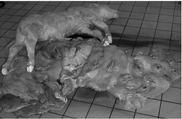
90 days of gestation. The remaining mature cross-bred recipient cow remained pregnant with a vi-able fetus at day 240 of gestation; however, this female died on day 252 of gestation while carry-ing a female fetus weighcarry-ing 62.7 kg (Fig. 3).
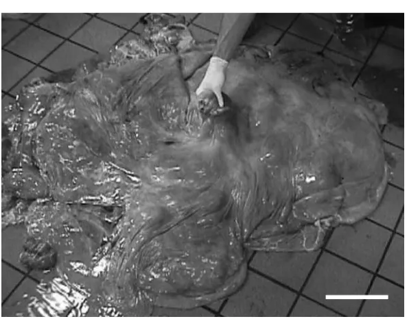
Gross pathology of the internal organs of re-cipient female, the fetus and placental tissues sug-gested that the fetus died following the death of the recipient. Furthermore, the gross pathology report from the Louisiana State University School of Veterinary Medicine’s Department of Pathol-ogy indicated that hydroallantois in addition to the massive size of the placental tissues (Fig. 4) and the mega-size fetus were involved (renal dys-function, hydronephosis, internal organ damage) in terminating the life of the recipient female.
MURAKAMI ET AL. 54
In the control group, a total of 61 NT-derived embryos were similarly nonsurgically transferred to 21 beef recipient cows (two to four embryos/ female). Of these recipients, eight (38.1%) females were pregnant on day 30 of gestation, but half of the pregnancies were lost prior to day 90 of ges-tation. Overall, three (14.3%) females remained pregnant at day 240 of gestation and each pro-duced a single calf by Caesarean section at 273–283 days of gestation. Each of these three pregnant re-cipient females remained healthy and viable dur-ing gestation up to and followdur-ing the Caesarean section.
Biopsy samples were obtained from the skin of the dead recipient cow that carried the recon-structed NT fetus, the large fetus and from the fe-tal cotyledonary tissue. Multiple fefe-tal and cotyle-donary samples were harvested from subsurface tissue biopsies. These samples and the NT donor cells were evaluated by ImmGen, Inc. (College
Station, TX ) using microsatellite DNA analysis. Results of the analysis established that the geno-type of the fetus was the same with that of the NT donor cow but different from that of the cipient cow (Table 1). Neither the fetus nor re-cipient cow had the same genotype as the fetal cotyledonary tissue.
DISCUSSION
The presence of noncloned trophoblastic tissue in bovine somatic cell NT conceptus has been pro-posed as a possible method to increase the chance of the fetal survival during pregnancy by pro-ducing a more normal placenta for the support of the developing NT fetus in sheep (De Sousa et al., 2001). NT embryo compensation with develop-mentally compromised tetraploid embryos has been suggested as another option for that
pur-TRANSFER OF INNER CELL MASS CELLS FROM BOVINE NT EMBRYOS 55
FIG. 2. Re-expanded reconstructed bovine em-bryos cultured for 3 h after microsurgery to intro-duce inner cell mass (ICM) cells from bovine nu-clear transfer (NT) embryos into the trophoblast of bovine in vitro–produced (IVP) embryos. Scale bar⫽50 m.
pose (Nagy et al., 1990). However, NT tetraploid embryo production has been reported not to be very efficient in cattle (Curnow et al., 2000; Iwasaki et al., 2000). Microsurgical methods to ex-change ICM cells and trophoblasts between blas-tocysts have been effectively demonstrated in mice (Papaioannou, 1982), and this approach was later used between mouse embryos that pos-sessed different genotypes (Gardner et al., 1999). In addition, blastocysts comprised of sheep ICM and goat tropholast were reconstructed using a similar approach to that reported herein, and live lambs were produced after transfer of the recon-structed embryos to a recipient caprine doe (Rorie et al., 1994).
In the present study, we reconstructed bovine blastocysts with ICM cells mechanically isolated from NT-derived embryos using the basic ap-proach reported a number of years earlier for mouse embryos by Gardner and Johnson (1972), then modified by Matta (1991), and introduced into the collapsed bastocoele cavity of IVP em-bryos using a method slightly modified from that previously described in this laboratory for sheep and goat embryos (Rorie et al., 1994). The approach to ICM isolation in bovine NT embryos was cho-sen for use because the immunosurgery method had been previously reported to reduce the via-bility of the post-treatment ICM cells when com-pared with those carefully isolated by the micro-manipulation procedure (Wells and Powell, 2000). When good quality IVP and NT embryos were selected for the embryo reconstruction
proce-dure, our success rate for trophoblast re-expan-sion and blastocyst formation was >90%. The transfer of these reconstructed embryos resulted in a viable near term somatic NT fetus with dif-ferent genotypes between the fetus and cotyle-donary tissue. This finding supports being able to produce a bovine somatic cell NT near term pregnancy with placental tissues derived from IVP-derived embryos. Unfortunately, in this study the recipient pregnancy rate of 10% from trans-ferring these IVF-NT-derived reconstructed em-bryos was lower than expected, and the one recipient female carrying the embryo recon-structed fetus to day 252 of pregnancy died be-fore delivery.
Correspondingly, 61 bovine control NT em-bryos were similarly cultured in vitroand trans-ferred to 21 recipients, resulting in a 38.1% preg-nancy rate for these females on day 30 of gestation; however, half of the pregnancies were lost prior to day 90 of gestation. Overall, only 14.3% of the recipient females remained pregnant on day 240 of gestation, and each of these delivered a single normal-size calf by Caesarean section at 273–283 days of gestation. Consequently, only one of these heifer calves survived the perinatal period after intense veterinary care, including oxygen therapy to assist in respiratory distress and follow up an-tibiotic treatment.
It is widely acknowledged that nuclear transfer in cattle results in increased rate of conceptus loss throughout pregnancy, exhibit abnormal placental tissues, often have larger than normal birth weights
MURAKAMI ET AL. 56
TABLE1. MICROSATELLITEDNA ANALYSIS OF THECELLDONOR, FETALTISSUE, COTYLEDONARYTISSUES ANDRECIPIENTCOWTISSUESa
Cell type ETH 10 ETH 225 ETH 3 BM 2113 BM 1824 SPS 115 TGLA 122 TGLA 227 TGLA 126 INRA 023 MGTG 4B SPS 113 TGLA 5
Donor cells 217 219 140 150 125 125 131 133 ND ND 248 252 154 170 81 85 123 123 208 214 135 135 139 153 154 170
Fetus 217 219 140 150 125 125 131 133 ND ND 248 252 154 170 81 85 123 123 ND ND 135 135 139 153 154 170
Cotyledon 217 217 ND ND 117 125 ND ND ND ND 248 252 154 162 81 85 123 123 ND ND 135 135 ND ND 154 162
Recipient 213 217 146 150 117 121 131 139 178 178 248 260 162 172 83 87 115 115 206 214 135 139 151 153 162 172
and increased perinatal deaths. These effects ap-pear more extreme with somatic cell nuclear trans-fer, and may relate to a deficiency or a combina-tion of deficiencies either in the nuclear transfer process itself or in the in vitroculture systems used prior to embryo transfer (Wells et al., 1999). It should be noted that the three NT control calves produced in this study originated from the same donor cell line and were cultured under the same
in vitro culture conditions; and these calves, al-though weak and stressed at the time of Caesarean section, did not have mega-size placental tissues and had normal birth weights. Previous studies have suggested that various aberrations, including abnormal expression of developmentally impor-tant genes and global methylation losses in mice and bovine somatic cell NT fetuses, as well as, dys-functional extra-embryonic membranes to be in-volved in the low overall success NT rates (Daniels et al., 2000; Kang et al., 2001; Wrenzycki et al., 2001; Humpherys et al., 2001, 2002).
Since there was an IVF-derived mega-size pla-centa attached to the 252-day NT bovine fetus at necropsy in the present study, one can not rule out that abnormal placental development, in this case, resulted from the IVF-derived bovine tro-phectoderm (Hasler et al.,1995; van Wagtendonk-de Leeuw et al., 1998; Farin et al., 2000), although this is not supported for both sheep and cattle by other researchers (Sinclair et al., 1998; Young et al., 1998; Barnes, 2000). Although the ICM was carefully removed mechanically from the NT em-bryos in our study, a few isolated trophoblast cells attached to the NT ICM could have re-mained at the time of embryo reconstruction and subsequently contributed to the trophoblastic tis-sues of the resulting reconstructed embryos. It should not be overlooked, however, that Young et al. (1998) have proposed that the cause of ab-normally large conceptuses is likely driven by the fetus and not the placental tissue.
It has been proposed that the developmental anomalies of cloned bovine embryos is likely due to incomplete epigenetic reprogramming of donor genomic DNA (Kang et al., 2001). Clearly, a mul-titude of other genes are involved in the success-ful development and implantation of mammalian embryos/fetuses. Therefore, more systematic ap-proaches, such as identification of genes and con-ditions critical in successful cloning, are needed for the current low cloning efficiency in cattle.
Results from this study indicate that bovine so-matic cell clone pregnancies with noncloned
pla-cental tissues can be produced after transfer of the embryos reconstructed by a microsurgical method to exchange ICM cells and trophoblast between different blastocysts. Unexpectedly, the NT-derived fetus and IVF-derived placental tis-sue were of mega-size and mega-weight at 252 days of gestation, so large that the weight and mass of these tissues were likely involved in ter-minating the life of the near term recipient female. Overtly large placentae have been commonly detected with cloned mice (Tanaka et al. 2001; Ogura et al., 2002), and abnormal placental tissue with fewer, larger cotyledonary structures and evidence of reduced vascularization of placental attachments have been reported in placentae from cloned sheep and cattle offspring (Hill et al., 2000, 2001; De Sousa et al., 2001; Hashizume et al., 2002). In addition, excessively large allantoic fluid volumes have been reported in the fetal al-lantois in cloned bovine pregnancies during late gestation (Wells et al., 1999; Heyman et al., 2002). In this study, the finding that the fetal placen-tal tissue was greatly enlarged with fewer but larger cotyledens (Bertolini et al., 2000, 2002) over that of calves resulting from natural matings brings into question the current hypothesis that placental tissue is the sole source and/or origin of the problems related to NT calf size, morbid-ity and mortalmorbid-ity that is presently associated with bovine somatic cell nuclear transfer. Obviously, further replications with improved skills, includ-ing the use of in vivo–derived embryos as host trophoblasts for embryo reconstruction, are needed to substantiate the unexpected effects of this approach for the production of bovine so-matic cell NT pregnancies in the future.
ACKNOWLEDGMENTS
This paper was approved for publication by the Director of the Louisiana Agricultural Experi-ment Station as manuscript no. 05-18-0319. This research was funded, in part, by the Louisiana Agricultural Experiment Station and the Federal Multistate Project W-1171.
REFERENCES
Barnes, F.L. (2000). The effects of the early uterine envi-ronment on the development of embryo and fetus. Theriogenology 53, 649–658.
TRANSFER OF INNER CELL MASS CELLS FROM BOVINE NT EMBRYOS 59
Bertolini, M., and Anderson, G.B. (2002). The placenta as a contribution to the production of large calves. Theri-ogenlogy 57, 181–202.
Bertolini, M. Famula, T.R., and Anderson, G.B. (2000). Ap-pearance of giant cotyledens in the large calf syndrome. Proc. West Section Am. Soc. Anim. Sci. 51, 65–68. Bertolini, M., Mason, J.B., Beam, S.W., et al. (2002).
Mor-phology and morphometry of in vivo– and in vitro–pro-duced bovine concepti from early pregnancy to term and association with high birth weights. Theriogenol-ogy 58, 973–994.
Brackett, B.G., and Oliphant G. (1975). Capacitation of rabbit spermatozoa in vitro. Biol. Reprod. 12, 260–274. Butler, J.E., Anderson, G.B., Bon Durrant, R.K., et al. (1987). Production of ovine chimerias by inner cell mass transplantation. J. Anim. Sci. 65, 317–324.
Curnow, E.C., Gunn, I.M., and Trounson, A.O. (2000). Electrofusion of two-cell bovine embryos for the pro-duction of tetraploid blastocysts in vitro. Mol. Reprod. Dev. 56, 372–377.
Daniels, R., Hall, V., and Trounson, A.O. (2000). Analysis of gene transcription in bovine nuclear transfer em-bryos reconstructed with granulosa cell nuclei. Biol. Re-prod. 63, 1034–1040.
De Sousa, P.A., King, T., Harkness, L., et al. (2001). Eval-uation of gestational deficiencies in cloned sheep fe-tuses and placentae. Biol. Reprod. 65, 23–30.
Farin, P.W., Stockenburger, E.M., Rodrigues, K.F., et al. (2000). Placental morphology following transfer of bovine embryos produced in vivo or in vitro. Therio-genology 53, 474(abst.).
Fehilly, C.B., Willadsen, S.M., and Tucker, E.M. (1984). In-terspecific chimaerism between sheep and goat. Nature 307, 634–636.
Gardner, R.L., and Johnson, M.H. (1972). An investigation of inner cell mass and trophoblast tissues following their isolation from the mouse blastocyst. J. Embryol. Exp. Morphol. 28, 279–312.
Gardner, R.L., Squire, S., Zaina, S., et al. (1999). Insulin-like growth factor–2 regulation of conceptus composi-tion: effects of the trophectoderm and inner cell mass genotypes in the mouse. Biol. Reprod. 60, 190–195. Han, Y.M., Kang, Y.K., Koo, D.B., et al. (2003). Nuclear
re-programming of cloned embryos produced in vitro. Theriogenology 59, 33–44.
Hashizume, K., Ishiwata, H., Kizaki, K., et al. (2002). Im-plantation and placental development in somatic cell clone recipient cows. Cloning Stem Cells 4, 197–209. Hasler, J.F., Henderson, W.B., Hurtgen, P.J., et al. (1995).
Production, freezing and transfer of bovine IVF em-bryos and subsequent calving results. Theriogenology 43, 141–152.
Heyman, Y., Chavatte-Palmer, P., LeBourhis, D., et al. (2002). Frequency and occurrence of late-gestation losses from cattle cloned embryos. Biol. Reprod. 66, 6–13. Hill, J.R., Burghardt, R.C., Jones, K., et al. (2000). Evidence
for placental abnormality as the major cause of mor-tality in first-trimester somatic cell cloned bovine fe-tuses. Biol. Reprod. 63, 1787–1794.
Hill, J.R., Edwards, J.F., Sawyer, N., et al. (2001).
Placen-tal anomalies in a viable cloned calf. Cloning Stem Cells 3, 83–88.
Hill, J.R., Schlafer, D.H., Fisher, P.J., et al. (2002). Abnor-mal expression of trophoblast major histocompatibility complex class I antigens in cloned bovine pregnancies is associated with a pronounced endometrial lympho-cytic response. Biol. Reprod. 67, 55–63.
Humpherys, D., Eggan, K., Akutsu, H., et al. (2001). Epi-genetic instability in ES cells and cloned mice. Science 293, 95–97.
Iwasaki, S., Campbell, K.H., Galli, C., et al. (2000). Pro-duction of live calves derived from embryonic stem-like cells aggregated with tetraploid embryos. Biol. Re-prod. 62, 470–475.
Kang, Y.K., Koo, D.B., Park, J.S., et al. (2001). Aberrant methylation of donor genome in cloned bovine em-bryos. Nat. Genet. 28, 173–177.
Kang, Y.K., Park, J.S., Koo, D.B., et al. (2002). Limited demethylation leaves mosaic-type methylation states in cloned bovine pre-implantation embryos. EMBO J. 21, 1092–1100.
Koo, D.B., Kang, Y.K., Choi, Y.H., et al. (2002). Aberrant allocations of inner cell mass and trophectoderm cells in bovine nuclear transfer blastocysts. Biol. Reprod. 67, 487–492.
Matta, C.A. (1991). Formation by aggregation of viable chimaeras between ICM and eight-cell mouse embryos with different genetic backgrounds. Funct. Dev. Mor-phol. 1, 35–39.
Murakami, M., Perez, O., Ferguson, C.E., et al. (2003). Use of in vivo–recovered oocytes and adult somatic cells from the same donor for nuclear transfer in cattle. Vet. Rec. 153, 713–714.
Murakami, M., Otoi, T., Sumantri, C., et al. (1998). Effects of centrifugation and lipid removal on the cryopreser-vation of in vitro–produced bovine embryos at the eight-cell stage. Cryobiology 36, 206–212.
Nagy, A., Gocza, E., Diaz, E.M., et al. (1990). Embryonic stem cells alone are able to support fetal development in the mouse. Development 110, 815–821.
Ogura, A., Inoue, K., Ogonuki, N., et al. (2002). Pheno-typic effects of somatic cell cloning in the mouse. Cloning Stem Cells 4, 397–405.
Patel, O.V., Yamada, O., Kizaki, K., et al. (2004). Expres-sion of trophoblast cell-specific pregnancy-related genes in somatic cell-cloned bovine pregnancies. Biol. Reprod. 70, 1114–1120.
Polzin, V.J., Anderson,D.L, Anderson, G.B., et al. (1987). Production of sheep-goat chimeras by inner cell mass transplantation. J. Anim. Sci. 65, 325–330.
Papaioannou, V.E. (1982). Lineage analysis of inner cell mass and trophectoderm using microsurgically recon-stituted mouse blastocysts. J. Embryol. Exp. Morphol. 68, 199–209.
Ravelich, S.R., Breier, B.H., Reddy, S., et al. (2004a). In-sulin-like growth factor-I and binding proteins 1, 2, and 3 in bovine nuclear transfer pregnancies. Biol. Reprod. 70, 430–438.
expres-MURAKAMI ET AL. 60
sion in placentomes from bovine nuclear transfer preg-nancies. Biol. Reprod. 71, 1862–1869.
Rorie, R.W., Pool, S.H., Prichard, J.F., et al. A simplified procedure for making reconstituted blastocysts for in-terspecific and intergeneric transfer. Vet. Rec. 135, 186– 187.
Roth, T.L., Anderson, G.B., Bon Durrant, R.H., et al. (1989). Survival of sheep ⫻ goat hybrid inner cell masses after injection into ovine embryos. Biol. Reprod. 41, 675–682.
Rosenkrans, C.F., Jr., and First, N.L. (1994). Effect of free amino acids and vitamins on cleavage and develop-mental rate of bovine zygotes in vitro. J. Anim. Sci. 72, 434–437.
Sinclair, K.D., McEvoy, T.G., Carolan, C., et al. (1998). Conceptus growth and development following in vitro culture of ovine embryos in media supplemented with bovine sera. Theriogenology 49, 218(abst).
Shiga, K., Fujita, T., Hirose, K., et al. (1999). Production of calves by transfer of nuclei from cultured somatic cells obtained from Japanese black bulls. Theriogenology 52, 527–535.
Stice, S.L., Strelchenko, N.S., Keefer, C.L., et al. (1996). Pluripotent bovine embryonic cell lines direct embry-onic development following nuclear transfer. Biol. Re-prod. 54, 100–110.
Summers, P.M., Shelton, J.N., and Bell, K. (1983). Synthe-sis of primary Bos taurus–Bos indicuschimaeric calves. Anim. Reprod. Sci. 6, 91–102.
Tanaka, S., Oda, M., Toyoshima, Y., et al. (2001). Placen-tomegaly in cloned mouse concepti caused by
expan-sion of the spongiotrophoblast layer. Biol. Reprod. 65, 1813–1821.
Van Wagtendonk-de Leeuw, A.M., Aerts, B.J.G., and den Daas, J.H.G. (1998). Abnormal offspring following in vitroproduction of bovine preimplantation embryos. A field study. Theriogenology 49, 883–894.
Wells, K.D., and Powell. A.M. (2000). Blastomeres from somatic cell nuclear transfer embryos are not allocated randomly in chimeric blastocysts. Cloning Stem Cells 2, 9–22.
Wells, D.N., Misica, P.M., and Tervit, H.R. (1999). Pro-duction of cloned calves following nuclear transfer with cultured adult mural granulosa cells. Biol. Reprod. 60, 996–1005.
Wrenzycki, C., Wells, D, Herrmann, D., et al. (2001). Nu-clear transfer protocol affects messenger RNA expres-sion patterns in cloned bovine blastocysts. Biol. Reprod. 65, 309–317.
Young, L.E., Sinclair, K.D., and Wilmut, I. (1998). Large offspring syndrome in cattle and sheep. Rev. Reprod. 3, 155–163.
Address reprint requests to:
Dr. Robert A. Godke Department of Animal Sciences J.B. Francioni Hall Louisiana State University Baton Rouge, LA 70803