Mavridis, et al.
54 Med J Indones
Spontaneous pneumothorax complicating lung emphysema.
So, what’s the catch?
Stylianos Mavridis, Hans-Georg Gnauk, Martina Schumacher, Roland H. Wagner
Vascular and Thoracic Surgery Department, Ernst von Bergmann Clinic, Potsdam, Germany
Abstrak
Pneumotoraks spontan sekunder dari karsinoma paru terjadi sangat jarang (0,05% dari seluruh pneumotoraks). Kami melaporkan sebuah kasus dari pria berusia 66 tahun dengan pneumotoraks spontan persisten sebelah kanan, yang pada mulanya dicurigai sekunder dari emisema bulosa. Penemuan intraoperasi berupa bula di bagian apeks dengan karniikasi non-spesiik pada bagian dasar dan sebuah bula pada lobus inferior. Secara mengejutkan, pemeriksaan histologi menampakkan karsinoma bukan sel kecil yang tidak terdeinisi. Meskipun pneumotoraks spontan yang berhubungan dengan karsinoma paru jarang terjadi, kecurigaan keganasan perlu ditingkatkan pada pasien di atas 40 tahun dengan pneumotoraks spontan, khususnya pasien risiko tinggi seperti perokok atau pasien dengan bronkitis kronik atau emisema. (Med J Indones. 2013;22:54-6)
Abstract
Spontaneous pneumothorax secondary to lung carcinoma is very rare (0.05% of all pneumothoraces). We report a case of a 66-year-old male with persistent right-sided spontaneous pneumothorax, initially suspected as secondary to bullous
emphysema. Intraoperative indings consisted of an apical bulla with a nonspeciic carniication at its base and a bulla at the lower lobe. Surprisingly, histological examination revealed an undeined non-small cell carcinoma. Although
spontaneous pneumothorax associated with lung carcinoma is rare, suspicion for malignancy should be raised in patients over 40 presenting with spontaneous pneumothorax, especially in high risk patients such as smokers or patients with chronic bronchitis or emphysema. (Med J Indones. 2013;22:54-6)
Keywords: Emphysema, lung cancer, pneumothorax
Correspondence email to: [email protected]
emphysema was referred to our institution in February 2011 due to progressive dyspnea worsening the last two
days. Chest pains or fever were denied. At the time of
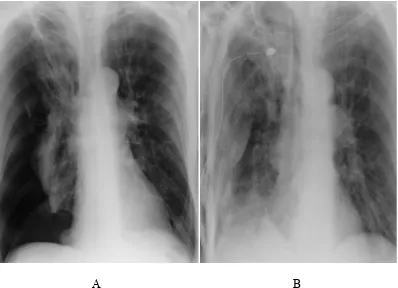
admission showed mild hypertension (150/80 mmHg), rhythmic heart beat of 110/min, normal breathing rate (24/min), oxygen saturation of 87% and normal body temperature (36.5oC). White blood cell count and C-reactive protein concentration were elevated to 15.5 Gpt/L and 228.80 mg/L, respectively. Chest radiograph revealed a right-sided large pneumothorax
without mediastinal shift (Figure 1A). Rupture of an
emphysematous bulla causing pneumothorax was suspected and a 24 Charrière chest tube was inserted after local anesthetic injection in the 4th intercostal space at anterior axillary line and attached to underwater
seal. The chest X-Ray taken afterwards demonstrated
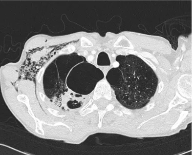
the incomplete expansion of the lung so consequently the drain was placed on suction (Figure 1B). CT scan on the fourth day exhibited the bullous emphysema, moderate subcutaneous emphysema, a 7 cm apical bulla
on the right side with an irregular ibrotic iniltration of
segment 1 of the right lung. On the left side, a 3.8 cm apical bulla was noticed (Figure 2).
Because of the persistent pneumothorax with air leak,
the patient was scheduled for the OR. Until now, no suspicion of a malignant disease was raised. Antero-lateral
thoracotomy was performed on the sixth post-tubing day.
At surgery the apical bulla was detected on segment 1, demonstrating a nonspeciic carniication of its base. The presence of air in the pleural cavity deines
the pneumothorax, which can be spontaneous, traumatic or iatrogenic. Spontaneous pneumothorax
(SP) can be classiied as primary or secondary.
The primary spontaneous pneumothorax (PSP) is common among young population especially thin, tall males without previous lung disease, whereas the secondary spontaneous pneumothorax (SSP) is often in the elderly with underlying parenchymal lung disease such as COPD, bullous emphysema, pulmonary tuberculosis and other pulmonary
infections, cystic ibrosis, idiopathic pulmonary
ibrosis, lymphangioleiomyomatosis etc.1,2 Primary
bronchogenic carcinoma or lung metastases have been sporadically described as a potential cause of SP, with an estimated rate of joint occurrence between 0.03 and 0.05% for primary bronchogenic carcinoma.3,4,5 Treatment of SP ranges from simple observation, needle aspiration, tube thoracostomy or chemical pleurodesis to video-assisted thoracoscopy or thoracotomy.
We report the case of a patient treated with thoracotomy for persistent SSP where unexpectedly a non-small cell
lung carcinoma of undeined histology was detected.
CASE ILLUSTRATION
A 66-year old male, heavy smoker with a previous
Vol. 22, No. 1, February 2013 Spontaneous pneumothorax related with lung carcinoma 55
A
B
Figure 1.A) Right sided pneumothorax with a slight mediastinal shift, B) post-tubing state
Therefore segmentectomy was performed. Furthermore another smaller bulla was observed on the right lower lobe, so wedge resection was carried out. Following this, partial pleurectomy completed the surgical procedure.
Chest drain was removed on the sixth postoperative day with patient’s course being uneventful. Hospital discharge was scheduled on the tenth day.
Surprisingly, histological examination revealed a low grade polymorphous and partially clear cell trabecular carcinoma (non-small cell cancer type) at the base of the bulla, with a maximal diameter
of 11 mm, resected in R0. A further histological
differentiation was not possible. No other metastases were detected and due to patient’s impaired lung-function and reduced clinical state no further treatment was performed.
DISCUSSION
Spontaneous pneumothorax remains a common pathology with an incidence between 18-28/100000 p.a. in males and 1.2 – 6/100000 p.a. in females.6 SSP is often from the end of the fourth decade due to progressive intrathoracic pathology in high risk population such tobacco smokers or COPD patients, resulting from a ruptured bleb or bulla into the visceral pleura.4 Lung cancer can also occur in the same high risk group.
SP as the irst manifestation of bronchogenic carcinoma
is extremely rare, and only 2% of all SP coincides with a malignant lung pathology, primary or secondary.3 Four possible mechanisms responsible for SP in lung carcinoma have been theorized. (1) The genesis of a bronchopleural communication secondary to tumor necrosis (e.g. after chemotherapy) and rupture into a bronchus or pleural cavity. (2) The spontaneous rupture of dilated alveoli or subpleural blebs distal to bronchial obstruction due to lung carcinoma. (3) The direct local
pleural iniltration by the neoplasm. (4) Coincidental
rupture of bullous emphysema either spontaneous or due to disturbance of the lung architecture caused by the neoplasm.3,4,5 Moreover, SP is also been described
in metastatic lung disease, mostly in afiliation with
metastatic sarcomas, germ cell tumors or lymphomas
with lung iniltration.5
In our patient, preoperative thorax CT scan didn’t raise any suspicion for an underlying malignant disease. In many cases of SSP secondary to lung carcinoma, diagnosis is late, delaying the proper and prompt treatment.
Mavridis, et al.
56 Med J Indones
Figure 2.Bilateral apical bullae with an irregular ibrotic iniltration of segment 1 of the right lung
(11.4%). The same authors found in 6.8%, of the 46
reviewed cases, carcinomas of undeined histology. In our case, our Pathology Division was unable to deine
the exact histology of the neoplasm at the ground of the resected bulla. Due to patients impaired status no other therapy was conducted.
To summarize, although SSP associated with lung carcinoma is a rare entity, we think that suspicion for malignancy should be raised in patients over 40 presenting with SP, especially in high risk patients such as smokers or patients with chronic bronchitis or emphysema.
Acknowledgments
Written informed consent was obtained from the patient for publication of this case report and any
accompanying images. A copy of the written consent
is available for review from the editorial ofice of
Medical Journal of Indonesia.
REFERENCES
1. Weissberg D, Rafaely Y. Pneumothorax. Chest.
2000;117:1279-89.
2. Kim SJ, Lee HS, Kim HS, et al. Outcome of video-assisted thoracoscopic surgery for spontaneous secondary pneumothorax. Koren J Thorac Cardiovasc Surg. 2011;44:225-8.
3. Vencevičius V, Cicėnas S. Spontaneous pneumothorax as a irst sign of pulmonary carcinoma. World Journal of
Surgical Oncology. 2009;7:57.
4. Steinhäuslin CA, Cuttat JF. Spontaneous pneumothorax. A
complication of lung cancer? Chest. 1985,88:709-13. 5. O’Connor BM, Ziegler P, Spaulding MB. Spontaneous
pneumothorax in small cell lung cancer. Chest. 1992;102:628-9.
6. Sousa C, Neves J, Sa N, et al. Spontaneous pneumothorax: