A rare case of cytomegalovirus papillitis in patient with immunodeficiency
Keywords: AIDS, cytomegalovirus, immunodeficiency, papillitis
pISSN: 0853-1773 • eISSN: 2252-8083 • http://dx.doi.org/10.13181/mji.v25i3.1364 • Med J Indones. 2016;25:190–4 • Received 26 Jan 2016 • Accepted 28 Aug 2016
Corresponding author: Made Susiyanti, [email protected]
Copyright @ 2016 Authors. This is an open access article distributed under the terms of the Creative Commons Attribution-NonCommercial 4.0 International License (http://creativecommons.org/licenses/by-nc/4.0/), which permits unrestricted non-commercial use, distribution, and reproduction in any medium, provided the original author and source are properly cited.
Dinda A. Devona, Made Susiyanti
Department of Ophthalmology, Faculty of Medicine, Universitas Indonesia, Cipto Mangunkusumo Kirana Hospital, Jakarta, Indonesia
C a s e Re p o r t
ABSTRAK
Seorang laki-laki berusia 26 tahun datang dengan penglihatan buram mendadak dan skotoma sentral pada mata kiri sejak 2 minggu sebelum ke rumah sakit. Visus hitung jari pada jarak 5 meter, tekanan intraokuler (TIO) normal, dan segmen anterior normal. Nervus optikus terlihat edema dengan eksudat dan perdarahan. Pasien terdiagnosis dengan AIDS dan memiliki hitung jenis CD4+ yang rendah sehingga dipikirkan kemungkinan infeksi oportunistik terkait infeksi HIV. Berdasarkan tampilan klinis dan pemeriksaan serologi, pasien diketahui menderita infeksi HSV akut sehingga diberikan asiklovir. Visus semakin memburuk menjadi no light perception sehingga pada pasien kemudian dikerjakan aqueous tap dan didapatkan DNA CMV positif. Papilitis CMV merupakan manifestasi tidak umum dari retinitis CMV. Pemeriksaan PCR dari aqueous atau vitreous tap perlu dilakukan sambil menunggu hasil pemeriksaan serologi, terutama untuk kasus yang memburuk dengan cepat. Dengan demikian, dokter dapat memberikan tata laksana yang sesuai dan cepat untuk mencegah kebutaan pada pasien.
ABSTRACT
Cytomegalovirus (CMV) papillitis is defined as greater than 270 degrees of disc edema/blurring of the disc margins as seen on direct examination and on color fundus photographs caused by CMV.1
CMV ocular infection will develop in 12% to 46% of patients with acquired immune deficiency
syn-drome(AIDS) in their lifetime. Of those patients
with AIDS in whom CMV retinitis develops, CMV papillitis reportedly develops in up to 4% as well.
Lestari2 reported the prevalence of CMV retinitis
in Cipto Mangunkusumo Hospital (CMH) is 5.8%, there is no papil involvement reported in those cases. The majority of these patients have CD4 counts of less than 50 cells/mm3.1-3
Diagnosis of CMV papillitis in the setting of human immunodeficiency virus (HIV)/AIDS is essential-ly clinical, based on the features just described. In CMV papillitis, the optic nerve head is edematous with focal hemorrhages, and an afferent pupil-lary defect usually is present. Polymerase chain reaction (PCR)-based analysis of the aqueous or
vitreous samples may provide critical diagnos-tic information of high sensitivity and specificity that allow the clinician to differentiate CMV from other herpetic causes of necrotizing retinitis and from toxoplasmic retinochoroiditis in immuno-compromised patients with atypical lesions.1,3 In
this case report, we demonstrated a rare case of ocular CMV infection that manifests as CMV papil-litis in HIV/AIDS.
CASE REPORT
A male, 26 years old, came to Cipto Mangunkusu-mo Hospital, Jakarta on January 2014 complain-ing sudden blurred vision in left eye and central scotoma since two weeks before he was admitted. It was not red, it did not swell, and it was pain-less. Patient did not get any treatment for his eye before. There was no history of spectacles usage, hypertension, and diabetes mellitus. In the past medical history, this patient had been diagnosed with pulmonary tuberculosis, cryptococcal men-ingitis, and positive HIV test result. From social history, patient was a transmigrant from Suma-tra Island. There was history of promiscuity with men and and intravenous drug user (IVDU).
Ophthalmological examination showed left eye’s visual acuity was 5/60, normal intraocular pres-sure (IOP), clear anterior segment, good right
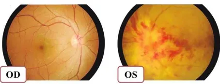
reflex with positive relative afferent pupillary defect (RAPD), and clear lens. In the posterior segment we found few cells in vitreous, and papil was hyperemic covered by exudates and peripap-illary hemorrhages. There were flame-shaped hemorrhages, with turtous vein, and macular re-flex hard to be evaluated. Right eye was in normal condition. We assessed this patient with specific opportunistic infection with several differential diagnosis including CMV, herpes simplex virus (HSV), cryptococcus, toxoplasmosis, and syphilis infection (Figure 1 and Figure 2).
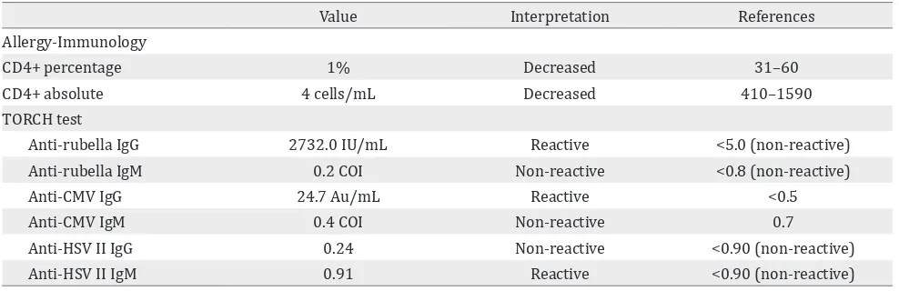
At the second and the third week after initial visit, the eye condition was getting worse, eventually leading to no light perception. Ancillary test re-vealed some abnormal result as shown in the Ta-ble 1. Based on clinical and laboratory results, we considered an active HSV infection, so we consult-ed the patient to Department of Internal Mconsult-edicine and the patient got 800 mg acyclovir four times daily (Figure 3).
Due to worsening condition and confusing diag-nosis, we performed PCR examination from
aque-Figure 1. Normal funduscopy (right); hyperemic papil covered by exudates and peripapillary hemorrhages, flame-shaped hemorrhages (left). At this intital visit, the visual acuity of the left eye is 5/60
ous tap to confirm the diagnosis. It was revealed that the patient had a positive CMV deoxyribo-nucleic acid (DNA). Therefore, we consulted to Department of Internal Medicine for a change to specific anti-CMV medication. The patient then received 900 mg valganciclovir twice a day for three weeks.
During the follow-up, five weeks after the first visit, the patient developed immune recovery uveitis with raising intraocular pressure up to 40 mmHg. He complained of pain of the left eye, accompanied with conjunctival and cilliary in-jection, the corneal edema and large keratitic precipitates were also noted. In anterior cham-ber, we found massive cells (+4) and flare (+2). The lenses were clear and the posterior seg-ment was still the same. The patient received anti-glaucoma medication and topical steroid (Figure 4).
In seventh week after the initial visit, patient came with subsided peripapillary hemorrhages and exudates after administration of oral ganci-clovir 900 mg twice daily in fourth week. This condition showed a good response to valganci-clovir treatment. Based on the clinical manifes-tation, we diagnosed this patient with CMV pap-illitis (Table 1).
Patient has been treated with highly-active an-ti-retroviral therapy (HAART) medication and also the anti-tuberculosis agent. HAART which consists of evafirenz, lamivudine, tenofovir diso-proxil fumarate.
DISCUSSION
Opportunistic infections (OIs), which have been defined as infections that are more frequent or more severe because of immunosuppression in HIV-infected persons. It is important to recognize
Figure 3. Immune recovery uveitis. Left eye shows conjunc-tival and ciliary injection, deep anterior chamber. Yellow ar-row shows massive cells (+4) and flare (+2) in the anterior chamber. Red arrow shows keratitic precipitate
Figure 4. Left eye was showing subsided peripapillary hem-orrhages and exudates after ad-ministration of oral valganci-clovir 2 x 900 mg in third week
Value Interpretation References
Allergy-Immunology
CD4+ percentage 1% Decreased 31–60
CD4+ absolute 4 cells/mL Decreased 410–1590
TORCH test
Anti-rubella IgG 2732.0 IU/mL Reactive <5.0 (non-reactive)
Anti-rubella IgM 0.2 COI Non-reactive <0.8 (non-reactive)
Anti-CMV IgG 24.7 Au/mL Reactive <0.5
Anti-CMV IgM 0.4 COI Non-reactive 0.7
Anti-HSV II IgG 0.24 Non-reactive <0.90 (non-reactive)
Anti-HSV II IgM 0.91 Reactive <0.90 (non-reactive)
Table 1. Laboratory examination of clinical importance
The laboratory results showed an active HSV type II infection so the patient got treated by acyclovir 5 x 800 mg. By the time, we observed that the ocular manifestation was not improved with the treatment, so we considered another etiology. Based on the literature, the most common opportunistic infection on AIDS with very low CD4+ count (<200 cells/mL) is CMV infection. To confirm the diagnosis, we did the aqueous tap and PCR examination and revealed a positive result CMV infection. TORCH: Toxo-plasma, rubella, cytomegalovirus, herpes simplex; IgG: immunoglobulin G; IgM: immunoglobulin M; anti CMV: anti
that the relationship between OIs and HIV infec-tion is bidirecinfec-tional.
Human immunodeficiency virus can cause sys-temic or organ diseases. Several syssys-temic OIs
in-clude Mycobacterium tuberculosis infection, Cryp-tococcus infection, cytomegalovirus disease,
toxo-plasmosis infection. At the initial visit, our patient was already diagnosed with pulmonary tubercu-losis and cryptococcal meningitis, still ongoing treatment. With the presenting systemic OIs, it is likely that this patient might have a specific organ disease.4,5
Iqbal6stated an association between CD4+ cells
count with ocular complication of HIV with the commonest infection is CMV. Cullen et al7 stated
that neuro-ophthalmic manifestations of HIV tend to present at an advanced stage of the disease when CD4 cell counts are depleted below 200 cells/µl. In HIV-infected patients, opportunistic infections such as cytomegalovirus, toxoplasmo-sis, syphilis, and tuberculosis are by far the most common causes of optic nerve disorders.
In our cases, we found that patient was complain-ing sudden blurred vision of his left eye, diag-nosed as AIDS since three months before came to this hospital (October 2013) with initial symp-toms were loss of consciousness due to crypto-coccal meningitis, starting HAART medication two months later (November 2013) and initial CD4+ count at first visit was 4 cells/mL (n: 410-1,590 cells/mL). Initial presentation showed vi-sual acuity 5/60, normal IOP, quiet eye with nor-mal anterior segment, positive RAPD, and pos-terior segment manifestation that is edematous papil, peripapillary hemorrhages and exudates, and hemorrhages in retina. From this condition, our primary consideration was opportunistic in-fection with differential diagnosis caused by CMV, HSV, Cryptococcus, Toxoplasma, and syphilis. The
laboratory examination and PCR testing show positive tendency to CMV infection. Based on this result we gave specific anti-CMV infection, val-ganciclovir 900 mg twice daily.
Cytomegalovirus papillitis has been described in immunocompromised patients and is usually a rapidly blinding disease.8 Many studies reported
the incidence of CMV papilitis found less than 10% in CMV retinitis cases. CMV retinitis ac-counts for 75–85% of CMV disease and over 90%
of blindness in AIDS patients. Freeman et al9 re
-port papillitis is present in up to 4% of patients with CMV retinitis. Gross et al10 noted 4.1%
pa-tients with CMV retinitis also had CMV papillitis at initial presentation. Similarly, Rosecan et al11
noted optic nerve involvement in 8.3% eyes with CMV retinitis. Roarty et al12also reported a patient
with severe CMV papillitis and visual acuity of no light perception vision, and Palestine et al13 noted
CMV papillitis in 7% eyes with CMV retinitis with no light perception vision upon initial presenta-tion. Until now, there is no report of isolated CMV papillitis cases in without retinal manifestation in AIDS patient.
Successful management of active CMV retinitis reported by Figueiredo14 in an active CMV
reti-nitis infection. In his report the patient had good visual outcome, from hand movement with good projection to 5/12 after induction doses of oral valganciclovir.
The Centers for Disease Control and Prevention (CDC), the National Institutes of Health, and the HIV Medicine Association of the Infectious Dis-eases Society of America15 give recommendation
for treating CMV retinitis. For sight threatening lesions which is adjacent to the optic nerve or fovea, the recommended initial therapy com-prised intravitreal injections of ganciclovir (2 mg/injection) or foscarnet (2.4 mg/injection) for 1–4 doses over a period of 7–10 days to pro-vide higher intraocular levels of drug and faster control of the infection until steady state intra-ocular ganciclovir concentrations are achieved. This initial therapy might be accompanied by valganciclovir 900 mg twice daily for two until three weeks then followed by once daily doses. In our case, he given a systemic valganciclovir 900 mg twice daily in first three weeks, to con-trol the presumably active CMV infection and to prevent the CMV involvement to the contralat-eral eye and also to improve patient’s systemic condition. Although the guideline of CMV papil-litis’ management has not been established yet, this CDC recommendation is applicable in this case.
or vitreous tap should be performed earlier while waiting for the serological testing result, espe-cially in doubtful cases which severity progress rapidly. An appropriate diagnosis and manage-ment can be established and performed earlier to prevent the irreversible visual loss.
Conflicts of interest
The authors affirm there is no conflict of interest in this study.
REFERENCES
1. Patel SS, Rutzen AR, Marx JL, Thach AB, Chong LP, Rao NA. Cytomegalovirus papilltis in patients with acquired immune deficiency syndrome. Visual prognosis of pa-tients treated with ganciclovir and/or foscarnet. Oph-thalmology. 1996;103(9):1476–82.
2. Lestari YD. Prevalensi manifestasi okular human im-munodeficiency virus/acquired imim-munodeficiency syn-drome di DKI Jakarta. [Thesis]. Jakarta: Univesitas Indo-nesia; 2009. p.3–5. Indonesian.
3. Moorthy RS, Rao PK, Read RW, Gelder RN, Vitale AT, Bo-daghi B, et al. Infectious ocular inflammatory diseases.
In: Skuta GL, Cantor LB, Weiss JS, editors. Intraocular inflamation and uveitis. San Fransisco: American Acad-emy of Ophthalmology; 2011-2012. p. 204–7.
4. Panel on Opportunistic Infections in HIV-Infected Adults and Adolescents. Guidelines for the preven-tion and treatment of opportunistic infecpreven-tions in HIV-infected adults and adolescents: recommendations from the Centers for Disease Control and Prevention, the National Institutes of Health, and the HIV Medi-cine Association of the Infectious Diseases Society of America. Available at http://aidsinfo.nih.gov/content-files/lvguidelines/adult_oi.pdf. Accessed (February 13, 2014). p N1–8.
5. Bentwich Z. Concurrent infections that rise the HIV viral load. J HIV Ther. 2003;8(3):72–5.
6. Iqbal TB. HIV-related eye condition. In: Menon A, Kama-rulzaman A, editors. Is it HIV? A hand-book for health
care provider Thailand: The Australian Society for HIV Medicine (ASHM);KP Marketing.Darlinghurst: 2009. p. 46–51.
7. Cullen C, Matlala B, Laher F, Pienaar A. Successful treat-ment of bilateral visual loss caused by idiopathic optic neuritis in an HIV-infected patient. The Southern Afri-can Journal of HIV Medicine 2011;12(4).
8. De Silva SR, Chohan G, Jones D, Hu M. Cytomegalovirus papillitis in an immunocompetent patient. J Neurooph-thalmol. 2008;28(2):126–7.
9. Freeman W, Lerner CW, Mines JA, Lash RS, Nadel AJ, Starr MB. A prospective study of the ophthalmologic findings in the acquired immune deficiency syndrome. Am J Ophthalmol. 1984;97(2):133–42.
10. Gross JG, Sadun AA, Wiley CA, Freeman WR. Severe vi-sual loss related to isolated peripapillary retinal and optic nerve head cytomegalovirus infection. Am J Oph-thalmol. 1989;108(6):691–8.
11. Rosecan LR, Stahl-Bayliss CM, Kalman CM, Laskin OL. Antiviral therapy for cytomegalovirus retinitis in AIDS with dihydroxy propoxymethyl guanine. Am J Ophthal-mol. 1986;101(4):405–18.
12. Roarty J, Fisher EJ, Nussbaum JJ. Long-term visual mor-bidity of cytomegalovirus retinitis in patients with ac-quired immune deficiency syndrome. Ophthalmology. 1993;100(11):1685–8.
13. Palestine AG, Stevens Jr G, Lane HC, Masur H, Fujikawa LS, Nussenblatt RB, et al. Treatment of cytomegalovirus
retinitis with dihydroxy propoxymethyl guanine. Am J Ophthalmol. 1986;101(1):95–101.
14. Figueiredo L, Rothwell R, Bilhoto M, Varandas R, Fon-seca S. Immune recovery uveitis masked as an endog-enous endophthalmitis in a patient with active CMV retinitis. Case Rep Ophthalmol Med. 2013;2013: Article ID 462968.p1–4