Prognostic factors of neuroinlammation and oxidative stress in brain
injury patients at Cipto Mangunkusumo Hospital Jakarta
Renindra A. Aman,1 Syaiful Ichwan,1 Alida Harahap,2 Yusuf Wibisono,1 Nia Yuliatri,1 Mohammad A. Aulia,1 Hans-Joachim Freisleben3
1 Department of Neurosurgery, Faculty of Medicine, Universitas Indonesia, Jakarta, Indonesia
2 Department of Clinical Pathology, Faculty of Medicine, Universitas Indonesia, Jakarta, Indonesia 3Medical Research Unit, Faculty of Medicine, Universitas Indonesia, Jakarta, Indonesia
Abstrak
Latar belakang: Penelitian ini bertujuan untuk menentukan hubungan antara neuroinlamasi dan stress oksidatif dengan
prognosis pasien cedera otak dan hubungan antara tindakan bedah saraf dengan kondisi neuroinlamasi dan stress oksidatif.
Metode: Desain penelitian adalah studi observasional prospektif pada 40 pasien cedera otak yang dioperasi. Pengukuran kadar IL-6, asam urat, MDA, antibodi NR2A dan GSH serum pasien cedera otak dilakukan sebelum dan sesudah operasi. Dilakukan pula penilaian hubungan seluruh variabel tersebut dengan GCS, GOS dan tindakan bedah saraf.
Hasil: Kadar IL6 pascaoperasi cenderung menurun bila dibandingkan dengan kadar IL6 praoperasi, dengan rerata penurunan -190,61 pg/mL. Kadar IL6 pascaoperasi berhubungan bermakna dengan GCS hari ke-7 (nilai p = 0,006), dengan OR 24. Kadar IL6 pascaoperasi berhubungan bermakna dengan GOS bulan ke-3 (nilai p = 0,016) dengan OR 11,6. Kadar asam urat pascaoperasi cenderung menurun bila dibandingkan dengan kadar praoperasi, dengan rerata penurunan -0,26 mg/dL. Terdapat perbedaan bermakna antara rerata kadar asam urat pascaoperasi pasien dengan GCS hari ke-7 ≤ 8 sebesar 4,16 mg/dL dengan rerata kadar asam urat pascaoperasi pada kelompok pasien dengan GCS hari ke-7 > 8 sebesar 2,71 mg/dL (nilai p = 0,042). Kadar MDA pasca operasi cenderung menurun bila dibandingkan dengan kadar praoperasi, dengan rerata penurunan -0,08 mmol/mL. Tidak ada hubungan bermakna antara kadar MDA dengan GCS dan GOS. Tidak ditemukan hubungan bermakna antara kadar antibodi NR2A dan GSH dengan GCS, GOS dan tindakan bedah saraf. Dari hasil analisis multivariat, variabel terkuat yang berhubungan dengan GCS dan GOS adalah IL-6.
Kesimpulan: Proses neuroinlamasi dan stres oksidatif memiliki nilai prognostik pada pasien cedera otak.
Tindakan bedah saraf dapat mengurangi proses neuroinlamasi. (Med J Indones. 2012;21:152-9)
Abstract
Background: This study aims to determine the association between neuroinlammation and oxidative stress with prognosis of brain injury patients and the association between neurosurgical procedure with neuroinlammation and oxidative stress condititons.
Methods: The study design is a prospective observation of 40 brain injury patients who underwent surgery. IL-6, uric acid, MDA, NR2A antibodies and GSH serum level of pre- and 1 day post-operation on brain injury patients were measured, and their association with GCS, GOS and neurosurgical procedures were analyzed.
Results: The post-operative IL-6 serum level showed a downward trend compared to pre-operative value (mean decrease: -190.61 pg/mL). The post-operative IL-6 level was signiicantly associated with GCS 7 days post-operation (p = 0.006), with OR 24. The post-operative IL-6 serum level was signiicantly associated with GOS 3 months post-trauma (p = 0.016) with OR 11.6. The post-operative uric acid serum level showed a downward trend compared to pre-operative value (mean decrease: -0.26 mg/dL). There was a signiicant difference between the mean value of post-operative uric acid serum level in patients with 7 days post-trauma with GCS ≤ 8 (mean: 4.16 mg/dL) and GCS > 8 (mean: 2.71 mg/dL), (p = 0.042). The
post-operative MDA serum level showed a downward trend compared to pre-operative value (mean decrease: -0.08 nmol/ mL). There is no signiicant association between MDA serum level, GCS and GOS and no signiicant association of NR2A antibody and GSH serum level with GCS, GOS and neurosurgical procedure. From the multivariate analysis, the most important neuroinlammatory variable associated with GCS and GOS is IL-6.
Conclusion: Neuroinlammation and oxidative stress may have prognostic values in brain-injured patients, in particular IL-6. Neurosurgical procedures may decrease the neuroinlammation process (Med J Indones. 2012;21:152-9)
Keywords: Neuroinlammation, oxidative stress, traumatic brain injury, neurosurgical procedures, interleukin 6, uric acid, malondialdehyde, NR2A antibody, glutathione
Correspondence email to: [email protected]
In developed countries such as U.S.A, mortality due to brain injury reaches 43% of all mortality from
trauma.2 During the last decades, 1.5 to 2.0 million
people suffered from brain injuries each year; about 250,000 of these patients required hospitalization and about 52,000 died. In 1985, prolonged post-traumatic In many developing and developed countries, trauma is
care became an economic burden, spending 4.5 billion USD in America, and currently causes health-care expenses of 11 billion USD every year in 11 Southeast Asian countries. The ”World report on trafic injury prevention” in the 2004 WHO publication titled ”Violence and injury prevention section” stated that by 2020, road trafic accidents will occupy the third rank in public health burden worldwide.3
Traumatic brain injury consists of primary injury related to biomechanical trauma and secondary damage. Secondary brain damage is triggered by post-traumatic hypoxia in brain tissue, which induces a cascade of destructive biochemical reactions on the cellular level, leading to additional mortality of nerve cells.4,5 This cascade of reactions comprises free radicals release, neuroinlammation and apoptosis, which are considered to worsen the primary brain injury and to inluence the outcome of patients.
Secondary brain damage as a result of neuroinlammation is due to the release of inlammatory mediators, especially interleukin 6 (IL-6), the primary regulator of neuroinlammatory reactions. High levels of IL-6 in acute phase of brain injury is allegedly associated with the degree of brain damage,4,5 including blood-brain barrier damage, which may lead to blood-brain tissue ischemia. Due to post-brain injury blood-brain barrier damage, it is assumed that IL-6 will be increased in the serum of patients with severe brain injury.6
Increased amount of free radicals that exceed the endogenous antioxidant capacity lead to oxidative stress, which can cause destruction of lipid, protein and DNA.7
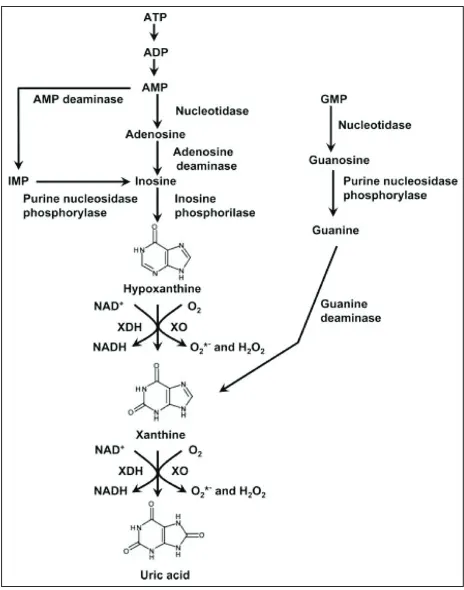
Uric acid is the product of endogenous purine breakdown from reactions catalyzed by xanthine oxidase. Increased activity of xanthine oxidase occurs in ischemic conditions and in response to the increased amounts of pro-inlammatory cytokines. A byproduct of the reaction catalyzed by xanthine oxidase is superoxide radical, which aggravates oxidative stress (Figures 1 and 2). Serum uric acid level measurement is expected to represent xanthine oxidase activity.8
Increased reactive oxygen species (ROS) as the product of serial enzymatic reactions or due to mitochondrial dysfunction in secondary brain injury can induce lipid peroxidation (LPO) and lead to post-traumatic neuronal degeneration. Because of the dificulty to measure free radicals directly, alternative method to assess their involvement in brain injury is by the LPO byproducts measurement, such as malondialdehyde (MDA).9
Glutathione (GSH), a tripeptide thiol, is an important substance in the defense mechanisms against free radicals and hydroperoxides. GSH functions in the storage and transportation of cysteine, in maintaining protein and thiol reduction status, and in protecting cells from toxic components such as ROS, drugs or heavy metals. GSH acts as an endogenous antioxidant.10,11
Ischemic condition may stimulate cascades of neurotoxic reactions, in which excitated
N-methyl-d-Figure 1.Reactions catalyzed by xanthine oxidase
aspartate (NMDA) receptor acts as the main regulator of neural cell membranes and may cause biochemical changes in brain tissues, blood brain barriers and vessels. Calcium (Ca2+) inlux through the channels of
NMDAR may potentiate mitochondrial depolarisation and increase superoxide radical production from mitochondria. Blood-brain barrier damage and activation of thrombin-activated serine protease can make a gap in synaptic NMDA receptors. Increased concentrations of peptides produced from a speciic NMDAR gap in the brain can be released into the systemic blood circulation, e.g. NR2A. The latter may act as antigen initiating the immune response that leads to the emergence of autoantibodies in the blood. NR2A antibody measurement was used to assess NMDAR activation and could thus have prognostic value in brain injury patients,9,12 similar to before-mentioned IL-6 (representing neuroinlammation) and uric acid, MDA, and GSH serum level (representing oxidative stress).
For assessment of brain injury patient awareness in the acute phase, the GCS scoring system (Glasgow Coma Scale) is universally used. Patients are categorized as in ’coma’ if the GCS score is under 8 (GCS < 8). For assessment of the patients’ outcome, the GOS scoring system (Glasgow Outcome Scale) is generally used. The GCS 7 days post-trauma and GOS at 1 month and 3 months post-trauma are the variables that represent the patient’s prognosis.
The neurosurgical procedures that are considered to be able to stop or slow down the secondary brain damage are decompression, either by decompressive craniectomy or hematoma evacuation.
METHODS
This prospective observational study was conducted on 40 patients with brain injury who underwent surgery.13,14 IL-6, uric acid, MDA, NR2A antibodies and GSH serum levels in post-traumatic and pre-operative patients (initial values) and 1 day after brain surgery (post-operative values) were measured. The prognostic association of the initial (pre-operative) and one day post-operative serum levels of these neuroinlammation and oxidative stress parameters with the initial GCS, GCS 7 days post-trauma, GOS 1 month and 3 months post-trauma were determined. Furthermore, the association between the neurosurgical procedures and the postoperative condition of the patients was analyzed.
IL-6 and NR2A antibodies serum levels were measured by ELISA. Uric acid serum level was measured by automated blood chemistry testing tool in the Clinical Pathology Laboratory of Cipto Mangunkusumo Hospital. The measurement of serum levels of MDA and GSH was done in collaboration with the laboratory of the Department of Biochemistry and Molecular Biology, Faculty of Medicine, Universitas Indonesia. Data was collected from August 2010 to April 2011.
RESULTS
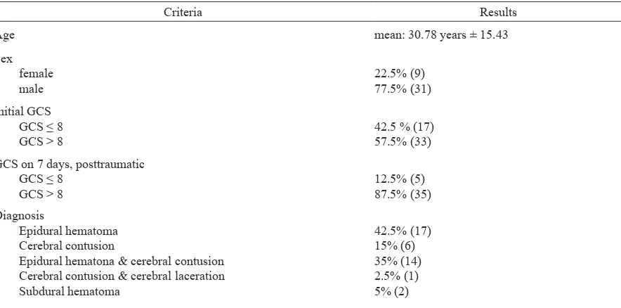
Forty subjects were categorized and divided according to sociodemographic criteria, consciousness status (GCS score), diagnosis, neurosurgical procedure, quality of life (GOS score), and IL-6, uric acid, MDA, NR2A antibody and GSH serum levels (Table 1).
Criteria Results
Age mean: 30.78 years ± 15.43
Sex female
male 22.5% (9)77.5% (31)
Initial GCS GCS ≤ 8
GCS > 8 42.5 % (17)57.5% (33)
GCS on 7 days, posttraumatic GCS ≤ 8
GCS > 8 12.5% (5)87.5% (35)
Diagnosis
Epidural hematoma Cerebral contusion
Epidural hematona & cerebral contusion Cerebral contusion & cerebral laceration Subdural hematoma
42.5% (17) 15% (6) 35% (14) 2.5% (1) 5% (2)
Neurosurgical procedure
Hematoma evacuation craniotomy Decompressive craniectomy
Depressed fracture correction & debridement
80% (32) 17.5% (7) 2.5% (1) GOS 1 month, posttraumatic
unfavorable
favorable 37.5% (15)62.5% (25)
GOS 3 months, posttraumatic unfavorable
favorable 15% (6)85% (34)
Initial IL-6 serum levels mean: 269.93pg/mL± 521.76median: 76.74 pg/mL minimum: 0.08 pg/mL maximum: 2734.65 pg/mL Initial IL-6 serum levels
≤100 pg/mL
>100 pg/mL 60% (24)40% (16)
Postoperative IL-6 serum levels mean: 79.31 pg/mL± 92.77
median: 35.55 pg/mL minimum: 5.11 pg/mL maximum: 353.19 pg/mL Postoperative IL-6 serumlevels
≤100 pg/mL
>100 pg/mL 77.5% (31)22.5% (9)
Initial uric acid serum levels
mean: 3.15mg/dL± 1.18 median: 2.90mg/dL minimum: 1.6 mg/dL maximum: 5.6 mg/dL
Postoperative uric acid serum levels
mean: 2.89 mg/dL± 1.16 median: 2.75 mg/dL minimum: 0.8 mg/dL maximum: 5.7 mg/dL
Initial MDA serum levels
mean: 0.47 nmol/L ± 0.24 median: 0.49 nmol/L minimum: 0.12 nmol/L maximum: 1.15 nmol/L
Postoperative MDA serum levels mean: 0.39 nmol/L ± 0.21median: 0.33 nmol/L minimum: 0.09 nMol/L maximum: 0.89 nmol/L
Initial NR2A antibody serum levels mean: 4.48 ng/mL± 3.24median: 4.29 ng/mL minimum: 0.1 ng/mL maximum: 11.07 ng/mL Initial NR2A antibody serum levels
≤ 2 ng/mL
< 2 ng/mL 67.5% (27)32.5% (13)
Postoperative NR2A antibody serum levels mean: 3.78 ng/mL± 2.74 median: 2.95 ng/mL minimum: 0.09 ng/mL maximum: 11.07 ng/mL Postoperative NR2A antibody serum levels
≤ 2 ng/mL
< 2 ng/mL 77.5% (31)22.5% (9)
Initial GSH serum levels
Initial GCS P value
(α < 0.05) PrevalenceRate
≤ 8 > 8
Initial IL-6 serum level
> 100 pg/mL (75%)12 (25%)4 16
≤ 100 pg/mL (20.8%)5 (72.9%)19 24 0.001 11.4
Total (42.5%)17 (57%)23 40
Mean of initial uric acid serum
levels 3.5 mg/dLN=17 2.89 mg/dLN=23 0.142
Median of initial MDA serum
levels 0.54 mmol/LN = 17 0.38 mmol/LN = 23 0.106
Initial NR2A antibody serum levels
≥ 2ng/mL (40.7%)11 (59.3%)16 27 1.0
< 2ng/mL (46.2%)6 (53.8%)7 13
Total (42.5%)17 (57.5%)23
Median of initial GSH serum
levels 0.59 mmol/LN = 17 0.52 mmol/LN = 23 0.511
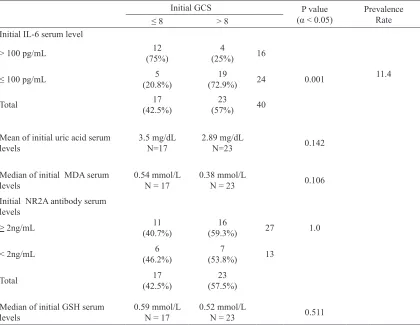
Table 2. Association between initial serum levels and initial GCS (pre-operative)
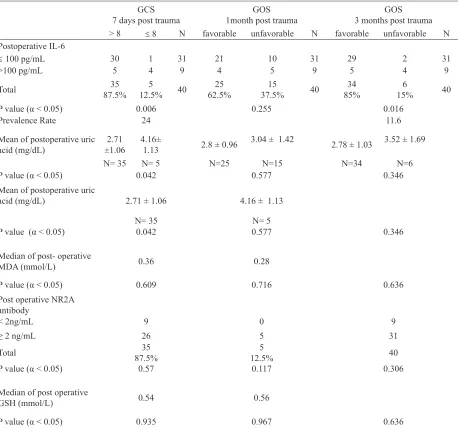
There is a signiicant association between the initial IL-6 serum level and the initial GCS score, p value: 0.001 and prevalence rate 11.4 (Table 2). The post-operative IL-6 serum level showed a downward trend compared to pre-operative value with a mean decrease of -190.61 pg/mL, p value: 0.001 (Table 3). The post-operative IL-6 serum level was signiicantly associated with GCS 7 days post-surgery (p = 0.006) with OR 24, meaning that patients with post-surgical levels of IL-6 < 100 pg/mL had a 24-times higher chance of getting the 7-days post-traumatic GCS > 8 (Table 4).
The post-operative IL-6 serum level was signiicantly associated with GOS 3 months post-trauma (p value: 0.016) with OR 11.6, meaning that patients with post-operative IL-6 levels < 100 pg/mL had a 11.6-times higher chance of reaching the 3 months post-traumatic ’favorable’ GOS (Table 4).
The post-operative uric acid serum level had a downward trend compared to pre-operative value, with a mean decrease of -0.26 mg/dL (Table 3). There was a signiicant difference between the mean post-operative uric acid serum levels in patients with 7-days post-trauma GCS ≤ 8 (mean: 4.16 mg/dL) and in patients
with 7-days post-trauma GCS > 8 (mean: 2.71 mg/dL; p = 0.042; Table 4).
The post-operative MDA serum levels had a downward trend compared to pre-operative value, with a mean decrease of -0.08 nmol/mL (p = 0.004; Table 3).
There was no signiicant association between surgery and GSH or NR2A antibody serum level (Table 3) and no signiicant association between MDA, GSH and NR2A antibody serum levels and GCS or GOS (Table 4).
From the multivariate analysis of the association between all variables with initial GCS, the neuroinlammation variable IL-6 was found to be related with the initial GCS (p value: 0.003) with OR 9.8 (95% CI: 2.1 – 45.1).
Variables Median Standard Deviation
P value (α < 0.05)
Median of initial IL-6 76.74 pg/mL 0.0001
Median of postoperative IL-6 35.55 pg/mL
Mean of initial uric acid levels 3.15 mg/dL 1.18 0.143 Mean of postoperative urid acid 2.89 mg/dL 1.16
Median of initial MDA levels 0.39 mmol/L 0.004
Median of postoperative MDA 0.33 mmol/L
Median of initial NR2A antibody levels 4.29 ng/mL 0.125 Median of postoperative NR2A antibody 2.95 ng/mL
Median of initial GSH levels 0.55 mmol/L 1.0
Median of postoperativeGSH 0.55 mmol/L Table 3. Pre- and post-operative differences of serum levels
GCS
7 days post trauma 1month post traumaGOS 3 months post traumaGOS > 8 ≤ 8 N favorable unfavorable N favorable unfavorable N Postoperative IL-6
≤ 100 pg/mL 30 1 31 21 10 31 29 2 31
>100 pg/mL 5 4 9 4 5 9 5 4 9
Total 87.5%35 12.5%5 40 62.5%25 37.5%15 40 85%34 15%6 40
P value (α < 0.05) 0.006 0.255 0.016
Prevalence Rate 24 11.6
Mean of postoperative uric
acid (mg/dL) ±1.062.71 4.16± 1.13 2.8 ± 0.96 3.04 ± 1.42 2.78 ± 1.03 3.52 ± 1.69
N= 35 N= 5 N=25 N=15 N=34 N=6
P value (α < 0.05) 0.042 0.577 0.346
Mean of postoperative uric
acid (mg/dL) 2.71 ± 1.06 4.16 ± 1.13
N= 35 N= 5
P value (α < 0.05) 0.042 0.577 0.346
Median of post- operative
MDA (mmol/L) 0.36 0.28
P value (α < 0.05) 0.609 0.716 0.636
Post operative NR2A antibody
< 2ng/mL 9 0 9
≥ 2 ng/mL 26 5 31
Total 87.5%35 12.5%5 40
P value (α < 0.05) 0.57 0.117 0.306
Median of post operative
GSH (mmol/L) 0.54 0.56
P value (α < 0.05) 0.935 0.967 0.636
Multivariate analysis of relations between all variables and GOS 3 months post-trauma showed that the neuroinlammation variable IL-6 was related with GOS 3 months post-trauma (p value: 0.031), with OR 10.6 (95% CI: 1.4 – 81.6), meaning that patients with post-operative IL-6 levels < 100 pg/mL had a 10.6-times better chance to achieve 3 months post-traumatic ’favorable’ GOS as compared to patients with a post-operative IL-6 level > 100 pg/mL.
DISCUSSION
In our study, the initial IL-6 level was signiicantly associated with initial GCS at a prevalence rate of 11.4. In other words, patients with an initial IL-6 level of 100 pg/mL or below had an 11.4-times higher chance of having an initial GCS over 8. This is consistent with the study of Woicienchowxky et al4 stating that initial IL-6 plasma levels are associated with brain injury severity. The higher the initial IL-6 serum level, the higher the patient’s risk of getting impaired consciousness (low GCS score). High IL-6 serum level of patients with pure brain injury showed neuroinlammation and damage of the blood brain barrier, which led to the release of IL-6 into systemic blood circulation.4,15
After surgery, IL-6 serum level had a downward trend. The post-operative IL-6 level was signiicantly associated with GCS 7 days post-surgery, meaning that surgery may reduce neuroinlammatory processes. Neurosurgical procedures aim to reduce intracranial pressure. In accordance with the study of Hergenroeder et al,16 high intracranial pressure is correlated with increased levels of IL-6. Intracranial pressure release after neurosurgical procedure can be ascribed to improvement of neuroinlammatory processes.16
After 3 months of follow up, lower post-operative IL-6 levels (under 100 pg/mL) have 11.6-times higher chance of reaching the 3 months post-trauma GOS ’favorable’.
Although there was no association between the initial levels of uric acid and the initial GCS score, post-operative uric acid serum level decreased signiicantly on the seventh post-traumatic day. This inding is consistent with the results of Kinuta et al17 that xanthine oxidase is located mainly in endothelial cells (3.7-fold compared to the cerebral cortex). The capillary is probably the primary site of oxygen delivery and free radical reactions, which are induced by the xanthine-metabolizing
system.17 Increased capillary permeability caused by
endothelial injury leads to brain edema (secondary brain damage).2,3,17-19 Neurosurgery procedures - conducted to decrease the elevated intracranial pressure - may also reduce intracranial oxidative stress.
Even though there is no signiicant difference in the mean value of uric acid levels 3 months post-trauma between patient groups with ’unfavorable’ and ’favorable’ GOS, there is a difference of 0.6 in the standard deviation. According to Sloan et al20 in practical guidelines for assessing the statistical signiicance in changes of health-related quality of life in a clinical trials, it may be assumed that there is still the possibility of clinical signiicance with a larger number of samples.20
There is no signiicant association between MDA serum levels and both, GCS and GOS. The reason might be that our measurements were not carried out in serial order to ind the highestMDA levels. In several studies, the measurement of MDA was performed in serial order in the irst 48 hours revealed high MDA levels in conditions with increased intracranial pressure.9,21-23
Nevertheless, the signiicant decrease of post-surgical MDA levels in our study compared to initial MDA levels showed that neurosurgical procedures may also reduce the intracranial oxidative stress.
The autoantibody reaction to the NMDA receptor (NR2A)24,25 is a humoral immune reaction. Dambinova et al26 measured NR2A antibody levels in serial order within the irst 72 hours, found that the peak of increase was at the irst 10 to 12 hours after ischemic cerebral vascular attack.26 Although the signiicant association between NR2A antibody serum levels and GCS, GOS and surgery were not found, there is still a possibility to obtain different results if NR2A antibody serum level is measured in serial order.
Highly reactive free radicals, such as hydroxyl or hydroperoxyl radicals, may be intercepted by the glutathione redox system including glutathione peroxidase, transferase and reductase. GSH transforms into oxidized glutathione (GSSG) during the glutathione peroxidase reaction and GSSG returns to GSH by glutathione reductase (redox cycle). GSH plays an important role in neutralizing free radicals and in maintaining protein thiols, in order to protect cells via the glutathione redox cycle against injury.27
We found no signiicant association between GSH serum levels with GCS, GOS and surgery. In traumatic brain injury, the leading mechanism is neuroinlammation,2,3 while glutathione and other antioxidants may play a major protective role in mutagenetic and apoptotic processes. On the other hand, it is dificult to separate mere anti-inlammatory from antioxidative protection because the one includes the other.
has signiicant association with initial and post-operative GCS score and 3-months post-traumatic GOS.
In conclusion, neuroinlammation and oxidative stress may have prognostic values in brain-injured patients, in particular IL-6. Neurosurgical procedures may decrease the neuroinlammation process.
Acknowledgment
The authors would like to extend their gratitude to the Directorate of Research and Community Service, Universitas Indonesia, that have supported this study through The Competitive Research Grant Universitas Indonesia 2010 (Hibah Riset Unggulan Universitas Indonesia).
REFERENCES
1. Selladurai B, Reilly P, editors. Initial management of head injury: a comprehensive guide. Brisbane: McGraw-Hill Australia Pty Ltd; 2007. p. 1-28.
2. Longhi L, Saatman K. Cellular basic of injury and recovery from trauma. In: Winn HR, editor. Youmans neurological surgery. Philadelphia: Saunders; 1996. p. 5025-33. 3. Zwienenberf-Lee M, Muizelaar JP. Clinical pathophysiology
of traumatic brain injury. In: Winn HR, editor. Youmans neurological surgery. Philadelphia: Saunders; 1996. p. 5039-58. 4. Woicienchowxky C, Schoning B, Cobanov J, Lanksch WR,
Volk HD, Docke WD. Early IL-6 plasma concentrations correlate with severity of brain injury and pneumonia in brain-injured patients. J Trauma. 2002;52:339-45.
5. Swartz KR, Liu F, Sewell D, Schocet T, Campbell I, Sandor M, et al. Interleukin-6 promotes post-traumatic healing in the central nervous system. Brain Res. 2001;896:86-95. 6. Schmidt OI, Infanger M, Heyde CE, Ertel W, Stahel F. The
role of neuroinlammation in traumatic brain injury. Eur J Trauma. 2004;30:135-49.
7. Bayir H, Kagan VE. Bench-to-bedside review: mitochondrial injury, oxidative stress and apoptosis – there is nothing more practical than a good theory. Crit Care. 2008;12:206. 8. Higgins P, Walters M, Dawson J. The potential for xanthine
oxidase inhibition in the prevention and treatment of cardiovascular and cerebrovascular disease. Cardiovascular Psychiatry and Neurology. 2009, Article ID 282059. 9. Paolin A, Nardin L, Gaetani P, Rodriguez Y, Baena R,
Pansarasa O, et al. Oxidative damage after severe head injury and its relationship to neurological outcome. Neurosurgery. 2002;51:949-55.
10. Dringen R, Gutterer JM, Hirrlinger J. Glutathione metabolism in brain metabolic interaction between astrocytes and neurons in the defense against reactive oxygen species. Eur J Biochem. 2000;267:4912-6. 11. Townsend DT, Tew KD, Tapiero AH. The importance of
glutathione in human disease. Biomed Pharmacother. 2003;57:145-55.
12. Bokesch PM, Izykenova GA, Justice JB, Easley KA, Dambinova SA. NMDA receptor antibodies predict adverse neurological outcome after cardiac surgery in high-risk patients. Stroke. 2006;37:1432-6.
13. Bullock MR, Chesnut R, Ghajar J, Gordon D, Servadei F, Hartl R, et al. Guidelines for the surgical management of traumatic brain injury. Neurosurgery. 2006;58(3):1-57. 14. Badiyono B, Moeslichan S, Sastroasmoro S, Budiman I, Purwanto
H. Perkiraan besar sampel. Dasar-dasar metodologi penelitian klinis. Jakarta: Sagung Seto; 2002. p. 259-86. Indonesian. 15. Baldwin SA, Fugaccia I, Brown DR, Brown LV, Scheff SW.
Blood-brain barrier breach following cortical contusion in the rat. J Neurosurg. 1996;85:476–81.
16. Hergenroeder GW, Moore AN, McCoy JP, Samsel W, Ward NH, Clifton GL, et al. Serum IL-6: a candidate biomarker for intracranial pressure elevation following isolated traumatic brain injury. J Neuroinlammation. 2010;7:19. 17. Kinuta Y, Kimura M, Itokawa Y, Ishikawa M,
Kikuchi H. Changes in xanthine oxidase in ischemic rat brain. J Neurosurg. 1989;71:417-20.
18. Pacher P, Nivorozhkin A, Szabo C. Therapeutic effects of xanthine oxidase inhibitors: renaissance half a century after the discovery of allopurinol. Pharmacol Rev. 2006;58(1):87-114.
19. Ayer RE, Zhang JH. Oxidative stress in subarachnoid haemorrhage: signiicance in acute brain injury and vasospasm.Acta Neurochir Suppl. 2008;104:33-41. 20. Sloan J, Symonds T, Vargas-Chanes D, Fridley B. Practical
guidelines for assessing the clinical signiicance of health-related quality of life changes within clinical trials. Drug Information Journal. 2003;37:23-31.
21. Shohami E, Gati I, Beit-Yannai E, Trembovler V, Kohen R. Closed head injury in the rat induces whole body oxidative stress: overall reducing antioxidant proile. J Neurotrauma. 1999;16:365-76. 22. Shohami E, Beit-Yannai E, Horowitz M, Kohen R.
Oxidative stress in closed-head injury: brain antioxidant capacity as an indicator of functional outcome. J Cereb Blood Flow Metab. 1997;17:1007-19.
23. Bullock MR, Merchant RE, Choi SC, Gilman CB, Kreutzer JS, Marmarou A, et al. Outcome measures for clinical trials in neurotrauma. Neurosurg Focus. 2002;13:1-11.
24. Brennan AM, Suh SW, Won SJ, Narasimhan P, Kauppinen TM, Lee H, et al. NADPH oxidase is the primary source of superoxide induced by NMDA receptor activation. Nat Neurosci. 2009;12(7):857-63.
25. Papadia S, Hardingham GE. The dichotomy of NMDA receptor signaling. Neuroscientist. 2007;13(6):572-9. 26. Dambinova SA, Khounteev GA, Izykenova GA, Zavolokov IG,
Ilyukhina AY, Skoromets AA. Blood test detecting autoantibodies to N-methyl-D-aspartate neuroreceptors for evaluation of patients with transient ischemic attack and stroke. Clin Chem. 2003;49(10):1752-62.