Vol 15, No 3, July – September 2006 Consequence of hypertension to pulmonary vein 173
Left ventricular hypertrophy are associated with increased ostial pulmonary
vein diameter
Yoga Yuniadi, Radityo Prakoso, Erika Maharani, Budi Nagawijaya, Muhammad Munawar
Abstrak
Fibrilasi atrium (FA) sering ditemui pada pasien hipertensi dengan hipertrofi ventrikel kiri (HVki). Vena pulmonalis (VP), yang berperan penting dalam terjadinya FA, mengalami peningkatan diameter ostialnya pada pasien FA. Tujuan penelitian ini untuk mengetahui perubahan vena pulmonalis pada HVki yang masih berirama sinus. Dari 70 subyek dengan hipertensi dan irama sinus, pada 42 subyek terdapat HVki. Ostium VP yang diukur memakai spiral multisliced CT scan, menunjukkan hasil berikut: kanan superior VP 19.62.78 vs 17.81.93 (p = 0.003), kanan inferior VP 18.43.12 vs 16.02.19 (p < 0.001), kiri superior VP 18.12.62 vs 16.02.16 (p < 0.001), dan kiri inferior VP 15.91.93 vs 15.41.85 mm (p = 0.284), pada pasien dengan dan tanpa HVki secara berturutan. Sekalipun dalam irama sinus, peningkatan diameter VP sudah terjadi pada HVki. Hasil ini mungkin dapat menerangkan tingginya prevalensi FA pada pasien dengan hipertensi. (Med J Indones 2006; 15:173-6)
Abstract
Atrial fibrillation (AF), which is called as a global epidemic disease, frequently found in hypertensive patients with left ventricular hypertrophy (LVH). Pulmonary vein (PV), which is known to have an important role in AF initiation and maintenance, increases in its diameter during AF. We sought to investigate PVs diameter changes in LVH with sinus rhythm. Of 70 hypertensive patients with sinus rhythm, 42 subjects demonstrated LVH. The mean ostial diameter of patient with and without LVH, assessed by doing spiral multisliced CT scan in the axial plane, were as follow: right superior (RSPV) of 19.62.78 vs 17.81.93 (p = 0.003), right inferior (RIPV) of 18.43.12 vs 16.02.19 (p < 0.001), left superior (LSPV) of 18.12.62 vs 16.02.16 (p < 0.001), and left inferior (LIPV) of 15.91.93 vs 15.41.85 mm (p = 0.284), respectively. Even during sinus rhythm, LVH causes PV dilation. This result might give an explanation of frequent AF prevalence in hypertensive patients. (Med J Indones 2006; 15:173-6)
Keywords: Pulmonary veins, Left ventricular hypertrophy
The huge incidence of atrial fibrillation (AF) in the world has been notified as a global epidemic disease. Pulmonary veins (PVs) were found to be important sources of ectopic beats for the initiation and maintenance of paroxysmal AF.1 Recent publication showed the special electrophysiology characteristics of PV including an isolation of special conducting cell in the PV that augment the likelihood of AF origin.2,3
Clinical data showed that AF is associated with hypertension especially if left ventricle hypertrophy (LVH) is present.4 The mechanism of hypertension induce AF still controversial. But, the dilation of PV has been found in AF patients and thought to be AF
risk factor.5 In addition, previous report revealed that diameter of arrhythmogenic PVs was significantly larger compared to non arrhythmogenic PVs.6 Since the PV anatomy in hypertensive patients with sinus rhythm has not been widely elucidated, we sought to elaborate the PV anatomy changes in hypertensive patients with evidence of left ventricle hypertrophy.
METHODS
Patients Selection
This is a case control study comparing the PVs ostial diameter of hypertensive patients with and without left ventricle hypertrophy using multislice CT scan. The study group consisted of 42 hypertensive patients with the evidence of LVH, and the control group consisted of sex matched 28 hypertensive patients without LVH. Department of Cardiology and Vascular Medicine, Faculty of
Yuniadi et al Med J Indones 174
CT Imaging
All subjects were underwent multi-sliced chest CT scan with 0.75 mm collimation and rapid administration of intravenous contrast media. The identification and measurement technique of PV has been noted elsewhere.7 In brief, the CT dataset was transferred to three-dimensional (3-D) workstation, where 3-D reconstruction of left atrium (LA) was performed to define the PV anatomy, ostial diameter, and its orientation at the junction of LA (Figure 1). Because PVs frequently do not make a 90o angle with the atrium but rather have funnel-shaped distal segments, the ostial diameter were measured at the point of the smallest angle with the atrial wall. Measurement of PVs was taken at the largest diameter on the axial plane (Figure 2). LVH is defined by thickening of interventricular septum wall more than 12 mm, and is determined by measuring axial plane of left ventricle (LV) by CT scan at the level of papillary muscle.8 (Figure 3) All measurements were performed by the single radiographer who was blinded to the patient history.
Figure 1. Three dimensional LA-PV reconstruction. The figure shows posterior view of LA-PV junction. LS = left superior PV, LI = left inferior PV, RS = right superior PV, RI = right inferior PV.
Figure 2. Measurement of ostial diameter. The figure shows largest ostial diameter measurement of right superior pulmonary vein. The measured diameter is 20.9 mm
Figure 3. LVH identification. The interventricle septum was measured from axial plane at the level of papillary muscle. Here, the measured thickness was 19.0 mm
Statistical Analysis
Continues variables are expressed as mean 1 SD. Independent t-test was used to analyze difference of PVs diameter between patients with and without LVH. A p value < 0.05 was considered statistically significant. All statistical analysis was performed using the SPSS 13.0 software.
RESULTS
Clinical Characteristics
Of 70 subjects eligible for this study comprised of 48 male. In the majority of patients, hypertension was controlled by calcium channel blocker (CCB) (13%). The rests were taken angiotensin converting enzyme (ACE) inhibitor (9%) and angiotensin II receptor blocker (ARB) (3%) or other antihypertensive drug in anytime during their medication period. The mean values of blood pressure were 13112.5/8210.3 and 13429.1/8211.3 mmHg in LVH and non-LVH group respectively. The mean interventricular septum thickness in subjects with LVH was 15.10.23 mm as compare to 10.20.18 mm in non-LVH group (p< 0.001). (Table 1) Sixteen percent of patients had coronary artery disease, 2% had diabetes mellitus, and 2% had arrhythmias.
Table 1. Clinical Characteristics
Characteristics LVH Non-LVH
Age 58 8.1 53 10.3
IVS 15.1 2.34 10.2 1.81 SBP 131 12.5 133 29.1
DBP 8210.3 8211.3
Vol 15, No 3, July – September 2006 Consequence of hypertension to pulmonary vein 175
Ostial Diameter
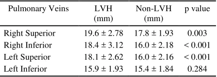
We measured 280 PVs from 70 subjects. All subjects had 4 PVs which were divided into right inferior PV (RIPV), right superior PV (RSPV), left inferior PV (LIPV), and left superior PV (LSPV). The mean ostial diameters of patient with and without LVH were as follow: RSPV of 19.62.78 vs 17.81.93 (p = 0.003), RIPV of 18.43.12 vs 16.02.19 (p < 0.001), LSPV of 18.12.62 vs 16.02.16 (p < 0.001), and LIPV of 15.91.93 vs 15.41.85 mm (p = 0.284), respectively. (Table 2) More than 60 percent of subject demonstrated oblong ostial morphology and the rests were circular.
Table 2. Ostial Diameter LVH. The increase of PVs diameter has been occurred even the cardiac rhythm still normal sinus rhythm.
Currently, either PV angiography, CT scan or magnetic resonance imaging (MRI) have been used to demonstrate the anatomy of LA and PVs. Using MRI, Kato et al9 showed that the PV ostia were more oblong than circular which give us reason to choose the largest diameter measurement in this study. The dilation of PVs in AF patients have been reported5 but its mechanisms is unclear. Herweg et al10 found that AF patients with hypertension and/or LVH had greater PVs diameter compare to that without hypertension and/or LVH. Our finding showed that the PVs dilation already happen in sinus rhythm patient with LVH. This new discovery gives away the possibility mechanism of AF in hypertensive patients. Since the prevalence of diastolic dysfunction is found approximately in 75 – 85% of mild to moderate hypertension and reached of 90% in LVH patients,11
our finding supports the mechanism of diastolic dysfunction as the cause of PVs dilation.
Ostial PVs – LA junction areas demonstrate special histological structure. Nathan et al12 are first to describe the presence of muscular sleeves continuing from LA into the PV with a mean extent of 13 mm. The longest sleeves were over the superior vein, and it was correlated with the relative distribution of PV effective and functional refractory periods (ERP and FRP). Interestingly, the LA ERP of both patients group were similar, hence there was ERP gradient between LA and PV which was shorter in PVs of AF patients but longer in PVs of control patients.13 The consistent finding of PVs dilation in AF along with its electrophysiological changes seem to be an inter-dependency process which have important role in AF occurrence. Mechano-electrical feedback due to stretch might impacts on the triggering substrate and on the atrial myocardial remodeling processes leading to perpetuation of AF. In animal models, acutely elevated atrial pressure increases the rate and organization of wavelets emanating from the PVs.11
The interesting results of our finding that the mean value in electrophysiological field, especially in era of PV isolation for AF management. This basic information will guide the electrophysiologist or physician in choosing or designing appropriate electrode catheter for PV isolation. Unfortunately we couldn’t perform statistical analysis to determine the significance of differences between those data.
Yuniadi et al Med J Indones 176
to be important for initiation and maintenance of AF. The mechanism of relation between complex myocardial arrangements of superior PVs and its larger diameter are unclear, but seminal work of Haissaguerre showed that the superior PVs are predominantly arrhythmogenic.1 Furthermore Yamane et al6 reported that diameters of arrhythmogenic PVs were significantly larger than those of nonarrhythmogenic.
Limitation: Lack of medication matching between LVH and non-LVH group in current study might have influence to diastolic dysfunction. ACE inhibitor and CCB have been proven to regress LVH. The regression of LVH by ARB results in improvement of LA dimension and function15 which in turn might be influence the PVs diameter.
CONCLUSION
PVs diameter enlarge in LVH patient with sinus rhythm. This finding might be one explanation of high prevalence rate of AF in hypertensive patients.
Acknowledgment
The investigators were acknowledging Mr. Irfan, the technician who gave great assistance in operating multi-sliced CT data base.
REFERENCES
1. Haissaguerre M, Jais P, Shah DC, Takahashi A, Hocini M, Quiniou G, Garrigue S, Le Mouroux A, Le Metayer P, Clementy J. Spontaneous initiation of atrial fibrillation by ectopic beats originating in the pulmonary veins. N Engl J Med. 1998; 339: 659 – 66
2. Chen YJ, Chen SA. Electrophysiology of pulmonary veins. J Cardiovasc Electrophysiol. 2006; 17: 220 – 4
3. Perez-Lugones A, McMahon JT, Ratliff NB, Saliba WI, Schweikert RA, Marrouche NF, et al. Evidence of specialized conduction cells in human pulmonary veins of
patients with atrial fibrillation. J Cardiovasc Electrophysiol. 2003; 14: 803 – 9
4. Fuster V, Ryden LE, Asinger RW, Cannom DS, Crijns HJ, Frye RL, et al. ACC/AHA/ESC guidelines for the management of patients with atrial fibrillation: executive summary. J Am Coll Cardiol. 2001; 38: 1231 – 65 5. Tsao HM, Yu WC, Cheng HC, Wu MH, Tai CT, Lin WS,
et al. Pulmonary vein dilation in patients with atrial fibrillation: Detection by magnetic resonance imaging. J Cardiovasc Electrophysiol. 2001; 12: 809 – 813
6. Yamane T, Shah DC, Jais P, et al. Dilatation as a marker of pulmonary veins initiating atrial fibrillation. J Interv Card Electrophysiol. 2002; 6: 245 – 9
7. Schwartzman D, Lacomis J, Wigginton WG. Characterization of left atrium and distal pulmonary vein morphology using multidimensional computed tomography. J Am Coll Cardiol. 2003; 41: 1349 – 57
8. Bruzzi JF, Rémy-Jardin M, Delhaye D, Teisseire A, Khalil C, Rémy J. When, why, and how to examine the heart during thoracic CT: Part 1, Basic Principles. Am J Roentgenol. 2006; 186: 324 – 32
9. Kato R, Lickfett L, Meininger G, et al. Pulmonary vein anatomy in patients undergoing catheter ablation of atrial fibrillation: Lessons learned by use of magnetic resonance imaging. Circulation. 2003; 107: 2004 – 10
10. Herweg B, Sichrovsky T, Polosajian L, Rozenshtein A, Steinberg JS. Hypertension and hypertensive heart disease are associated with increased ostial pulmonary vein diameter. J Cardiovasc Electrophysiol. 2005; 16: 2 – 5 11. Kalifa J, Jalife J, Zaitsev AV, Bagwe S, Warren M,
Moreno J, et al. Intra-atrial pressure increases rate and organization of waves emanating from the superior pulmonary veins during atrial fibrillation. Circulation. 2003; 108: 668 – 71
12. Nathan H, Eliakim M. The junction between the left atrium and the pulmonary veins. An anatomic study of human hearts. Circulation. 1966; 34: 412 – 22
13. Jais P, Hocini M, Macle L, et al. Distinctive electro-physiological properties of pulmonary veins in patients with atrial fibrillation. Circulation. 2002; 106: 2479 – 85 14. Cirillo S, Tosetti I, Gaita F, Bianchi F, Gandini G, Regge
D. Magnetic resonance angiography of the pulmonary veins before and after radiofrequency ablation for atrial fibrillation. Radiol Med. 2005; 109: 488 – 99