CARIES AND TOOTH ERUPTION IN ELEMENTARY SCHOOL CHILDREN IN AREAS OF GOITER ENDEMIC IN JEMBER
Ari Tri Wanodyo Handayani
Departement of Dental Public Health, Faculty of Dentistry Jember University
ABSTRACT
Nutrition are needed for growth and development processes, including teeth. The need of nutrition for tooth development have to be supplied during a period of pregnancy because the tooth development process was started intra uteri. Thyroid hormone as growth hormone, its secretion depends on the presence of iodine. Iodine deficiency can be associated with caries and tooth eruption. This research was done in Jember, an endemic area of goiter.
The aim of this research was to analyze a relationship between goiter and secondary tooth eruption of elementary school goitrous children in Jember and analyze a relationship between goiter and primary tooth caries of elementary school goitrous children in Jember.
This research was an analytical-observational study with cross sectional method. The population was the first and the second grade of elementary school children in Mayang District and Sumbersari District in Jember. Sampel size on this research was 100, consist of the first and the second grade of elementary school children age 6 up to 7,5 year. The sampling technique was simple random sampling. Examination of secondary tooth eruption, primary tooth caries, and nutritional status was done. The t-test, Mann-Whitney, and Chi-Square were used for statistical analysis. Regression test was also done to determine the correlation.
The result of relation analysis indicate that in general possibility there are relation between tooth eruption and caries with goiter. Percentage of tooth which have eruption at children who suffering goiter (73%) significantly (p<0.05) more lower than normal (88%). Mean of def-t at children who suffering goiter significantly (p<0.05) more higher (5.09) than normal children (3.94). Iodine deficiencies result decreasing of growth hormone secretion which play important role in osteoblast and osteoclast stimulation. Osteoclast is needed for making of eruption channel. The trouble that happened can pursue tooth eruption. Low of thyroid hormone also can cause trouble in process of odontogenesis. Thyroid hormone, its receptor and its binding protein play a part in every tooth forming phase. The trouble can result to low of enamel and dentin quality, so that tooth become easy be caries.
Conclusion of this research are most of secondary tooth eruption of goitrous children were delayed compared to normal children and the number of primary tooth caries in goitrous children was higher compared to normal child.
Correspondence : Ari Tri Wanodyo Handayani, Departement of Dental Public Health, Faculty of Dentistry Jember University, Jl. Kalimantan 37 Jember 68121, Indonesia, Phone: (0331) 333536, e-mail: [email protected]
INTRODUCTION
Nearly 2 billion or more than one-third of people worldwide have insufficient iodine intake, with those in south Asia particularly affected byiodine deficiency that cause of goiter and affects one-third of school-age children worldwide. 1,2,3,4 According to WHO report (1990) in developing countries almost 1 billion people possesed risk of iodine deficiency disorder (GAKY), among them 200 million suffering goiter, more than 5 million have cretinism with mental retardation and more than 15 million have severe mental retardation. 5,6
In Indonesia, iodine deficiency is still a major public health nutrition problem. According to Health Department (2005) the prevalence of GAKY in 2001 was 9,8% in Indonesia, increased to 11,1% in 2003. Sub-Province of Jember was one of the goiter endemic area in East Java. According GAKY survey in 2003, its prevalence reached 21,94%. Consist of 8 districts as severe endemic area, 7 districts as moderate endemic area, 11 districts as mild endemic area, 4 districts as early endemic area and only 1 district as non endemic area.6,7,8
Various factor can be associated to tooth eruption in mouth for example race, gender, environment, nutrition and endocrine (hyperfunction or hypofunction), including thyroid hormone. The point of time for the secondary tooth eruption are more varying than the primary tooth eruption because of the balance of genetic and environment factor.9,10
Have reported that hypothitoidism cause delay in eruption of teeth and in shedding of primary teeth. There was a delayed tooth eruption in children who lived in goiter endemic area and founded that people suffering from goiter are more susceptable to dental caries.11,12,13 In the Balinese people who lived in the area lack of iodine have smaller dental arch compared to iodine sufficient area.5
According to Bloom theory, many factors influenced in health, including oral and dental health, were behavioral factor, environment, health service and genetic factors.14 Based on the theory, this research will clarify the correlation between goitrous children with caries and tooth eruption.
MATERIAL AND METHODS
This research was an analytical-observasional study with cross sectional method. The population were the first and the second grade of elementary school children in Mayang District and Sumbersari District, Sub-Province of Jember. The sample taken were the first and the second grade of elementary school children age 6 up to 7,5 years old in Mayang District and Sumbersari District, Sub-Province of Jember resided in 3 chosen villages mapped in GAKY 2003. The sampling technique was simple random sampling. A cross-sectional survey was carried out on 100 subject used formula by Lemeshow. 15
Data collected were the severity of goiter measured by palpation method, examination of primary tooth caries (def-t) and secondary tooth eruption by clinical examination of mandibular first incisor using the explorer with sufficient lighting, nutritional status in the form of body height measured by microtoise and body weight measured by digital scale.
RESULT
[image:4.595.115.511.313.524.2]In this research, the examination was done on permanent mandibular central incisor. The primary tooth caries examination used def-t index and the assessment of nutritional status was done based on anthropometric, using parameter of TB/U and BB/TB. Then the assessment of nutritional status based on Z-Score index. The result of the examination and assessment shown on Table 1 and the result of different and correlation test showed on Table 2 and Table 3.
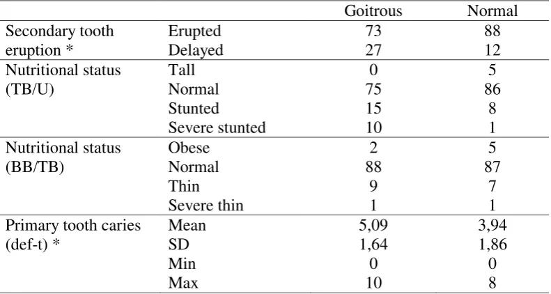
Table 1. Distribution of Secondary Tooth Eruption, Nutritional Status and Primary Tooth Caries of Goitrous and Normal Children in Sub-Province of Jember.
Goitrous Normal Secondary tooth eruption * Erupted Delayed 73 27 88 12 Nutritional status (TB/U) Tall Normal Stunted Severe stunted 0 75 15 10 5 86 8 1 Nutritional status (BB/TB) Obese Normal Thin Severe thin 2 88 9 1 5 87 7 1 Primary tooth caries
(def-t) * Mean SD Min Max 5,09 1,64 0 10 3,94 1,86 0 8 Note :
* = significant (used different test between goiter and normal groups) TB/U = body height of age
Table 2. Result of the different test between goitrous and normal children. p
Secondary tooth eruption
0,012 *
TB/U 0,107
BB/TB 0,795
Primary tooth caries 0,000 * Note :
* = Significant
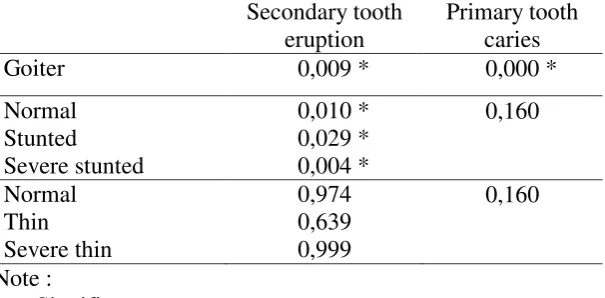
Table 3. Result of the correlation test between secondary tooth eruption and primary tooth caries with goiter and nutritional status.
Secondary tooth eruption
Primary tooth caries
Goiter 0,009 * 0,000 *
Normal Stunted
Severe stunted
0,010 * 0,029 * 0,004 *
0,160
Normal Thin Severe thin
0,974 0,639 0,999
0,160
Note :
* = Significant
Pursuant to Table 1, showed that number of goitrous children have experienced of secondary tooth eruption were 73 and delayed eruption were 27. This number was significant different (p=0,000) from normal children where there were 88 children have tooth eruption and 12 children have delayed tooth eruption (Table 2). Secondary tooth eruption has correlation with goiter (p=0,009) and nutritional status (TB/U) (Table 3).
[image:5.595.117.420.304.453.2]nutritional status with normal category (p=0,795) and has no correlation with secondary tooth eruption or primary tooth caries (Table 2 and 3).
And then about primary tooth caries we can see on Table 1-3 that the number of def-t of goitrous children have mean 5,09 ± 1,64. This number was significant different (0,000) from normal children (3,94 ± 1,86) and there are correlation between goiter and primary tooth caries (p=0,000)
DISCUSSION
Anthropometri parameter as base of nutritional status assessment. The combination of some parameter were called Anthropometric Index. This study used TB/U index and BB/TB index. The calculation of TB/U index based on Z-Score were categorized as tall, normal, stunted, and severe stunted showed a condition of chronic malnutrition, while the calculation of BB/TB index based on Z-Score were categorized as obese, normal, thin and severe thin showed a condition of acute malnutrition.
The result of statistical analysis indicate the nutritional status (TB/U and BB/TB) in goitrous children and normal children were almost the same, most of them were in normal nutritional status. In this study, the calculation of Z-Score to determined the normal nutritional status had a wide range, from - 2 SD up to + 2 SD. The wide span range enabling many sample to be included in normal category. The nutritional status in goitrous children were mostly in lower position of normal category. It position on - 2 up to 0 SD. That are different from normal children that their position on -1 up to + 2 SD.
Two methods are available for measuring goitre: neck inspection and palpation, and thyroid ultrasonography. By palpation, a thyroid is considered goitrous when each lateral lobe has a volume greater than the terminal phalanx of the thumbs of the subject being examined. Goitre surveys are usually done in school age children.16
Iodine has long been known as an essential element for humans, and for mammals in general, where it is concentrated in the thyroid gland, being a vital component of the thyroid hormones, they are triiodothyronine (T3) and tetraiodothyronine or thyroxine (T4). So iodine is essential to the production of these two hormones of the master gland of metabolism. Deprivation of iodine results in a series of iodine deficiency disorders, the most commonly recognized of which is endemic goiter, a condition where the thyroid gland becomes enlarged, the earliest clinical sign of hypothyroidism.17,18
Goitrous children can be relatively said had lack of micronutrient (iodine) disorders for long times. The hypothyroid condition can degraded the secretion of T3 and T4. The low secretion of T3 and T4 as growth hormone, the linear growth (body height) can be disrupted. Iodine deficiency during fetal development and in the first year of life can result in endemic cretinism, a disease that causes stunted growth and general development.19,20
Based on previous study, various factor can be associated with tooth eruption, such as race, gender, nutrition, endocrine hormone, environmental factor and genetic.9,10 The result of the research indicated that the tooth eruption in goitrous children more less compared to normal children. Possibly caused by the influence of nutrition and secretion of thyroid hormone. Dietary lack of iodine causes endemic goiter and hypothyroidism. Low intake of protein can disturb metabolism of iodine. Finnaly can disturb of stimulating cell metabolism, including tooth eruption process.11
Thyroid hormone promotes GH secretion andmodulates the effects of GH at its receptor.21 Hypothyroidism in goiter patient can cause disorders of growth factor stimulus like hormone growth (GH), insulin-like growth. factor-I (IGF-I), epidermal growth factor (EGF), and interleukin-I alpha (IL-1α). Those factors influenced the stimulation of osteoclast from osteoblast for the resorption of alveolar bone. Dental follicle is needed for eruption is because it initiates and regulates the required osteoclastogenesis and osteogenesis, at least for the intraosseous phase of eruption leading to tooth emergence. As a result, there was a disorder in making channel for the tooth eruption so there is not enough space for the tooth eruption.22,23,24,25
EGF can exercise a physiological role in the dental eruption, and the dental follicle can be the structure in which the growth factor acts. The forming of periodontal ligament were expanded after the tooth eruption. The development of periodontal ligament was not needed in eruption process. The periodontal ligament were not a part of the tooth eruption process.26,27
The result of this research also indicates there is a correlation between nutritional status especially TB/U (stunted and severely stunted) children with tooth eruption. It can explaining the correlation of chronic malnutrition with tooth eruption. Chronic malnutrition usually include calori-protein deficiency. Protein deficiency inhibit the growth of incisors and molars and cause delayed eruption. It seems there is a physiologic correlation between skeletal growth and tooth eruption.28,29,30
and secondary tooth caries.Malnutrition could influence the tooth forming process and increasing the susceptibility to dental caries. In rats, calorie-protein malnutrition showed reduction of saliva flow, influencing composition of saliva and immune system and also increasing enamel solubility. 29 Malnutrition affected all tissues, including enamel. Enamel was an epithel tissue with special development characteristic and susceptible in the process of amelogenesis. The clinical effect of malnutrition in tooth were enamel hypoplasia with the image of hollow white spot or even without enamel.28,32
Besides tooth susceptance factor to caries influenced during the tooth development, caries was also influenced by environmental factor of oral cavity. Oral hygiene factor was also associated with the incidence of dental caries. In normal condition, tooth is always wetted by saliva. Low level of oral hygiene caused the acidic oral condition. Saliva plays as protector and cleaner of tooth, but in that way saliva was also play important role in forming tooth plaque. Saliva was also a good media for the growth of certain microorganism related to caries. The decrease of salivary pH can increased the susceptibility to caries.31,33
The permanent or continuous reduction of thyroid hormone will cause hypertropi and hyperplasi of gland, whereas it was the start of goiter enlargement process. The thyroid hormone activity was correlate inversed with forming lesion of caries in studied rats. Less of thyroid hormone increased the caries.13
Some experts explained that if there is any disorders of thyroid hormone, can caused protein metabolism disorders. Beside that protein was required by the tooth enamel and plays important role in forming of tooth enamel. In goitrous people, they had degradation of protein bounding iodine. 13
EGF were found in cells and tissues participating at odontogenesis process. Its presence was constant in dental follicle during the odontogenesis process. The growth hormone and IGF-I also have important role in embryonic growth of tooth with regulation of interaction of mesenchyme epitel influencing the growth and cell differentiation.34,35,36
described on ameloblast, odontoblasts and cementoblasts at various stages of dental development. It is detected at bud stage, cap stage and bell stage and located in tooth epitel, differensiated mesenchyme cell, pre odontoblast and odontoblast of tooth. Recently GH found to influence crown width, root length, and dentin thickness. Its role as paracrine or autocrine in tooth development in a period of intra uterin or post uterin. 37
The main changes in thyroid function associated with pregnancy are due to an increase in hormone requirements that begin in the first trimester of gestation. This increase can only be met by a proportional increase in hormone production, something that depends directly upon the availability of iodine. When dietary iodine is lacking, an adequate physiological adaptation is difficult to achieve and is progressively replaced by pathological alterations that occur in parallel with the degree and duration of iodine deprivation. Iodine deficiency has devastating neurological effectson the fetus.38,39
Based on the research conducted by Smid et.al (2007) that GH disorders can caused anomaly of crown width, root length and thickness of dentin. GH level were the only significant factors associated with dental maturity. 34,40
The presence of protein and iodine were needed for normal secretion of thyroid hormone. Thyroid hormone influenced body cell activities and also metabolism of energy. The disorders of secretion of thyroid hormone, will also effect on protein metabolism, while protein needed for forming the tooth. A low iodine diet resulted in decreased absolute amounts of circulating triiodothyronine and protein-bound iodine (PBI). In goiter patient, there is a degradation of PBI. Decreasing of protein caused irregularity of predentin and decreased of interglobular space which caused susceptance of caries.6,13,33,34
condition disturbed the forming process of enamel and dentin. As the result, enamel and dentin were imperfect formed.
REFERENCES
1. Zimmermann, MB, Pieter LJ, Ngoako SM, Serena S, Ralf B, Linda FM and Xikombiso M. Vitamin A supplementation in iodine-deficient African children decreases thyrotropin stimulation of the thyroid and reduces the goiter rate American Journal of Clinical Nutrition, Vol. 86, No. 4, 2007. p: 1040-1044
2. Zimmermann, MB, Pieter LJ, Chandrakant SP. Iodine-deficiency disorders. The Lancet.Vol. 372, Iss. 9645; 2008.p: 1251, 12 pgs
3. Ralf Biebinger, Ralf, Myrtha A, Wolfgang L, Richard FH, Michael BZ. Vitamin A Repletion in Rats with Concurrent Vitamin A and Iodine Deficiency Affects Pituitary TSH[beta] Gene Expression and Reduces Thyroid Hyperstimulation and Thyroid Size. The Journal of Nutrition. Vol. 137, Iss. 3; 2007. p: 573, 5 pgs
4. Zimmermann, MB. Research on Iodine Deficiency and Goiter in the 19th and Early 20th Centuries1,2. The Journal of Nutrition. Vol. 138, Iss. 11; 2008.p: 2060, 4 pgs
5. Wijaya, M dkk. Efficacy of daily and weekly multiple micronutrient food-like tablets for the correction of iodine deficiency in Indonesian males aged 6-12 mo. American Journal of Clinical Nutrition, Vol. 85, No. 1, 2007. p: 137-143. 6. Almatsier. S. Prinsip Dasar Ilmu Gizi. PT Gramedia. Jakarta. 2002. p:
305-307
7. Dinas Kesehatan Jember, Laporan Tahunan Program Perbaikan Gizi Kabupaten Jember. Dinas Kesehatan Kabupaten Jember. 2005. p: 8-21
8. Campbell, AA, Andrew TL, Kai S, Saskia dP, Klaus K, Regina MP, Mayang S, Nasima A, Martin WB,Richard DS. Greater Household Expenditures on Fruits and Vegetables but Not Animal Source Foods Are Associated with Decreased Risk of Under-Five Child Mortality among Families in Rural Indonesia. J. Nutr. Vol. 138, 2008.p: 2244-2249
9. Djoharnas, H, Rata-rata Umur Erupsi Gigi Geligi Permanen Anak di Indonesia Dibandingkan dengan Anak di Negara Maju. Jurnal Kedokteran Gigi Universitas Indonesia. Edisi 7 No. 3. 2000.p: 37-43
10. Primasari A, Waktu Erupsi Gigi Molar Satu dan Incisivus Satu Permanen pada Murid-murid Sekolah Taman Kanak-kanak dan Sekolah Dasar di Kotif Rantau Prapat. Majalah Kedokteran Gigi USU. Edisi Januari No. 2. 1997.p: 28-34
11. Foley, TP. Hypothyroidism. Pediatrics in Review. Vol.25 No.3. 2004.p: 138-152
Yodium (GAKY), Stomatognatik (Majalah Kedokteran Gigi) FKG-Unej, 3 (1). 2004. p: 6-10
13. Musaikan, SW. Hubungan Gondok Endemik dengan Konsentrasi Yodium dalam Air Minum, Karies Gigi dan Penyakit Periodontal di Kecamatan Ngambon. Tesis. Universitas Airlangga. Surabaya. 1985. p: 65-84
14. Prasetyo RA, Setijanto D, Hapsoro A. Hubungan antara Tingkat Pendidikan dan Pengetahuan Ibu dengan Gambaran Kebersihan Gigi. Majalah Kedokteran Gigi (Dental Journal). Vol. 33 No. 4 Okt : 2000. p: 140-144 15. Lemeshow S et.al, Besar Sampel dalam Penelitian Kesehatan. Penerjemah:
Dibyo Pramono. Gadjah Mada University Press. Yogyakarta. 1997.
16. Zimmermann, MB. Methods to assess iron and iodine status. The British Journal of Nutrition.Vol. 99, Iss. S3; 2008. p: S2, 8 pgs
17. Fuge, Ron. Iodine Deficiency: An Ancient Problem in a Modern World Vol. 36, Iss. 1; 2007. p: 70, 3 pgs
18. Zimmermann, MB. The impact of iodised salt or iodine supplements on iodine status during pregnancy, lactation and infancy. Public Health Nutrition.Vol. 10, Iss. 12A; 2007. p: 1584, 12 pgs
19. Semba, RD, Saskia dP, Sonja YH, Kai S, Mayang S,Martin WB. Child malnutrition and mortality among families not utilizing adequately iodized salt in Indonesia. American Journal of Clinical Nutrition, Vol. 87, No. 2, 2008. p: 438-444
20. Zimmermann, MB, Pieter LJ, Ngoako SM, Xikombiso M, Serina S, Ralf B, Noureddine C, Maksim B, Lindita G and John B. Treatment of Iodine Deficiency in School-Age Children Increases Insulin-Like Growth Factor (IGF)-I and IGF Binding Protein-3 Concentrations and Improves Somatic Growth. The Journal of Clinical Endocrinology & Metabolism.Vol. 92, No. 2. 2007. p: 437-442
21. Shomon M. Iodine and Thyroid. Medical Review Board. 2004.
22. Wise GE et.al, Cellular, Molecular, and Genetic Determinants of Tooth Eruption. Critical Reviews in Oral Biology & Medicine. Vol. 13 (4). 2002. p: 323-335
23. Wise, GE. Mechanisms of Tooth Eruption and Orthodontic Tooth Movement. J Dent Res. 87(5). 2008. p: 414-434
24. Symons AL. Reduced Growth Hormone Receptor Immunoreactivity in Osteoclasts Adjacent to The Erupting Molar in The Incisor-absent (Osteopetrotic) Rat. Eur J Oral Sci. Vol 111. 2003. p: 503–509
25. Heinrich J et.al, CSF-1, RANKL and OPG Regulate Osteoclastogenesis during Murine Tooth Eruption. Arch Oral Biology. Vol. 50. Oktober. 2005.p: 897-908
26. Peixoto BC, Santos CRR, Canuto CE. The expression of the epidermal growth factor (EGF) during odontogenesis: Immunohistochemical study in mice (Mus musculus). Cienc Odontol Bras. 6 (4): 2003. p: 25-31
27. Maltha JC, Tooth Eruption, physiology. Ned Tijdschr Tandheelkd. Vol. 113. Agustus (8). 2006. p: 322-5
29. Alvarez JO. Nutrition, Tooth Development, and Dental Caries. Am J Clin Nutr. Vol. 61 (suppl). 1995. p: 410S-416S.
30. Diorio LP, Miller SA, and Navia AM. The Separate Effects of Protein and Calorie Malnutrition on the Development and Growth of Rat Bones and Teeth. J. Nutr.103: 2003. p: 856-865
31. Navia JM. Carbohydrates an Dental Health. Am J Clin Nutr. 59 (suppl). 1994. p: 719S-27S.
32. Goodman AH, Martinez C, Chavez A. Nutritional Supplementation and The Development of Linear Enamel Hypoplasias in Children from Tezonteopan,Mexico. Am J Clin Nutr. Vol. 53. 1991. p: 773-81
33. Suwelo IS. Karies Gigi pada Anak dengan Pelbagai Faktor Etiologi. Kajian pada Anak Usia Prasekolah. EGC. Jakarta. 1992. p: 15-20
34. Volpert EM and Werner SC. Serum triiodothyronine concentration in the iodine-deficient rat. American Journal of Anatomy.Vol 135, Issue 2 , 2005.p: 187 - 190
35. Cherfên B.P, Santos CRR, Canuto, CE, The expression of the epidermal growth factor (EGF) during odontogenesis:Immunohistochemical study in ice (Mus musculus). Cienc Odontol Bras. Vol: 6 (4). 2003. p: 25-31
36. Smid JR, Rowland JE, Young WG, Coschigano KT, Kopchick JJ, Waters MJ. Mouse Molar Dentin Size/Shape is Dependent on Growth Hormone Status. J Dent Res 86(5). 2007. p: 463-468,
37. Cazaux SL, etal. Growth Hormone Stimulates Proliferation and Differentiation in M2H2 Odontoblasts. European Cells and Materials. Vol. 14. Suppl. 2, 2007. p: 68
38. Glinoer, Daniel. The importance of iodine nutrition during pregnancy. Public Health Nutrition. Vol. 10, Iss. 12A; 2007. p: 1542, 5 pgs
39. Girling, Joanna. Thyroid disease in pregnancy. The Obstetrician & Gynaecologist. Vol. 10:4, 2008. p: 237-243