ELSEVIER
Animal Reproduction Science 48 (1997) 187- 195REPEON
SCIENCE
The effect of sperm-oocyte incubation time on
in vitro embryo development using sperm from a
tetraparental chimeric bull
C. Sumantri *, A. Boediono, M. Ooe, M. Murakami, S. Saha,
T. Suzuki
United Graduate School of Veterinary Sciences, Yamaguchi Unilaersity, Yamaguchi 753, Japan
Accepted 27 May 1997
Abstract
The present study was designed as 5 X 4 factorial to investigate the effects of using sperm from 5 bulls, and varied sperm-oocyte incubation times (5, 10, 15 and 20 h) on the fertilization, cleavage rates and blastocyst formation on an in vitro bovine embryo production system. The bulls included a tetraparental Chimera, its sires (Japanese Black and Limousin), its maternal grand-sires (Japanese Brown and Holstein). The proportion of polyspermy, 2-pronuclei formation, fertiliza- tion, cleavage and development to blastocyst were affected (p < 0.01) by the duration of sperm-oocyte incubation, as well as by the interaction between bulls and their corresponding sperm-oocyte incubation time. Blastocyst rate observed after 5 h in oocytes inseminated with Chimera, Japanese Black and Limousin were higher (p < 0.05) than those observed at 20 h incubation. The proportion of blastocysts from oocytes inseminated with Japanese Black observed at 10 h of incubation did not differ from that of Chimera, but both were higher (p < 0.05) than those observed for the Limousin, Japanese Brown and Holstein sires. The present study showed that there was an effect by the duration of sperm-oocyte incubation on in vitro embryo development. The optimal time of sperm-oocyte incubation for the Chimera was similar to that of its sires (Japanese Black and Limousin) but differed from its maternal grand-sires (Japanese Brown and Holstein). The fertilization rates for the sperm from the Holstein bull increased up to
15 h suggesting that this might be the only bull that would benefit from a long incubation period for insemination. 0 1997 Elsevier Science B.V.
Keywords: Sperm-oocyte incubation time; Tetraparental; Embryology; In vitro fertilization
* Corresponding author. Tel.: + 81 839 33 5935; fax: +81 839 33 5935; e-mail: [email protected] u.ac.jp.
1. Introduction
Sperm from individual bulls have been reported to differ in their ability to fertilize
matured oocytes in vitro and in the development to the pre-implantation stage
(Leibfried-Rutledge et al., 1987, 1989; Shi et al., 1990, 1991). Similar findings were
also reported in rams (Fukui et al., 1988). Spermatozoa from a single bull had
fertilization and cleavage rates affected with different heparin concentrations and
incubation periods (Iritani et al., 1986; Fukui et al., 1990). Also, spermatozoa from
different semen lot and straws within the same semen lot from a single bull affected the
developmental capacity of embryos (Otoi et al., 1993).
In vitro maturation of bovine oocytes incubated with sperm for 18-20 h after in vitro
fertilization has been reported (Long et al., 1993). It has been suggested that the optimal
time of sperm oocytes incubation for achieving maximum fertilization rate after IVM-
IVF is 24 h (Rehman et al., 1994). In some IVF systems, a significantly higher incidence
of polyspermy has been shown in oocytes cultured with sperm for 24 h compared to
those of 8 h (Chian et al., 1992).
Tetraparental chimeric cattle were successfully produced by aggregating IVF em-
bryos of Fl(Holstein
XJapanese Black) and Fl(Japanese Brown
XLimousin) and cul-
turing in vitro without the zona pellucida (Boediono et al., 19931, and the sperm from
this tetraparental chimeric bull used for producing IVF bovine embryos (Sumantri et al.,
1997). However, the optimal time of sperm-oocyte incubation required for tetraparental
chimeric bull was not determined.
Therefore, the present study was conducted to examine the effect of sperm-oocyte
incubation time on in vitro fertilization and later developmental stages of IVF embryos
using sperm from tetraparental Chimera, its sires (Japanese Black and Limousin) and its
maternal grand sires (Japanese Brown and Holstein).
2.
Materials and methods
2.1.
In vitro maturation of oocytes
C. Sumantri et al. /Animal Reproduction Science 48 (1997) 187-195 189
2.2.
In vitro fertilization
Frozen-thawed
semen from a 17 month-old tetraparental
Chimera (CH) was used for
in vitro fertilization.
Frozen-thawed
semen obtained from its sires (Japanese Black/JB
and Limousin/L),
and from its maternal grand-sires
(Japanese Brown/JBr
and Hol-
stein/H)
were also used for comparison.
Frozen semen was thawed in a water bath
(37”(Z), washed twice using 2.5 mM caffeine
in Brackett
and Oliphant’s
medium
(Caff-BO),
as previously
described
by Brackett
and Oliphant
(1975), followed
by
centrifugation
at 500 g for 5 min. Then, the semen was resuspended
in caff-BO
supplemented
with
1%
bovine serum albumin (BSA, Sigma) and 20 ( pg/ml)
heparin
(Shimizu Pharmaceutical,
Shimizu, Japan) to yield a sperm concentration
of 5 X 1 06/ml.
A 100 ,ul aliquot of the sperm suspension
was covered with mineral oil and pre-in-
cubated
for 1 h at 38.5”C in humidified
5% CO,
in air. Matured
oocytes
were
transferred into sperm microdrops (20 to 25 oocytes per microdrop) separately for each
bull: Chimera
(0-CH),
Japanese
Black (0-JB),
Limousin
(O-L), Japanese
Brown
(0-JBr) and Holstein (O-H) followed by incubation
for different times (5, 10, 15 and 20
h).
2.3.
Experiment 1: Assessing pronuclear formation
and fertilization
rates
A total of 761 matured inseminated
oocytes from different sperm-oocytes
incubation
time (5, 10, 15, and 20 h) treatments were washed and cultured for 15, 10, 5, and 0 h,
respectively;
followed
by staining
to observe
fertilization
rate. The cumulus
cells
surrounding
the embryos
were removed by several pipettings
in PBS (Gibco, Grand
Island, NY, USA) supplemented
with 5% SCS, before being fixed in camoy solution
(ethanol:acetic
acid = 3: 1) for 72 h and stained in 1% aceto-orcein
to examine
the
formation of pronuclei. The fertilization
rate was calculated as the percentage of stained
ova that had 2 or more pronuclei.
2.4.
Experiment 2: Assessing cleavage and blastocyst rate
The inseminated
oocytes with cumulus cells from all treatments
were washed and
transferred separately into culture medium for further development.
The culture medium
consisted
of TCM-199
supplemented
with 5% SCS, 5 (pug/ml)
insulin (Wako Pure
Chemicals
Osaka, Japan) and 50 (pg/ml)
gentamicin.
At 48 h after fertilization,
the
cumulus cells surrounding
the embryos were removed by several pipettings,
while the
cumulus cell layer attached to the bottom of the culture dish was undisturbed,
and used
as a co-culture.
The culture medium was replaced with a new one after 96 h.
The cleavage (2-, 4- and 8 cell stage) rate was calculated from the number of total
ova inseminated
after 48 h of insemination
(IVF = Day 0). The blastocyst
rate was
calculated from the total cleavage that had developed to the blastocyst
stage by day 9
after insemination.
2.5.
Embryo evaluation
All experiments
were repeated 3 times. Data were analyzed as 5 X 4 factorial (5 types
h). Mean proportions were subjected to least-square analysis of variance with arcin
transformation. Duncan’s Multiple Range Test was used for specific comparisons.
3. Results
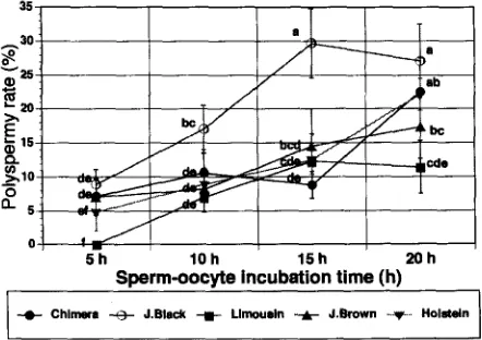
The proportion of polyspermy observed after 5 h of sperm-oocyte incubation was
significantly lower
( p
< 0.05) than that observed after 20 h (Fig. 1). The proportion of
polyspermy gradually increased when oocytes were incubated with sperm for longer
periods with each of the bulls (7.1 vs 22.5%, 8.9 vs 27.1%, 0.0 vs 11.4%, 7.0 vs 17.3%
and 4.8 vs 22.5% for 0-CH, 0-JB, O-L, 0-JBr and O-H, respectively). Nonetheless, J.
Black had a higher (p < 0.05) polyspermy rate after 10 and 15 h of incubation than the
other bulls.
The proportion of 2-pronuclei formation observed after 5 h sperm-oocyte incubation
for oocytes inseminated with Chimera sperm did not differ from that observed after 20 h
(Fig. 2). However, the proportion of 2-pronuclei formation observed at this point for the
oocytes inseminated with J. Black and Limousin were higher (p < 0.05) than those
observed after 15-20 h. In contrast, oocytes inseminated with J. Brown and Holstein
had lower formation of 2-pronuclei at 5 h incubation compared to that of 20 h
(p < O.OS>,
(74.9 vs. 73.8%; 70.1 vs. 56.8%; 69.5 vs. 51.2%; 39.5 vs. 54.3% and 17.7
vs. 63.8% for 0-CH, 0-JB, O-L, 0-JBr and O-H; respectively).
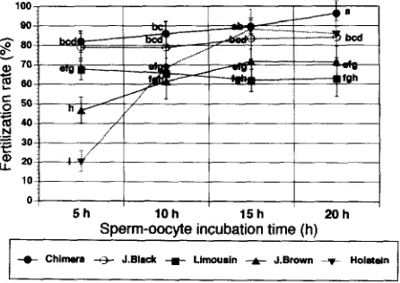
Oocytes inseminated with J. Black and Limousin sperm retained the same fertiliza-
tion rate in each time treatment (Fig. 3). However, the fertilization rates observed after 5
h sperm-oocyte incubation in the Chimera, Japanese Brown and Holstein were lower
(p < 0.05) than those observed after 20 h of incubation (81.9 vs 96.3%, 79.1 vs 83.9%,
0 ,
5h 10h 15h 20h
Sperm-oocyte incubation time (h)
f Chim e ra +I - J .Bla dc + Llm m m in -A- J .Brow n -.f. H oldn
Fig. 1. The effect of sperm-oocyte incubation time on polyspermy rate. Chimera (n = 159), J. Black
[image:4.505.140.361.404.560.2]C. Sumantri et al./Animal Reproduction Science 48 (1997) 187-195 191
5h IOh 15 h ZOh
Sperm-oocyte incubation time (h)
Fig. 2. The effect of sperm-oocyte incubation time on 2-pronuclei formation rate. Chimera (n = 159), J. Black (n = 1441, Limousin (n = 158). J. Brown (n = 145) and Holstein (n = 155). II (No. of stained oocytes). Each treatment was replicated 3 times. Mean with different superscripts are significantly different (ANOVA, Duncan’s multiple range test, a vs. b, c, d, e; b vs. c, d. e; c vs. d, e, f, g; d vs. e, f, g; e vs. f, g; f vs. g; p<0.05anda,bvs.f,g;p<0.01).
67.8 vs 62.9%, 46.6 vs 7 1.6%, and 20.5 vs 85.9% for 0-CH, 0-JB, O-L, 0-JBr and O-H
sperm, respectively).
A total of 4057 matured oocytes were inseminated
for producing
IVF embryos to
study the effect of sperm-oocyte
incubation
time on cleavage rate. The cleavage rate for
oocytes inseminated
with Limousin
sperm remained the same for each incubation
time
100
0
5h 10h 15 h 20h
Sperm-oocyte incubation time (h)
t Chimera -++ J.Bladc + Llmousin t J.Brown -. HoleWn
I I
[image:5.504.136.368.72.243.2] [image:5.504.141.364.405.563.2]IO]
I I I II
0: I
I
I
I5h
10
h
15h 20 hSpermoocyte incubation time (h)
Fig. 4. The effect of sperm-oocyte incubation time on cleavage rate. Chimera (n = 917). J. Black (n = 763), Limousin (n = 825), J. Brown (n = 771) and Holstein (n = 781). n (No. of inseminated oocytes). Each treatment was replicated 3 times. Mean with different superscripts are significantly different (ANOVA, Duncan’s multiple range test, a vs. b, c, d, e, f, g, h, i; b vs. c, d, e, f, g, h, i; c vs. d, e, f, g, i, j; d vs. e, f, g, h, i, j; e vs. f, g, h, i, j; f vs. g, h, i, j; g vs. h, i, j; i vs. j; p < 0.05, a, b, vs. j; p < 0.01).
(Fig. 4). Only small changes occurred using sperm from J. Black and Chimera. The
trends with sperm from J. Brown more greater (45.7, 53.9, 59.9 and 60.5% for 5, 10, 15
and 20 h, respectively). Cleavage rates using Holstein sperm to inseminate oocytes
increased from 18.3% at 5 h to 68.6% at 20 h (p < 0.01; Fig. 4).
The results of blastocyst formation are summed in Fig. 5. In this case, a total of 2550
cleaved (2-, 4-, and 8-cell) embryos were used. In general terms, as the time of
sperm-oocyte incubation increased, the blastocyst rate decreased. Also, the Limousin,
5h 1Oh 15h 20h
Sperm-oocyte incubation tlme (h)
(
-e- Chlnwa -@- J.EInck + Llmousln -A- J.Brown -v- Holstdn ( [image:6.506.142.359.70.224.2] [image:6.506.140.358.399.560.2]C. Summztri et al./Animal Reproduction Science 48 (1997) 187-195 193
Holstein and J. Brown sperm inseminated
oocytes showed a lower blastocyst formation
than the other bulls (Chimera, and J. Black) for all incubation
times.
Blastocyst rate for the Chimera and J. Black sperm inseminated
oocytes were similar
at 5 h, but different
from Limousin,
J. Brown
and Holstein
inseminated
oocytes
(p < 0.05) which did not differ from each other. Subsequent
ranges and differences
tended to be smaller. At 20 h of incubation,
Chimera inseminated
oocytes were superior
(p < 0.05) on blastocyst
rate (18.8%) compared
to J. Black (10.0%) and Limousin
(7.6%) bulls, but similar to J. Brown (17.5%).
4.
Discussion
The incidence of polyspermy
among the bulls used in this study showed a tendency
to increase
with time of sperm-oocyte
incubation
but the magnitude
of this effect
differed from one bull to another. J. Black seemed to be the most susceptible,
while the
Limousin
bull had the lowest rate. The extent of these differences
among the bulls in
terms of the sperm capacitation
times and acrosome reactions
has been reported by
Parrish et al. (1986).
The fertilization
patterns of matured oocytes inseminated
with Chimera was similar to
its sires (J. Black and Limousin),
but different from that of its maternal grand sires
(J. Brown and Holstein).
The latter two bulls had lower fertilization
rates after 5 h of
incubation.
Increasing
the sperm-oocyte
incubation
time improved
the fertility rate.
Similar differences
among the bulls have been documented
by others (Iritani et al.,
1986; Leibfried-Rutledge
et al., 1987; and Shi et al., 1990); and in sheep (Fukui et al.,
1988).
The results in Fig. 5, indicate that the rate of blastocyst
formation
decreases with
increased
sperm-oocyte
incubation
time for each of the 5 bulls. These observations
contrast with those reported by Long et al. (1993) and Rehman et al. (1994). They found
that the blastocyst
rate from IVM-IVF
oocytes was not affected by sperm-oocyte
incubation
period. Similarly, Long et al. (1993) showed that sperm concentration
had no
effect on monospermic
fertilization.
These differences
with our results may be due to
differences between the bulls used in each study. There are two possibilities
contributing
to the decline in rate of blastocyst
formation
at 20 h in this experiment:
First, the
increasing
incidence of polyspermy
could have lead to an increased rate of mortality in
pre-implantation
embryos;
and second,
the bull effect in our case seemed
to be
dependent
on the interrelationship
among them (that is, Chimera has its sires, J. Black
and Limousin,
and its grand sires, J. Brown and Holstein).
The similarity between Chimera and its sire (Japanese Black) in fertilization,
cleav-
age and blastocyst rate might be associated with the significantly
higher proportion
of
the cell in the gonads derived from Japanese Black than from the Limousin
which
contributed
in the formation of these chimeric cattle. Sumantri et al. (1996) reported that
78.6% of microsatellite
DNA present in the tetraparental
chimeric cattle were uniquely
contributed
from the Japanese
Black and only 21.4% from the Limousin.
Detailed
analysis of mosaicism in interspecific
Chimeras between
Mus
musculusand
Mus
carolitissues (Rossant and Chapman, 1983). These findings indicate that there may be a
paternal effect on fertilization, cleavage and blastocyst rate. Shire and Whitten (1980a,b)
and Goldbard and Warner (1982) reported similar cases in the mouse embryo, where the
cleavage rate and speed of development were dependent upon genetic factors, including
maternal and paternal effects. There is also a strong paternal effect on pre-implantation
development and blastocyst formation in the human embryo (Janny and Menezo, 1994).
In conclusion, incubation time did affect in vitro embryo development. Although
there were inherent differences with the Chimera and its sire (Japanese Black) having
the highest rate of blastocyst production, there were no significant differences between 5
and 10 h for the Chimera, J. Black and Limousin. Only the Chimera and Japanese Black
decreased at 15 h and 4 of the 5 sires decreased at 20 h.
Acknowledgements
We would like to thank Dr. R. Rajamahendran, Prof. of the Department of Animal
Science, University of British Columbia, and Dr. Varisanga M.D., Yamaguchi Univer-
sity for their helpful evaluation of the manuscript. Dr. T. Otoi, Tokushima Prefecture
Beef Cattle and Swine Experiment Station, for statistical analysis. The Hiroshima and
the Kita Kyusu slaughterhouses for giving the ovaries and the Yamaguchi Zootechnical
Experiment Station for providing the superovulated cow serum.
References
Boediono, A., Ooe, M., Yamamoto, M., Takagi, M., Saha, S., Suzuki, T., 1993. Production of Chimeric calves by aggregation of in vitro-fertilized bovine embryos without zonae pellucidae. Theriogenology 40,
1221-1230.
Brackett, B.G., Oliphant, G., 1975. Capacitation of rabbit spermatozoa in vitro. Biol. Reprod. 12, 260-274. Chian, R.C., Nakahara, H., Niwa, K., Funahashi, H., 1992. Fertilization and early cleavage in vitro of aging
bovine oocytes after maturation in culture. Theriogenology 37, 666-672.
Fukui, Y., Glew, A.M., Gandolfi, F., Moor, R.M., 1988. Ram specific effects on in vitro fertilization and cleavage of sheep oocytes matured in vitro. J. Reprod. Fert. 82, 337-340.
Fukui, Y., Sonoyama, T., Mochizuki, H., Ono, H., 1990. Effect of heparin dosage and sperm capacitation time on in vitro fertilization and cleavage of bovine oocytes matured in vitro. Theriogenology 34, 579-591. Goldbard, S.B., Warner, C.M., 1982. Genes affect the timing of early mouse embryo development. Biol.
Reprod. 27,419-424.
Iritani, A., Utsumi, K., Miyake, M., Yamaguchi, Y., 1986. Individual variation in the in vitro fertilizing ability of bull spermatozoa. Devel. Growth Differ. 45 (Suppl. 28). 35, abstract.
Janny, L., Menezo, Y.J.R., 1994. Evidence for a strong paternal effect on human preimplantation embryo development and blastocyst formation. Mol. Rep. Develop. 38, 36-42.
Leibfried-Rutledge, M.L., Critser, E.S., Eyestone, W.H., Northey, D.L., Frist, N.L., 1987. Development potential of bovine oocytes maturated in vitro or in vivo. Biol. Reprod. 36, 376-383.
Leibfried-Rutledge, M.L., Critser, E.S., Parrish, J.J., First, N.L., 1989. In vitro maturation and fertilization of bovine oocytes. Theriogenology 31, 61-74.
Long, CR., Chase, C.N., Balise, J.J., Duby, R.T., Robl, J.M., 1993. Effect of sperm removal time, sperm concentration and motility enhancer on fertilization parameters and development of bovine embryos in vitro. Theriogenology 39, 261, abstract.
C. Sumntri et al./Animal Reproduction Science 48 (1997) 187-195 195
Otoi, T., Tachikawa, S., Kondo, S., Suzuki, T., 1993. Effect of different lots of semen from the same bull on in vitro development of bovine oocytes fertilized in vitro. Theriogenology 39, 713-718.
Parrish, J.J., Susko-Parrish, J., Leibfried-Rutledge, M.L., Critser, E.S., Eyestone, W.H., First, N.L., 1986. Bovine in vitro fertilization with frozen-thawed semen. Theriogenology 25, 591-600.
Rehman, N., Collins, A.R., Suh, T.K., Wright, R.W. Jr., 1994. Effect of sperm exposure time on in vitro fertilization and embryo development of bovine oocytes maturated in vitro. Theriogenology 41, 1447- 1452. Rossant, J., Chapman, V.M., 1983. Somatic and germline mosaicism in interspecific chimaeras between Mus
musculus and Mus caroli. J. Embryo]. Exp. Morp. 73, 193-205.
Shi, D.S., Lu, K.H., Gordon, I., 1990. Effect of bulls on fertilization of bovine oocytes and their subsequent development in vitro. Theriogenology 33, 324, abstract.
Shi, D.S., Lu, K.H., Gordon, I., Polge, C., 1991. Variation in cleavage and embryonic development of bovine oocytes in vitro fertilized with different bull ejaculates. Theriogenology 35, 271, abstract.
Shire, J.G.M., Whitten, W.K., 1980a. Genetic variation in the timing of first cleavage in mice: Effect of paternal genotype. Biol. Reprod. 23, 363-368.
Shire, J.G.M., Whitten, W.K., 1980b. Genetic variation in the timing of first cleavage in mice: Effect of maternal genotype. Biol. Reprod. 23, 369-376.
Sumantri, C., Sugimoto, Y., Watanabe, T., Morita, M., Boediono, A., Suzuki, T., 1996. Blood typing and microsatellite DNA genotyping of tetraparental chimeric cattle. Proc. 8th Anim. Sci. Congr. Asian Australasian Assoc. Anim. Prod. Sot. 2, pp. 34-35 (abstract).
ELSEVIER
Animal Reproduction Science 48 (1997) 187- 195REPEON
SCIENCE
The effect of sperm-oocyte incubation time on
in vitro embryo development using sperm from a
tetraparental chimeric bull
C. Sumantri *, A. Boediono, M. Ooe, M. Murakami, S. Saha,
T. Suzuki
United Graduate School of Veterinary Sciences, Yamaguchi Unilaersity, Yamaguchi 753, Japan
Accepted 27 May 1997
Abstract
The present study was designed as 5 X 4 factorial to investigate the effects of using sperm from 5 bulls, and varied sperm-oocyte incubation times (5, 10, 15 and 20 h) on the fertilization, cleavage rates and blastocyst formation on an in vitro bovine embryo production system. The bulls included a tetraparental Chimera, its sires (Japanese Black and Limousin), its maternal grand-sires (Japanese Brown and Holstein). The proportion of polyspermy, 2-pronuclei formation, fertiliza- tion, cleavage and development to blastocyst were affected (p < 0.01) by the duration of sperm-oocyte incubation, as well as by the interaction between bulls and their corresponding sperm-oocyte incubation time. Blastocyst rate observed after 5 h in oocytes inseminated with Chimera, Japanese Black and Limousin were higher (p < 0.05) than those observed at 20 h incubation. The proportion of blastocysts from oocytes inseminated with Japanese Black observed at 10 h of incubation did not differ from that of Chimera, but both were higher (p < 0.05) than those observed for the Limousin, Japanese Brown and Holstein sires. The present study showed that there was an effect by the duration of sperm-oocyte incubation on in vitro embryo development. The optimal time of sperm-oocyte incubation for the Chimera was similar to that of its sires (Japanese Black and Limousin) but differed from its maternal grand-sires (Japanese Brown and Holstein). The fertilization rates for the sperm from the Holstein bull increased up to
15 h suggesting that this might be the only bull that would benefit from a long incubation period for insemination. 0 1997 Elsevier Science B.V.
Keywords: Sperm-oocyte incubation time; Tetraparental; Embryology; In vitro fertilization
* Corresponding author. Tel.: + 81 839 33 5935; fax: +81 839 33 5935; e-mail: [email protected] u.ac.jp.