A mitochondrial protein homologous to the mammalian
peripheral-type benzodiazepine receptor is essential for
stress adaptation in plants
Wolfgang Frank1,*, Kim-Miriam Baar1,†, Enas Qudeimat1, Mayada Woriedh1, Ali Alawady2, Diah Ratnadewi1,‡, Louis Gremillon1, Bernhard Grimm2and Ralf Reski1
1
Plant Biotechnology, Faculty of Biology, University of Freiburg, Schaenzlestr. 1, 79104 Freiburg, Germany, and 2
Institute of Biology, Plant Physiology, Humboldt University Berlin, Philippstraße 13, 10115 Berlin, Germany
Received 19 December 2006; revised 26 April 2007; accepted 15 May 2007.
*For correspondence (fax +49 (0) 761 203 6945; e-mail [email protected]).
†Present address: Department of Cardiology, Faculty of Medicine, University of Freiburg, Breisacher Str. 33, 79106 Freiburg. ‡Present address: Department of Biology, Bogor Agricultural University, Jl. Raya Pajajaran, Bogor, Indonesia.
Summary
The cloning of abiotic stress-inducible genes from the mossPhyscomitrella patensled to the identification of the gene PpTSPO1, encoding a protein homologous to the mammalian mitochondrial peripheral-type benzodiazepine receptor and the bacterial tryptophane-rich sensory protein. This class of proteins is involved in the transport of intermediates of the tetrapyrrole biosynthesis pathway. Like the mammalian homologue, the PpTSPO1 protein is localized to mitochondria. The generation ofPpTSPO1-targeted moss knock-out lines revealed an essential function of the gene in abiotic stress adaptation. Under stress conditions, thePpTSPO1 null mutants show elevated H2O2levels, enhanced lipid peroxidation and cell death, indicating an important role of PpTSPO1 in redox homeostasis. We hypothesize that PpTSPO1 acts to direct porphyrin precursors to the mitochondria for heme formation, and is involved in the removal of photoreactive tetrapyrrole intermediates.
Keywords: Physcomitrella patens, abiotic stress tolerance, mitochondria, tetrapyrroles, reactive oxygen species.
Introduction
In animals, tetrapyrrole biosynthesis starts in the mito-chondria, continues in the cytoplasm up to the formation of coproporphyrinogen III, and finishes in the mitochondria with the synthesis of protoheme. In contrast, the subcellular localization of the tetrapyrrole biosynthesis pathway in plants is distributed to plastids and mitochondria. All tetra-pyrrole end products are synthesized in plastids, with the exception that the last two steps of heme synthesis are additionally found in mitochondria (Cornah et al., 2003; Papenbrock and Grimm, 2001). The most abundant tetra-pyrrole end product in plants is chlorophyll. Heme serves as a co-factor for proteins in diverse cellular processes such as respiration (cytochromes) and oxygen metabolism (cata-lases, peroxidases and NADPH oxidases) (Beale and Weinstein, 1990; Grimm, 1998). The plant tetrapyrrole metabolic pathway is also needed for the synthesis of siroheme, which is a co-factor for the nitrite and sulphite reductases required for the assimilation of inorganic
nitrogen and sulphur. Finally, the linear tetrapyrrole phyto-chromobilin is derived from protoheme and is assembled with phytochrome.
Although chlorophyll is confined to the chloroplast, as is siroheme, heme is present in all cellular compartments, and phytochrome operates in the cytosol and in the nucleus (Kircheret al., 2002; Moulin and Smith, 2005). The subcel-lular partition of the plant tetrapyrrole pathway, leading to the targeting of diverse end products into different subcel-lular compartments, necessitates a coordinated distribution of tetrapyrrole intermediates between the two organelles, plastids and mitochondria. Moreover, the pool of tetrapyr-role intermediates and end products has to be tightly controlled, as they can be excited by light and their photoreactivity can generate radicals and singlet oxygen species. The tetrapyrrole biosynthesis pathway is substan-tially regulated by environmental stimuli, and during plant development at different levels of gene expression for
certain enzymes of this pathway (Grimm, 1998; Reinbothe and Reinbothe, 1996).
Recently, the mammalian adenosine 5¢
-triphosphate-binding cassette transporter ABCB6, which is located at the outer mitochondrial membrane, was found to be required for mitochondrial porphyrin uptake and heme biosynthesis (Krishnamurthy et al., 2006). A previously reported key element in the regulation of steroid biosyn-thesis in mammals is the 18-kDa peripheral-type benzodia-zepine receptor, which mediates mitochondrial cholesterol import (Li and Papadopoulos, 1998; Papadopoulos, 1998). Recently, a new nomenclature for this protein family was suggested based on the protein structure and molecular function (Papadopoulos et al., 2006) designating these proteins translocator proteins (TSPO). The mammalian TSPO is localized in the outer mitochondrial membrane (Papadopouloset al., 1994) where it is closely associated with other proteins, such as the 34-kDa voltage-dependent anion channel and the inner membrane adenine nucleotide carrier (McEneryet al., 1992). Moreover, TSPO was shown to possess the highest binding affinity for protoporphyrin IX, indicating an involvement in the tetrapyrrole metabo-lism in mouse erythroleukemia cells (Taketaniet al., 1994). It was suggested that TSPO is involved in porphyrin transport, and is a critical factor in erythroid-specific induction of heme biosynthesis genes. More than ten years ago Yeliseev and Kaplan (1995) reported on the tryptophane-rich sensory protein (TspO) from the a
-pro-teobacterium Rhodobacter sphaeroides, with significant similarities to the mammalian TSPO. TspO is involved in the negative regulation of photosynthesis genes in response to oxygen. Nevertheless, evidence that TspO directly regulates the expression of photosynthesis genes inR. sphaeroides was not provided. The TspO protein is localized at the outer membrane of R. sphaeroides cells (Yeliseev and Kaplan, 1995) and functions in the export of excessive intermediates of the tetrapyrrole pathway (Yeliseev and Kaplan, 1999). Furthermore, the rat TSPO homologue was able to substitute for TspO, indicated by the suppression of photosynthesis genes in response to oxygen (Yeliseevet al., 1997). This suggests an evolution-ary and functional relationship between the bacterial TspO and TSPO. Additionally, Lindemannet al.(2004) provided evidence that a homologous protein to the mammalian TSPO and the R. sphaeroides TspO is also present in
Arabidopsis thaliana and other plants. Transport studies with the recombinant Arabidopsis TSPO inEscherichia coli
revealed a benzodiazepine-stimulated high-affinity uptake of protoporphyrin and cholesterol, leading to the hypo-thesis that the Arabidopsis homologue functions in the transport of potoporphyrinogen IX to the mitochondrial site of protoheme formation. Immunogold staining using an antimouse TSPO peptide antibody led to signals both at the outer thylakoid membrane of plastids and in the
mitochondria ofDigitalis lanataleaves. An antibody raised against the Arabidopsis TSPO homologue recognized proteins with a molecular mass of 19–20 kDa in mitochon-dria of Arabidopsis, potato and D. lanata. Even though substrate binding properties of the Arabidopsis TSPO were initially determined, its role in transport and regulation of plant metabolism is still unknown.
Here, we report on the isolation of an abiotic stress-induced TSPO homologue from the moss Physcomitrella patens. Functional studies with generated targeted moss knock-out lines suggest an essential role of the protein in plant stress adaptation.
Results
Isolation of PpTSPO1fromP. patens
Comparison of mRNA pools of the untreated and dehy-drated moss P. patens by differential display reverse transcription-polymerase chain reaction (DDRT-PCR) facili-tated the identification of genes induced upon dehydration. One arbitrary primer (see Experimental procedures) gave rise to a cDNA fragment derived from the cDNA pool of dehydrated tissue, which was absent in the RT-PCR reaction using the cDNA from untreated plants. The PCR fragment was cloned, sequenced and used for the identification of a 1041-bp full-length cDNA sequence, containing an open reading frame coding for a protein of 198 amino acids, with a predicted molecular mass of 21.8 kDa. BLAST searches with the predicted amino acid sequence revealed homology to proteins from bacteria, animals and plants, including TspO fromR. sphaeroides, (Yeliseev and Kaplan, 1995), the human mitochondrial TSPO (Papadopoulos, 1998) and the TSPO-like transporter from Arabidopsis (Lindemannet al., 2004) (Table S1). Based on the similarity with the well-characterized TspO protein from R. sphaeroides, the isolated gene from P. patens was designated PpTSPO1
(P. patens tryptophane-rich sensory protein 1). An align-ment of homologous proteins from different kingdoms performed with theCLUSTALWmultiple sequence alignment
program (Thompson et al., 1994) is shown in Figure S1. BLAST searches performed in the Genbank expressed sequence tag (EST) database revealed that PpTSPO1 shares the highest similarity to a deduced protein sequence from the desiccation-tolerant moss Tortula ruralis. Protein topology prediction for PpTSPO1 using the TMHMM 2.0
that these proteins exert their function as integral mem-brane proteins.
PpTSPO1is induced by abiotic stresses and abscisic acid (ABA)
To verify the induction ofPpTSPO1by drought stress, RNA blot analysis was performed with RNA from dehydrated
P. patens plants. As many dehydration-responsive genes are also induced by other abiotic stresses, we investigated the PpTSPO1expression pattern in response to cold and salt. We have previously shown that the induction of stress-responsive genes in P. patens is mediated by the phyto-hormone ABA, which complies with the role of ABA as a second messenger in stress-induced gene expression in vascular plants (Frank et al., 2005b). Thus, PpTSPO1
expression was also examined for its ABA-dependent con-trol. After 1 h of the various stress treatments and applica-tion of ABA thePpTSPO1mRNA levels increased rapidly and
strongly, whereas only weak quantities ofPpTSPO1mRNA could be detected in untreated control plants (Figure 1). Thus, PpTSPO1 expression is induced by different ABA-dependent abiotic stress-related pathways. PpTSPO1
expression analyses 15 and 30 min after the application of different stimuli revealed a rapid induction of the gene in response to ABA, whereas the expression in response to NaCl and dehydration was not markedly altered (Figure 1). Until now, a stress-induced expression of this class of genes in plants was not reported. Based on the specific expression pattern ofPpTSPO1in response to abiotic stress, we suggest a functional role of the encoded protein in abiotic stress adaptation inP. patens.
PpTSPO1 is localized to mitochondria
The topology prediction for PpTSPO1 indicated the presence of five membrane-spanning regions, suggesting that PpTSPO1 represents an integral membrane protein. Attempts to predict the subcellular localization of PpTSPO1 by making use of common target prediction tools did not lead to conclusive results. For a genuine proof of sub-cellular localization of PpTSPO1, we have generated a
PpTSPO1::GFPfusion construct containing the open reading frame ofPpTSPO1and the green fluorescent protein (GFP) under the control of the CaMV35Spromoter. The resulting construct was used for transfection ofP. patensprotoplasts, which were analysed by confocal laser scanning microscopy 48 h after the transfection (Figure 2). GFP fluorescence was detected in mitochondria of transformed protoplasts and the observed GFP pattern was identical to already descri-bed mitochondria-localized proteins in P. patens, like the
(a)
(b)
(c)
Figure 1.Expression analysis ofPpTSPO1in response to abiotic stress and abscisic acid (ABA).
(a)Physcomitrella patensplants were dehydrated, treated with 20lMABA or treated with 250 mMNaCl for the indicated time periods. Untreated plants served as controls. A 10-lg sample of each RNA was loaded for RNA gel blot. The RNA blot was hybridized with aPpTSPO1cDNA probe and anEF1a control sample to verify equal loading of the gel.
(b)P. patensplants were kept on ice for the indicated time periods. A 10-lg sample of each RNA was loaded for RNA gel blot. The blot was hybridized with aPpTSPO1cDNA fragment. The lower panel shows ethidium bromide stained rRNA bands to indicate equal loading of the samples.
(c)P. patensplants were dehydrated, treated with 20lMABA or treated with 250 mMNaCl for 15 and 30 min, respectively. Untreated plants served as controls. A 10-lg sample of each RNA was loaded for RNA gel blot and hybridized with aPpTSPO1cDNA probe. The lower panel shows ethidium bromide stained rRNA bands to indicate equal loading of the samples.
(a)
(b)
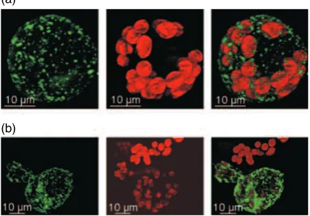
Figure 2.Subcellular localization of PpTSPO1.Physcomitrella patens proto-plasts were transfected with aPpTSPO1::GFPC-terminal fusion construct. At 48 h after transfection protoplasts were analysed by confocal laser scanning microscopy.
(a) and (b) Two independent protoplasts transfected with thePpTSPO1::GFP construct. Left panels, green fluorescent protein (GFP) fluorescence; middle panels, chlorophyll autofluorescence; right panels, overlay of red chlorophyll and green GFP fluorescence.
phage-type RNA polymerases PpRpoT1 and PpRpoT2 (Richteret al., 2002).
Generation of targetedPpTSPO1knock-out mutants
Based on its unique ability to integrate DNA into its nuclear genome by means of homologous recombination (Schaefer, 2001),P. patenshas become a versatile plant model system for reverse genetics approaches (Streppet al., 1998). For the functional analysis of PpTSPO1 we prepared a PpTSPO1
gene disruption construct by inserting an nptII selection marker cassette into thePpTSPO1cDNA (Figure 3a). After the transfection ofP. patensprotoplasts with the gene dis-ruption construct and subsequent selection, plants were
screened for the disruption of thePpTSPO1genomic locus. PCR was performed on genomic DNA of transgenic lines and wild-type plants with primers spanning the insertion site of the nptII selection marker cassette (Figure 3b). Genomic DNA of those transgenic lines, which generates a PCR product identical to the one obtained from wild-type plants, was considered to be subjected to an illegitimate integration of the knock-out construct. Alternatively, transgenic lines containing genomic DNA that could not be amplified, or generated a 1.5-kb bigger PCR product according to the size of the nptII cassette, were considered to be putative
PpTSPO1knock-out lines. A total of 82 transgenic lines were screened, and 20 lines (24.3%) failed to give rise to a PCR product. To validate the generation of loss-of-function mutants, six of these lines were chosen to analyse the
PpTSPO1transcript by RT-PCR and RNA gel blot analysis (Figure 3c,d). For RT-PCR studies primers were used that spanned the integratednptII cassette. As thePpTSPO1gene was found to be induced by abiotic stress and ABA, wild-type control plants and the transgenic lines were treated with 20lMABA for 2 h. PCR reactions using cDNA derived
from the six knock-out lines failed to give rise to an ampli-fication product, whereas the control PCR with cDNA derived from wild-type plants produced an amplicon of the expected size. Additionally, thePpTSPO1transcript was not detected by RNA gel blot analysis using RNA from ABA-treated knock-out lines, confirming that the integration of thePpTSPO1
disruption construct led to PpTSPO1 null mutants.
(a)
(b)
(c)
(d)
(e)
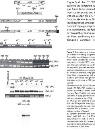
Figure 3.Generation and molecular analysis ofPpTSPO1knock-out lines. (a) Scheme illustrating the generation of thePpTSPO1knock-out construct. The middle panel illustrates the resultingPpTSPO1knock-out construct. The lower panel depicts the genomic structure of thePpTSPO1 locus after integration of thePpTSPO1knock-out construct by homologous recombina-tion. The primers that were used for molecular analyses of the transgenic lines are indicated by arrows. White box,nptII cassette; grey boxes,PpTSPO1cDNA fragments; black boxes, genomicPpTSPO1locus.
(b) Molecular analysis using genomic DNA to identifyPpTSPO1knock-out lines. One representative gel showing polymerase chain reaction (PCR) reactions performed with primers Fwd and Rev (c.f. a). Six transgenic lines that failed to give rise to PCR products are indicated (KO 1–6); WT, wild-type control.
(c) Analysis of thePpTSPO1transcript in wild type andPpTSPO1knock-out lines by RT-PCR. PCR reactions were performed using cDNA prepared from abscisic acid (ABA)-treated plants. Upper panel: RT-PCR using the primers Fwd and Rev. Verified null-mutants are indicated by KO 1–6. Lower panel: control PCR using primers derived from the ABA-induced genePpSHP1to indicate integrity of the cDNA.
(d) RNA gel blot analysis of wild-type (WT) andPpTSPO1knock-out lines (KO 1–6). RNA gel blot using 5lg of total RNA hybridized withPpTSPO1and a hybridization probe derived from an ABA-induced dehydrin gene to monitor effective ABA treatment. Lower panel: ethidium bromide stained rRNA to indicate loading of the gel.
Independently, the six knock-out lines were analysed by flow cytometry to exclude the generation of diploid lines by protoplast fusion during the transformation process. All six knock-out lines were shown to be haploid (data not shown). To exclude additional integration sites of the PpTSPO1
knock-out construct within the nuclear DNA, two of the knock-out lines were subjected to genomic Southern blot analysis (Figure 3e). A DNA fragment comprising the com-pletenptII selection marker cassette present in the knock-out construct was used as a hybridization probe. The resulting hybridization pattern demonstrates a single integration event in both mutant lines. These two lines were used for further experimental studies.
PpTSPO1 is essential for salt stress adaptation
Under standard growth conditions thePpTSPO1knock-out lines did not show any phenotypic differences compared withP. patenswild-type plants (Figure 4a), suggesting that under favourable growth conditions the encoded protein does not contribute to phenotypic distinction. However, the stress-induced expression ofPpTSPO1 suggests a role of this protein during the abiotic stress response inP. patens. The tolerance ofP. patensplants to various abiotic stress conditions was determined previously (Franket al., 2005b), where we could demonstrate thatP. patensplants are able to tolerate elevated NaCl concentrations. To obtain func-tional data on the role ofPpTSPO1during the stress adap-tation, we compared the twoPpTSPO1knock-out lines with wild-type plants for their ability to withstand enhanced NaCl concentrations. Wild-type plants and both knock-out lines were grown on standard medium supplemented with 400, 500 and 600 mMNaCl, respectively. After only 8 h of growth
on salt medium, the twoPpTSPO1knock-out lines showed phenotypic deviations compared with wild-type plants, like shrinking of gametophores and protonema filaments, and bleaching of the green tissue. The most prominent differ-ences between the knock-out lines and wild-type plants during further growth on the NaCl plates were observed at
(a)
(b)
(c)
(d)
Figure 4.NaCl treatment of Physcomitrella patens wild-type plants and PpTSPO1knock-out lines.
(a)P. patenswild-type plants (WT) and plants of thePpTSPO1knock-out lines (KO 1, KO 2) were grown on standard growth medium supplemented with 600 mMNaCl. From each line, 10 plants were analysed in two independent experiments. Pictures from representative plants were taken after the indicated time periods.
(b)P. patenswild-type (WT) andPpTSPO1knock-out lines (KO 1, KO 2) were grown in standard liquid medium (untreated) and liquid medium supple-mented with 500 mMNaCl and photographed after 72 h post-inoculation. (c) Cell death was measured spectrophotometrically by Evans blue staining in wild-type plants andPpTSPO1knock-out lines. Plants were grown in standard growth medium (untreated) or in medium supplemented with 500 mMNaCl for the indicated time periods. Error bars indicate SD (n¼3).
(d) Growth ofPpTSPO1knock-out lines in the dark (D) and under standard growth conditions (L/D 16 h/8 h) for 72 h in the presence of 500 mMNaCl.
the 600 mM NaCl concentration (Figure 4a). Thirteen days
after transfer onto 600 mM NaCl all plants from the two
knock-out lines were almost completely bleached, whereas the wild-type plants tolerated the elevated NaCl concentra-tion without phenotypic changes. We observed similar res-ponses of wild-type andPpTSPO1knock-out lines in liquid medium supplemented with 500 mMNaCl. Only 3 days after
inoculation of the plants in the NaCl-containing medium, the two mutant lines were completely bleached, whereas the wild-type plants did not show macroscopic differences (Figure 4b). The observed bleaching of thePpTSPO1 knock-out lines in response to elevated NaCl concentrations sug-gests enhanced photooxidative damage of photosynthetic pigments, which are often accompanied by irreversible deleterious effects leading to cell death. As an indication for loss of membrane integrity and resulting cell death, cultures were stained with Evans blue. Cell death was measured from wild-type plants and the two PpTSPO1 knock-out lines grown in liquid standard medium or medium supplemented with 500 mMNaCl (Figure 4c). Interestingly, the two
knock-out lines already showed enhanced Evans blue staining under normal growth conditions, which could be indicative for the need ofPpTSPO1expression under normal culture conditions. The values determined at 500 mMNaCl indicate
that high NaCl concentrations severely affect thePpTSPO1
knock-out lines, thus confirming the results obtained from the growth experiment on NaCl-containing medium. To study the influence of light, which might cause photo-oxidative damage, the twoPpTSPO1knock-out lines were grown in liquid medium in the presence of 500 mM NaCl
either in constant darkness or under normal light conditions. After 3 days the plants that were grown under normal light conditions were completely bleached, whereas the plants grown in darkness were unaffected and remained green (Figure 4d). The decreased tolerance of the twoPpTSPO1
knock-out lines reveals that PpTSPO1 is an essential protein required for salt stress adaptation inP. patens.The light-dependent susceptibility to enhanced concentrations of NaCl observed for thePpTSPO1knock-out lines suggests an involvement of the PpTSPO1 protein in the control of photooxidative damage.
PpTSPO1knock-out lines produce elevated levels of hydrogen peroxide
The elevated chlorophyll bleaching in thePpTSPO1 knock-out lines compared with wild-type plants observed during growth on medium containing NaCl suggested an enhanced generation of reactive oxygen species (ROS) in the mutant lines. Quantitative H2O2 measurements and
detection of H2O2by staining with 3, 3¢-diaminobenzidine
(DAB) were performed with wild-type plants andPpTSPO1
knock-out lines after exposure to 500 mMNaCl. The H2O2
(a)
(b)
(c)
(d)
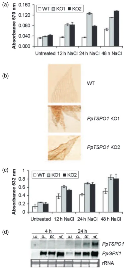
Figure 5.H2O2detection, measurement of lipid peroxidation and analysis of
PpTSPO1expression in response to oxidative stress.
(a)Physcomitrella patenswild-type plants (WT) andPpTSPO1knock-out lines (KO 1, KO 2) were grown in standard liquid medium (untreated) and liquid medium supplemented with 500 mM NaCl. H2O2 was measured at the indicated times. Absorbance values indicate absorption of the produced oxidation product resofurin at 570 nm. Error bars indicate SD (n¼3).
(b) H2O2levels were visualized with 3, 3¢-diamonobenzidine in wild-type plants (WT) andPpTSPO1mutants grown in the presence of 500 mMNaCl for 24 h. (c) Wild-type plants andPpTSPO1mutants were grown in the presence of 500 mMNaCl and lipid peroxidation was determined at the indicated time points by measuring malone dialdehyde levels, as described in Experimental procedures. Error bars indicate SD (n¼3).
levels were elevated in both PpTSPO1 knock-out lines compared with wild-type plants after 12, 24 and 48 h of the NaCl treatment (Figure 5a). These measurements were further confirmed by the DAB staining of PpTSPO1 mutants as well as wild-type plants 24 h after exposure to 500 mM
NaCl (Figure 5b). ROS are harmful compounds that cause a number of adverse modifications within the cell, like protein oxidation, DNA damage and lipid peroxidation (Mittler, 2002). To analyse the effect of the elevated ROS levels detected in thePpTSPO1knock-out lines, lipid per-oxidation after exposure to 500 mM NaCl was determined
(Figure 5c). The two PpTSPO1 knock-out lines showed enhanced levels of lipid peroxidation, even under standard growth conditions. Upon NaCl stress the degree of lipid peroxidation increased in both the wild-type andPpTSPO1
knock-out lines. However, the degree of lipid peroxidation measured in bothPpTSPO1knock-out lines was above the levels detected in wild-type plants. These findings are in agreement with the observed elevated levels of H2O2in the
PpTSPO1 mutants, and imply a role of PpTSPO1 in the prevention of ROS formation or scavenging of ROS. As ROS could act as a direct stimulus to induce PpTSPO1
expression, we asked if the site of ROS production may influencePpTSPO1gene expression. Wild-type plants were treated with paraquat (methylviologen), an inhibitor of photosynthetic electron transport (Dodge, 1971), and the two mitochondrial electron transport inhibitors rotenone and antimycin A (Ohnishi et al., 1966). These inhibitors impair the electron transport in the photosynthetic and respiratory electron chains, respectively. In consequence, ROS will be generated by the transfer of electrons to oxygen. To monitor the generation of ROS by the diffe-rent inhibitors, a P. patens gene homologous to plant glutathione peroxidases (PpGPX1; accession number DQ645821), which are known to be upregulated by eleva-ted ROS levels, was used. The mRNA levels of thePpGPX1
control were upregulated 4 h after application of the three inhibitors, indicating elevated ROS levels caused by the inhibitor treatments (Figure 5d). At this time point increasedPpTSPO1mRNA levels were not observed. The same results were obtained after 8 and 12 h of inhibitor treatments (data not shown). However, after 24 h the
PpTSPO1mRNA levels increased in plants treated with the mitochondrial inhibitors rotenone and antimycin A, but not in response to the photosynthetic inhibitor paraquat. In contrast to the rapid accumulation ofPpTSPO1mRNA by abiotic stress and ABA, the induction ofPpTSPO1by oxi-dative stress seems to be less effective when compared with other oxidative stress-responsive genes likePpGPX1. However, the PpTSPO1 expression is dependent on the site of ROS generation. In agreement with the subcellular localization of the PpTSPO1 protein, elevated PpTSPO1
mRNA levels were caused only by mitochondrial respira-tory electron chain inhibitors. This finding suggests a
specific role for PpTSPO1 in the control of ROS in mitochondria.
PpTSPO1knock-out lines show elevated heme and protoporphyrin IX levels in response to abiotic stress
The Arabidopsis TSPO-like protein is able to transport pro-toporphyrin IX (Lindemannet al., 2004). We hypothesized that the loss of PpTSPO1 will result in a decreased proto-porphyrinogen transport into the mitochondria, and in turn would lead to decreased mitochondrial heme levels. As
PpTSPO1mRNA levels are markedly increased upon dehy-dration, wild-type plants and the twoPpTSPO1knock-out lines were subjected to dehydration and subsequently heme concentrations were analysed (Figure 6a). The heme levels in wild-type plants as well as inPpTSPO1knock-out lines increased in response to dehydration, which may reflect an enhanced demand for heme as prosthetic group upon stress conditions. Intriguingly, the twoPpTSPO1knock-out lines showed higher heme levels compared with the wild-type plants, which could result from elevated heme pools in plastids and an altered heme turnover in the PpTSPO1
mutants. Heme is an allosteric inhibitor of the glutamyl tRNA reductase that catalyses the second step of the 5-amino-levulinic acid (ALA) biosynthesis (Papenbrock and Grimm, 2001). If the elevated heme levels in thePpTSPO1knock-out lines can be ascribed to an increased plastidic heme con-centration, the allosteric inhibition of the glutamyl tRNA reductase should result in reduced ALA synthesis rates. Indeed, measurement of ALA synthesis rates in wild-type plants and onePpTSPO1knock-out line in the presence of 250 mMNaCl revealed a reduced ALA synthesis rate in the
PpTSPO1 mutant line (Figure 6b). These data support enhanced plastidic heme accumulation as a consequence of PpTSPO1 deficiency. Furthermore, we applied ALA to liquid cultures of P. patens wild-type plants and one PpTSPO1
knock-out line, which should result in a boost of the porphyrin synthesizing part of the tetrapyrrole biosynthesis pathway, because the synthesis of ALA is known to be the rate-limiting step in this pathway (Papenbrock and Grimm, 2001). Subsequently, the plant cultures were either grown in standard growth medium or in growth medium supple-mented with 250 mMNaCl. To assess the role of PpTSPO1
for protoporphyrinogen allocation to mitochondria followed by heme synthesis, the accumulation of tetrapyrrole inter-mediates was determined after 6 and 12 h of ALA feeding under standard and salt-stress conditions. It is expected that upon a defective transport of protoporphyrinogen IX into the mitochondria, protoporphyrin IX will accumulate in the plastids and/or leak out into the cytoplasm (Jacobset al., 1990; Leeet al., 1993; Liet al., 2003). Between 6 and 12 h after ALA feeding of cultures in standard growth medium without NaCl, we measured increased but similar pro-toporphyrin IX concentrations in the wild type and the
PpTSPO1knock-out line compared with the untreated con-trol plants. Upon salt and ALA incubation, protoporphyrin IX levels increased even more in wild type and in thePpTSPO1
knock-out line, but the protoporpyrin IX levels of the mutant line were two times higher compared with wild type (Fig-ure 6c). This finding indicates an enhanced accumulation of the metabolic intermediate protoporphyrin IX in the mutant as a result of PpTSPO1 deficiency. It is assumed that this accumulated metabolite cannot be sufficiently directed to
mitochondria, and remains non-metabolized in plastids and cytoplasm. To further prove this hypothesis, liquid cultures of thePpTSPO1knock-out lines and wild-type plants were treated with 30lM acifluorfen in the presence of 350 mM
NaCl. The diphenyl-ether herbicide acifluorfen inhibits plastidic as well as mitochondrial protoporphyrinogen IX oxidases, resulting in elevated levels of protoporphyrin IX (Becerril and Duke, 1989; Camadroet al., 1991; Matringe and Scalla, 1988; Matringe et al., 1989; Sherman et al., 1991; Witkowski and Halling, 1988, 1989), which may accumulate in the plastids and/or leak into the cytosol. After 7 days of growth in the presence of acifluorfen and NaCl thePpTSPO1
knock-out lines were completely bleached, whereas the wild-type plants remained green (Figure 6d). Furthermore,
PpTSPO1knock-out lines grown in the presence of 350 mM
NaCl without acifluorfen remained green, indicating that the bleaching was specifically caused by the inhibition of the protoporphyrinogen IX oxidases. From these data we con-clude that PpTSPO1 is required to control protoporphyrin IX levels within the cytosol. Upon defective translocation of protoporphyrin IX into the mitochondria, protoporphyrin IX accumulates and elevated protoporphyrin IX concentrations induce photooxidative reactions including photobleaching within the plastids.
Identification ofPpTSPO1homologues
The Arabidopsis genome contains only one TSPO-like pro-tein (Lindemannet al., 2004). An EST database search with the PpTSPO1 protein sequence as query revealed two additional homologues inP. Patens, which were designated
PpTSPO2 and PpTSPO3. Interestingly, compared with
PpTSPO1the two identified genes encode shorter proteins of 175 and 180 amino acids, respectively, and lack the N-terminal region present in PpTSPO1. Moreover, PpTSPO2 and PpTSPO3 are more closely related to each other than to PpTSPO1 (Table 1; Figure S3). The mRNA levels ofPpTSPO2
andPpTSPO3were analysed by RNA gel blots with RNA
(a)
(b)
(c)
(d) Figure 6.Measurements of heme and protoporphyrin IX concentrations,
5-aminolevulinic acid (ALA) synthesis rates and acifluorfen treatment in wild-type plants andPpTSPO1knock-out lines.
(a) Heme concentrations were determined after dehydration treatments at the indicated time points. Untreated plants served as controls. Error bars indicate SD (n= 3).
(b) ALA synthesis rates in wild-type plants (WT) and thePpTSPO1knock-out line 1 (KO 1).
from stressed and untreated P. patens wild-type plants (Figure 7a,b).PpTSPO3showed an abiotic stress and ABA-induced expression pattern similar toPpTSPO1. However,
PpTSPO2was detected in all RNA samples, including RNA derived from untreated P. patens plants, indicating a constitutive expression.
Discussion
The existence of TSPO homologues in different kingdoms suggests a long-standing functional significance of this class of proteins during evolution. Consistent with the fact that mitochondria have originated from photosynthetic a
-pro-teobacteria (Grayet al., 2001), the eukaryotic TSPO homo-logues analysed so far were found to reside in mitochondria (Anholtet al., 1986; Lindemannet al., 2004), including the
P. patenshomologue PpTSPO1. Considering the common evolutionary background, the question arises if these pro-teins are also functionally related. In fact, TSPO homologues from plants, animals and bacteria bind benzodiazepine derivatives and PK11195, an isoquinilone carboxamide, with high affinity at nanomolar concentrations. In mammals,
porphyrins are the predominant endogenous molecules with high affinity for the TSPO (Vermaet al., 1987). The TspO protein fromR. sphaeroidesis involved in the transport or efflux of tetrapyrrole intermediates of the heme and bacte-riochlorophyll biosynthetic pathway (Yeliseev and Kaplan, 1999). Studies with the Arabidopsis TSPO-like protein indi-cated a functional role of this protein in the transport of protoporphyrin IX to mitochondria (Lindemannet al., 2004). The isolation of a stress-responsive homologue of the
TSPOgene family fromP. patensprovides evidence for the involvement of this class of transporters in plant stress adaptation. The closest plant homologue of PpTSPO1 is encoded by a gene from the desiccation-tolerant moss
T. ruralis. The corresponding mRNA was found to be among the most abundant transcripts identified from aT. ruralis
cDNA library prepared from rapidly dried and subsequently rehydrated plants (Oliveret al., 2004). Detailed expression data of TSPO homologues from seed plants have not yet been reported. Therefore, Arabidopsis gene expression data were analysed using the GENEVESTIGATOR microarray database (Zimmermannet al., 2004). As forPpTSPO1, the microarray data reveal a stress-responsive expression pat-tern of the Arabidopsis homologue, with highest mRNA levels detected in response to salt and osmotic stress treatments. Most likely the transcriptional regulation depends on ABA, as indicated by increased mRNA levels upon treatment with ABA. However, mRNA levels in Arabidopsis do not considerably increase during dehydra-tion and cold treatments, as observed forPpTSPO1. Elevated mRNA levels of the ArabidopsisTSPO-like gene were also detected during seed maturation, implying a role in desiccation processes and dormancy during seed matura-tion, as well as in stress adaptation.
The functional analysis of thePpTSPO1moss knock-out lines confirms an essential role of the encoded protein in abiotic stress adaptation. Based on its mitochondrial local-ization and presumable protoporphyrin transport activities, we hypothesize that PpTSPO1 plays a role in directing tetrapyrrole intermediates to mitochondria for heme pro-duction. We have indirect indications that PpTSPO1 is capable of transporting porphyrins, as we observed reduced chlorophyll autofluorescence in chloroplasts of cells transfected with thePpTSPO1::GFPoverexpression constr-uct. The reduced chlorophyll fluorescence observed in
PpTSPO::GFP-overexpressing cells could be assigned to a possible disturbance of the porphyrin allocation between plastids and mitochondria.
It is proposed that an increased protoporphyrin import into mitochondria occurs in the adaptive response upon stress conditions for the delivery of substrates for heme bisosynthesis. Heme is the required co-factor for the ROS scavenging enzymes catalases and peroxidases. In animal cells, mitochondria are major sites of ROS formation, and major targets of ROS-induced damage (Kowaltowski and
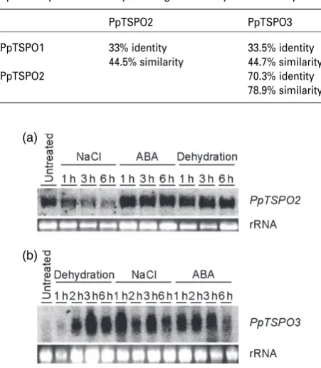
Table 1Protein sequence comparison of PpTSPO1, PpTSPO2 and PpTSPO3 presented as a percentage of identity and similarity
PpTSPO2 PpTSPO3
PpTSPO1 33% identity
44.5% similarity
33.5% identity 44.7% similarity
PpTSPO2 70.3% identity
78.9% similarity
(a)
(b)
Figure 7.Expression analysis of PpTSPO2and PpTSPO3.Physcomitrella patensplants were dehydrated, treated with 20lMabscisic acid or treated with 250 mMNaCl for the indicated time periods. Untreated control plants served as controls. A 10-lg sample of each RNA was loaded for RNA gel blots. (a) RNA gel blot hybridized with thePpTSPO2probe.
(b) RNA gel blot hybridized with thePpTSPO3probe. The lower panel in each shows ethidium bromide stained rRNA bands to indicate equal loading of the samples.
Vercesi, 1999; Liuet al., 2002). Also in plants, mitochondria have emerged to be a major source for ROS production caused by the interaction of oxygen with reduced forms of electron transport components (Moller, 2001). In particular, under stress conditions physical changes in membrane components may lead to constraints on the mitochondrial electron transport chain, resulting in enhanced ROS pro-duction (Wagner, 1995; Wagner and Krab, 1995). Studies on wheat indicated that mitochondria are the main target for oxidative damage. Even under normal growth conditions, mitochondria showed higher levels of oxidative damage compared with chloroplasts and peroxisomes. These levels were markedly increased upon drought conditions (Bartoli
et al., 2004). To overcome enhanced ROS production in mitochondria, plants possess specific alternative respiratory pathways that play a role in the control of ROS formation and scavenging. Among these, non-proton-pumping NAD(P)H dehydrogenases bypass complex I, and the alter-native oxidase (AOX) accepts electrons directly from the ubiquinone pool without the intervention of the cytochrome
coxidase pathway through complexes III and IV (Rasmusson
et al., 1998). Nevertheless, once formed, ROS must be detoxified efficiently to minimize the consequential detri-mental effects. Detoxifiying enzymes include superoxide dismutases (SODs) that convert the superoxide anion radical (O2–Æ) to H2O2and O2(Scandalios, 1993). MnSODs are found
in mitochondria, and MnSOD expression often is upregu-lated by stress conditions (Bowleret al., 1989; Tsanget al., 1991). The main enzymatic H2O2scavengers in plants are
catalases and ascorbate peroxidases, which convert H2O2to
H2O and O2(Asada, 1992; Willekenset al., 1995). The mRNA
levels of genes encoding catalases and ascorbate peroxi-dase increase upon different stress treatments (Datet al., 2000; Shigeokaet al., 2002), and both enzymes need heme as a co-factor. Until now there was only limited knowledge about the role of catalases and ascorbate peroxidases in plant mitochondrial ROS scavenging. The existence of the ascorbate/glutathione cycle in plant mitochondria was shown in pea (Jimenez et al., 1997), and, recently, the targeting of rice ascorbate peroxidase OSAPX6 to mito-chondria was demonstrated (Teixeiraet al., 2006). Further-more, the activity of several mitochondrial isoforms of ascorbate peroxidase increased upon salt treatment in the salt-tolerant tomato speciesLycopersicon pennellii(Mittova
et al., 2004), suggesting a function in mitochondrial ROS scavenging. However, although the localization of catalase in mitochondria has been shown in animals (Radi et al., 1991), its presence in plant mitochondria is still an open question. One plant catalase, CAT3 from maize, was found in mitochondrial fractions (Scandalioset al., 1980), but further evidence for mitochondrial localization is missing. Interest-ingly, in potato both catalase and AOX are involved in the suppression of membrane potential breakdown, which triggers programmed cell death (Mizunoet al., 2005).
How-ever, localization studies indicating mitochondrial targeting of the catalase have not been performed.
The measurement of heme contents in dehydratedP. pat-enswild-type plants indicated increasing heme levels during the dehydration process, which could reflect an enhanced demand for heme prosthetic groups upon abiotic stress conditions. However, during dehydration the twoPpTSPO1
mutant lines showed even higher heme levels compared with wild-type plants. These results could be interpreted as a stress-responsive induction of PpTSPO1 that leads to enhanced heme biosynthesis in mitochondria. We believe that the elevated heme levels in thePpTSPO1knock-out lines result from increased plastidic heme synthesis in conse-quence of a block in mitochondrial uptake, and, accordingly, a redirection of plastid-remained protoporphyrin IX for the synthesis of plastid heme. This hypothesis was supported by the reduced ALA synthesis rate measured in thePpTSPO1
knock-out line. In accordance with the effect of heme as an allosteric inhibitor of the glutamyl tRNA reductase, the elevated heme levels observed in the PpTSPO1 mutants most likely can be ascribed to increased plastidic heme concentrations. Yet, an altered heme turnover in the
PpTSPO1mutants can also not be excluded. However, in spite of the elevated heme levels, thePpTSPO1knock-out lines are more susceptible to abiotic stress conditions. Therefore, not the total quantity of heme, but rather its concentration in specific cellular compartments could be the critical factor for stress adaptation. In this case, the lack of heme groups in mitochondria could lead to reduced ROS scavenging and enhanced damage of mitochondrial compo-nents. In turn, mitochondrial dysfunction will result in elevated ROS levels affecting the redox homeostasis of the whole plant cell. The contribution of mitochondrial heme biosynthesis to the total intracellular heme pool is contro-versially discussed. In pea, the total mitochondrial heme biosynthetic activity was reported to be less than 10% compared with the activity detected in plastids, suggest-ing that plastids are the major site of heme biosynthesis (Cornahet al., 2002). However, these studies did not include measurements of mitochondrial and plastidic heme biosyn-thesis under abiotic stress conditions. In vivosubcellular localization studies of the two cucumber ferrochelatases CsFeC1 and CsFeC2 revealed that both proteins are solely targeted to plastids (Masuda et al., 2003). Based on this observation it must be considered that the mitochondrial heme biosynthetic pathway is lacking in particular plant species, and that the complete synthesis of heme is accom-plished in the plastids. Nevertheless, the results obtained from our studies support the scenario of an enhanced demand for heme under stress conditions comprising the mitochondrial heme biosynthesis pathway.
the cytosol. Tetrapyrrole intermediates are highly photo-reactive, and, consequently, are a major source for the generation of ROS, especially under different abiotic stress conditions (Apel and Hirt, 2004; Mittler, 2002). The application of diphenylether herbicides, which inhibit protoporphyrino-gen oxidase, led to the accumulation of protoporphyrin IX as a consequence of protoporphyrinogen IX oxidation in the cytoplasm (Jacobset al., 1990; Leeet al., 1993; Liet al., 2003). In turn, protoporphyrin reacts with light to produce singlet oxygen leading to lipid peroxidation, membrane disruption and cell death. In contrast, overexpression of a proto-porphyrinogen oxidase gene in rice, and dual targeting of the enzyme into chloroplasts and mitochondria resulted in reduced oxidative stress after herbicide treatment (Jung and Back, 2005). Several experiments performed in this study support a function of PpTSPO1 in the distribution of tetra-pyrrole intermediates inP. patens. Compared with wild-type plants, thePpTSPO1mutant lines were more susceptible to the herbicide acifluorfen, which causes increased proto-porphyrin IX levels. Furthermore, the deleterious effect of NaCl in thePpTSPO1knock-out lines was more pronounced when the PpTSPO1 knock-out lines were grown in light, which suggests a role of PpTSPO1 in the removal of photoreactive porphyrin intermediates. Finally, we detected elevated protoporphyrin IX levels in thePpTSPO1knock-out line in response to salt stress, which most likely is caused by the defective step of loading protoporphyrinogen into the mitochondria. Moreover, the resulting effects observed in the
PpTSPO1 knock-out lines are consistent with the studies mentioned above, where protoporphyrin IX accumulation was found to be a major cause for lipid peroxidation, membrane disruption and cell death. The efficient transport of porphyrins into mitochondria under abiotic stress condi-tions could result in reduced levels of photoreactive com-pounds, thereby limiting the generation of ROS.
Contrary to the situation in Arabidopsis,P. patens pos-sesses two additional TSPO homologues. PpTSPO2 and
PpTSPO3encode shorter proteins, which lack the N-terminal region present in PpTSPO1, and could suggest a different subcellular targeting of both proteins. The use of bioinfor-matics protein localization prediction tools did not reveal unambiguous results for the localization of both proteins. To obtain a deeper insight into the function of this class of proteins, the subcellular targeting of both proteins must be analysed. However, the stress-responsive expression pat-tern ofPpTSPO3suggests a role in the adaptation to adverse environmental conditions, as shown forPpTSPO1.
Experimental procedures
Plant material
Physcomitrella patensplants were cultured as previously described (Franket al., 2005a). Dehydration of plants, treatment with 250 mM
NaCl and exogenous application of 20lMABA were performed as
described by Frank et al.(2005b). Cold treatment of plants was performed by incubation of liquid cultures on ice. Treatments of liquid cultures with 0.5lMmethylviologen, 4lMrotenone and 1lM
antimycin A, and 0.1% ethanol (control), were carried out at 25C under constant light conditions of 110lmol m)2sec)1. The ALA feeding was performed in standard growth medium supplemented with 500lMALA. For the salt treatment, NaCl was added at the
appropriate time point at a concentration of 250 mM. For the
aci-fluorfen treatment liquid cultures with 100 mg of plant material were grown in standard growth medium supplemented with 350 mMNaCl and 30lMacifluorfen under normal growth condi-tions. Detection of the PpTSPO1::GFP fusion protein was performed 48 h after transformation ofP. patensprotoplasts, using 25lg of
the PpTSPO1::GFP fusion construct. Confocal laser scanning microscopy was carried out with an LSM510 Meta confocal micro-scope (Zeiss, http://www.zeiss.com) using 488-nm excitation and two-channel measurement of emission from 500–560 nm (green/ GFP) as well as >590 nm (red/chlorophyll).
Measurement of ALA synthesis rates
Measurement of ALA synthesis upon treatment with levulinic acid (LA), a specific competitive inhibitor of ALA-dehydratase, was determined according to the method described by Goslingset al.
(2004) with minor modifications.P. patenswild-type plants and a
PpTSPO1knock-out line were grown in standard medium supple-mented with 250 mM NaCl for 1 h in light, and then shifted to
darkness with the addition of 80 mM LA for 6 h. Samples were harvested and immediately frozen in liquid nitrogen and stored at
)80C. Accumulated ALA was quantified as follows: samples were
grinded and homogenized in 20% Trichloroacetic acid (TCA) (w/v), boiled for 15 min and centrifuged for 10 min (>14 000g). One vol-ume of acetate buffer was added and the samples were centrifuged again for 10 min (>14 000g). After addition of 0.2 volumes of ace-tylacetone the samples were boiled for 10 min. After cooling on ice, one volume of freshly prepared modified Ehrlich’s reagent was added. The samples were centrifuged for 5 min (>14 000g) and afterwards kept at room temperature (25C) for 10 min. OD was determined at 553 nm. ALA concentrations were calculated according to an ALA calibration curve. The measurements were performed as three independent replications.
Molecular cloning
Differential display reverse transcription PCR (DDRT-PCR) was per-formed with total RNA derived from untreated, and 1-, 2-, 4- and 8-h dehydratedP. patensplants, respectively, using the GeneFishing DEG Kit 101 (Seegene, http://www.seegene.com). ThePpTSPO1
cDNA fragment was amplified with the provided arbitrary primer ACP4. APpTSPO1full-length cDNA was identified from an internal EST database by BLASTN searches (Altschulet al., 1997) and was sequenced. For the construction of aPpTSPO1knock-out construct, aPpTSPO1cDNA region was amplified using the following primers: 5¢-GGATCCTTCAGAAGAGGAGCAGGGAC-3¢ and 5¢ -GGATCCCA-AATGTAAGGAAAGGATTGC-3¢(BamHI restriction sites added to the primer sequences are underlined). A selection marker cassette (CaMV35S promoter::neomycin phosphotransferase::CaMV35S
terminator) was amplified from the vector pRT101neo (Girkeet al., 1998) with the primers 5¢-ACCGGTAACATGGTGGAGCACGACAC-3¢
and 5¢-ACCGGTACTGGATTTTGGTTTTAGGAA-3¢(AgeI restriction sites added to the primers are underlined) and cloned into anAgeI
restriction site present in thePpTSPO1cDNA. Before transforma-tion, thePpTSPO1knock-out construct was released from the PCR4 TOPO vector by digestion withBamHI. Primers used to identify
PpTSPO1knock-out lines were: 5¢-GTTCCACAGCGTCACTCTTG-3¢
and 5¢-CCAATAGCGATGGAATTCTCC-3¢. The same primers have been used to confirm the loss ofPpTSPO1transcript by PCR. RT-PCR primers for the amplification of aP. patens EF1ahomologue were: 5¢-AGCGTGGTATCACAATTGAC-3¢and 5¢ -GATCGCTCGATC-ATGTTATC-3¢. For the generation of aPpTSPO1::GFPfusion con-struct, thePpTSPO1open reading frame was amplified by PCR with the primers 5¢-GGATCCATGAATTCCGAGGGTCTT-3¢and 5¢ -GGTA-CCATGACCACCACGACTATTC-3¢(BamHI andKpnI restriction sites added to the primers are underlined) and cloned into theBamHI/
KpnI restriction sites of the GFP expression vector pMAV4 (Kircher
et al., 1999). The describedPpTSPOknock-out lines are deposited in the International Moss Stock Center with the accessions IMSC-40110 and IMSC-40111.
Identification of homologues ofPpTSPO1and glutathione peroxidases
The following protein sequences were used to identifyP. patens
homologues by TBLASTN search of a P. patens EST database: PpTSPO1 protein sequence and the Arabidospsis glutathione peroxidase AAB52725.
RNA and DNA blot hybridization
Total RNA isolation and RNA blot hybridization were carried out as described by Franket al.(2005b) using the following radioactively labelled cDNA probes: the dehydrin homologuePpCOR47(Frank
et al., 2005b); the PpTSPO1 cDNA fragment, amplified by PCR using the primers 5¢-GTTCCACAGCGTCACTCTTG-3¢and 5¢ -CCAA-TAGCGATGGAATTCTCC-3¢; and cDNAs ofPpTSPO2,PpTSPO3and the glutathione peroxidase homologuePpGPX1were all amplified with M13 primers present in the vector backbone. The cDNA fragment of the constitutively expressed geneEF1awas amplified using the primers described above. Genomic DNA was isolated as described by Bierfreundet al.(2003) and digested with the indi-cated restriction enzymes. Genomic blots of thePpTSPO1 knock-out lines were hybridized with the completenptII selection marker cassette.
H2O2measurement
H2O2was extracted as described by Raoet al.(2000). Briefly, 50 mg of plant material was ground to a powder under liquid nitrogen, and homogenized with 1 ml of 0.2M HClO4, held on ice for 6 min, centrifuged at 14 000gfor 15 min at 4C and then neutralized to pH 7.0–8.0 with 0.2MNH4OH, pH 9.5. H2O2was measured using the Amplex red H2O2/peroxidase assay kit (Invitrogen, http://www. invitrogen.com). The absorption of the oxidation product resorufin was measured spectrophotometrically at 570 nm, subtracting the value for non-specific absorbance at 595 nm. All experiments were performed as three independent replications.
Histochemical staining for H2O2
Production of H2O2in wild-type plants andPpTSPO1knock-out lines was monitored by staining plants with 3, 3¢-DAB, as previously described (Reaet al., 2004), and then boiling in 96% ethanol for 10 min.
Cell-death measurement
Cell death was measured spectrophotometrically by Evans blue staining, indicating loss of plasma membrane integrity as described by Guo and Crawford (2005). Briefly, 50 mg of plant material was submerged in a 0.1% (w/v) aqueous solution of Evans blue (Sigma-Aldrich, http://www.sigmaaldrich.com) for 30 min followed by two 2-min cycles of vacuum. The plants were then washed three times with distilled water. Dye bound to dead cells was solubilized in 50% (v/v) methanol and 1% (w/v) sodium dodecyl sulphate at 60C for 30 min and then quantified by measuring the absorbance at 600 nm. All experiments were performed as three independent replications.
Detection of lipid peroxidation
Lipid peroxidation in plants was analysed by measuring the level of malone dialdehyde, a decomposition product of the oxidation of polyunsaturated fatty acids, as described previously (Havaux
et al., 2003). Briefly, 50 mg of plant material was ground in 1 ml of chilled reagent [0.25% (w/v) thiobarbituric acid in 10% (w/v) tri-chloroacetic acid]. After incubation at 90C for 20 min, the extracts were cooled at room temperature and centrifuged at 14 000gfor 20 min. The absorbance of the supernatant was measured at 532 nm, subtracting the value for non-specific absorbance at 600 nm. All experiments were performed as three independent replications.
Heme and protoporpyrin IX analysis
Heme was extracted from dried and frozen P. patens tissue as described by Weinstein and Beale (1984) with slight modification. The free heme and chlorophyll were removed from plant tissue with alkaline acetone. The non-covalently bound heme was extracted from the insoluble pellets with a mixture of one volume DMSO, five volumes of icy acetone and a quarter volume of concentrated HCl. The supernatants were transferred to ether, purified and concentrated on a CL-6B DEAE-Sepharose column (Amersham Biosciences, http://www.amersham.com). The level of heme was measured spectrophotometerically at 398 nm using the extinction coefficient 144 mMcm)1. Protoporphyrin and Mg-proto-porphyrins were extracted from frozen tissue with methanol, KH2PO4buffer (pH 7.8) and PEX mixture [acetone, methanol, 0.1 N NH4OH (10:9:1 v/v)]. Aliquots of the supernatant were separated by HPLC (Agilent, http://www.home.agilent.com) on an RP 18 column (Novapak C18, 4-lm particle size, 3.9·150 mm; Waters, http://
www.waters.com) as described before (Alawady and Grimm, 2005). The porphyrins were identified and quantified by authentic standards purchased from Frontier Scientific (http:// www.frontiersci.com). All measurements were performed as three independent replications.
Acknowledgements
Supplementary material
The following supplementary material is available for this article online:
Figure S1.Multiple protein sequence alignment ofPpTSPO1 homo-logues from bacteria, animals and plants.
Figure S2.Topology prediction of PpTSPO1 and its homologues. Figure S3.Protein sequence alignment of PpTSPO1, PpTSPO2 and PpTSPO3.
Table S1.Homologues of thePhyscomitrella patensPpTSPO pro-tein from bacteria, plants and animals.
References
Alawady, A.E. and Grimm, B.(2005) Tobacco Mg protoporphyrin IX methyltransferase is involved in inverse activation of Mg porphyrin and protoheme synthesis.Plant J.41, 282–290. Altschul, S.F., Madden, T.L., Schaffer, A.A., Zhang, J., Zhang, Z.,
Miller, W. and Lipman, D.J. (1997) Gapped BLAST and PSI-BLAST: a new generation of protein database search programs.
Nucleic Acids Res.25, 3389–3402.
Anholt, R.R., Pedersen, P.L., De Souza, E.B. and Snyder, S.H.(1986) The peripheral-type benzodiazepine receptor. Localization to the mitochondrial outer membrane.J. Biol. Chem.261, 576–583. Apel, K. and Hirt, H.(2004) Reactive oxygen species: metabolism,
oxidative stress, and signal transduction.Annu. Rev. Plant Biol.
55, 373–399.
Asada, K. (1992) Ascorbate peroxidase: a hydrogen peroxide scavenging enzyme in plants.Physiol. Plant,85, 497–504. Bartoli, C.G., Gomez, F., Martinez, D.E. and Guiamet, J.J.(2004)
Mitochondria are the main target for oxidative damage in leaves of wheat (Triticum aestivumL.).J. Exp. Bot.55, 1663–1669. Beale, S.I. and Weinstein, J.D.(1990) Tetrapyrrole metabolism in
photosynthetic organisms. InBiosynthesis of Heme and Chloro-phylls(Dailey, H.A., ed). New York: McGeaw-Hill, pp. 287–391. Becerril, J.M. and Duke, S.O. (1989) Protoporphyrin IX content
correlates with activity of photobleaching herbicides. Plant Physiol.90, 1175–1181.
Bierfreund, N., Reski, R. and Decker, E.L.(2003) Use of an inducible receptor gene system for the analysis of auxin distribution in the moss Physcomitrella patens.Plant Cell Rep.21, 1143–1152. Bowler, C., Alliotte, T., De Loose, M., Van Montagu, M. and Inze,
D.(1989) The induction of manganese superoxide dismutase in response to stress inNicotiana plumbaginifolia.EMBO J.8, 31– 38.
Camadro, J.M., Matringe, M., Scalla, R. and Labbe, P.(1991) Kinetic studies on protoporphyrinogen oxidase inhibition by diphenyl ether herbicides.Biochem. J.277 (Pt 1), 17–21.
Cornah, J.E., Roper, J.M., Pal Singh, D. and Smith, A.G. (2002) Measurement of ferrochelatase activity using a novel assay suggests that plastids are the major site of haem biosynthesis in both photosynthetic and non-photosynthetic cells of pea (Pisum sativumL.).Biochem. J.362, 423–432.
Cornah, J.E., Terry, M.J. and Smith, A.G.(2003) Green or red: what stops the traffic in the tetrapyrrole pathway?Trends Plant Sci.8, 224–230.
Dat, J., Vandenabeele, S., Vranova, E., Van Montagu, M., Inze, D. and Van Breusegem, F.(2000) Dual action of the active oxygen species during plant stress responses.Cell Mol. Life Sci.57, 779– 795.
Dodge, A.D.(1971) The mode of action of the bipyridylium herbi-cides, paraquat and diquat.Endeavour,30, 130–135.
Frank, W., Decker, E.L. and Reski, R.(2005a) Molecular tools to study
Physcomitrella patens.Plant Biol. (Stuttg)7, 220–227.
Frank, W., Ratnadewi, D. and Reski, R. (2005b) Physcomitrella patensis highly tolerant against drought, salt and osmotic stress.
Planta,220, 384–394.
Girke, T., Schmidt, H., Zahringer, U., Reski, R. and Heinz, E.(1998) Identification of a novel delta 6-acyl-group desaturase by targeted gene disruption inPhyscomitrella patens.Plant J.15, 39–48. Goslings, D., Meskauskiene, R., Kim, C., Lee, K.P., Nater, M. and
Apel, K.(2004) Concurrent interactions of heme and FLU with Glu tRNA reductase (HEMA1), the target of metabolic feedback inhi-bition of tetrapyrrole biosynthesis, in dark- and light-grown Arabidopsis plants.Plant J.40, 957–967.
Gray, M.W., Burger, G. and Lang, B.F.(2001) The origin and early evolution of mitochondria.Genome Biol.2, REVIEWS1018. Grimm, B. (1998) Novel insights in the control of tetrapyrrole
metabolism of higher plants.Curr. Opin. Plant Biol.1, 245–250. Guo, F.Q. and Crawford, N.M. (2005) Arabidopsis nitric oxide
synthase1 is targeted to mitochondria and protects against oxidative damage and dark-induced senescence.Plant Cell,17, 3436–3450.
Havaux, M., Lutz, C. and Grimm, B.(2003) Chloroplast membrane photostability in chlP transgenic tobacco plants deficient in tocopherols.Plant Physiol.132, 300–310.
Jacobs, J.M., Jacobs, N.J., Borotz, S.E. and Guerinot, M.L.(1990) Effects of the photobleaching herbicide, acifluorfen-methyl, on protoporphyrinogen oxidation in barley organelles, soybean root mitochondria, soybean root nodules, and bacteria.Arch. Bio-chem. Biophys.280, 369–375.
Jimenez, A., Hernandez, J.A., Del Rio, L.A. and Sevilla, F.(1997) Evidence for the presence of the ascorbate-glutathione cycle in mitochondria and peroxisomes of pea leaves.Plant Physiol.114, 275–284.
Jung, S. and Back, K.(2005) Herbicidal and antioxidant responses of transgenic rice overexpressing Myxococcus xanthus proto-porphyrinogen oxidase.Plant Physiol. Biochem.43, 423–430. Kircher, S., Wellmer, F., Nick, P., Rugner, A., Schafer, E. and Harter,
K.(1999) Nuclear import of the parsley bZIP transcription factor CPRF2 is regulated by phytochrome photoreceptors.J. Cell Biol.
144, 201–211.
Kircher, S., Gil, P., Kozma-Bognar, L., Fejes, E., Speth, V., Husselstein-Muller, T., Bauer, D., Adam, E., Schafer, E. and Nagy, F. (2002) Nucleocytoplasmic partitioning of the plant photoreceptors phytochrome A, B, C, D, and E is regulated dif-ferentially by light and exhibits a diurnal rhythm.Plant Cell,14, 1541–1555.
Kowaltowski, A.J. and Vercesi, A.E.(1999) Mitochondrial damage induced by conditions of oxidative stress.Free Radic. Biol. Med.
26, 463–471.
Krishnamurthy, P.C., Du, G., Fukuda, Y., Sun, D., Sampath, J., Mercer, K.E., Wang, J., Sosa-Pineda, B., Murti, K.G. and Schuetz, J.D.(2006) Identification of a mammalian mitochondrial porphy-rin transporter.Nature,443, 586–589.
Krogh, A., Larsson, B., von Heijne, G. and Sonnhammer, E.L.(2001) Predicting transmembrane protein topology with a hidden Markov model: application to complete genomes.J. Mol. Biol.
305, 567–580.
Lee, H.J., Duke, M.V. and Duke, S.O.(1993) Cellular localization of protoporphyrinogen-oxidizing activities of etiolated barley (Hordeum vulgareL.) leaves (relationship to mechanism of action of protoporphyrinogen oxidase-inhibiting herbicides). Plant Physiol.102, 881–889.
Li, H. and Papadopoulos, V.(1998) Peripheral-type benzodiazepine receptor function in cholesterol transport. Identification of a putative cholesterol recognition/interaction amino acid sequence and consensus pattern.Endocrinology,139, 4991–4997.
Li, X., Volrath, S.L., Nicholl, D.B., Chilcott, C.E., Johnson, M.A., Ward, E.R. and Law, M.D.(2003) Development of protoporphyri-nogen oxidase as an efficient selection marker for Agrobacterium tumefaciens-mediated transformation of maize. Plant Physiol.
133, 736–747.
Lindemann, P., Koch, A., Degenhardt, B., Hause, G., Grimm, B. and Papadopoulos, V.(2004) A novelArabidopsis thalianaprotein is a functional peripheral-type benzodiazepine receptor. Plant Cell Physiol.45, 723–733.
Liu, Y., Fiskum, G. and Schubert, D.(2002) Generation of reactive oxygen species by the mitochondrial electron transport chain.
J. Neurochem.80, 780–787.
Masuda, T., Suzuki, T., Shimada, H., Ohta, H. and Takamiya, K. (2003) Subcellular localization of two types of ferrochelatase in cucumber.Planta,217, 602–609.
Matringe, M. and Scalla, R.(1988) Studies on the mode of action of acifluorfen-methyl in nonchlorophyllous soybean cells: accumu-lation of tetrapyrroles.Plant Physiol.86, 619–622.
Matringe, M., Camadro, J.M., Labbe, P. and Scalla, R.(1989) Pro-toporphyrinogen oxidase as a molecular target for diphenyl ether herbicides.Biochem. J.260, 231–235.
McEnery, M.W., Snowman, A.M., Trifiletti, R.R. and Snyder, S.H. (1992) Isolation of the mitochondrial benzodiazepine receptor: association with the voltage-dependent anion channel and the adenine nucleotide carrier.Proc. Natl. Acad. Sci. USA,89, 3170– 3174.
Mittler, R.(2002) Oxidative stress, antioxidants and stress tolerance.
Trends Plant Sci.7, 405–410.
Mittova, V., Guy, M., Tal, M. and Volokita, M.(2004) Salinity up-regulates the antioxidative system in root mitochondria and peroxisomes of the wild salt-tolerant tomato species Lycopers-icon pennellii.J. Exp. Bot.55, 1105–1113.
Mizuno, M., Tada, Y., Uchii, K., Kawakami, S. and Mayama, S.(2005) Catalase and alternative oxidase cooperatively regulate pro-grammed cell death induced by beta-glucan elicitor in potato suspension cultures.Planta,220, 849–853.
Moller, I.M. (2001) Plant mitochondria and oxidative stress: electron transport, NADPH turnover, and metabolism of react-ive oxygen species. Annu. Rev. Plant Physiol. Plant Mol. Biol.
52, 561–591.
Moulin, M. and Smith, A.G. (2005) Regulation of tetrapyrrole biosynthesis in higher plants. Biochem. Soc. Trans. 33, 737– 742.
Ohnishi, T., Sottocasa, G. and Ernster, L.(1966) Current approaches to the mechanism of energy-coupling in the respiratory chain. Studies with yeast mitochondria.Bull. Soc. Chim. Biol. (Paris)48, 1189–1203.
Oliver, M.J., Dowd, S.E., Zaragoza, J., Mauget, S.A. and Payton, P.R. (2004) The rehydration transcriptome of the desiccation-tolerant bryophyteTortula ruralis: transcript classification and analysis.
BMC Genomics,5, 89.
Papadopoulos, V.(1998) Structure and function of the peripheral-type benzodiazepine receptor in steroidogenic cells.Proc. Soc. Exp. Biol. Med.217, 130–142.
Papadopoulos, V., Boujrad, N., Ikonomovic, M.D., Ferrara, P. and Vidic, B. (1994) Topography of the Leydig cell mitochondrial peripheral-type benzodiazepine receptor.Mol. Cell Endocrinol.
104, R5–R9.
Papadopoulos, V., Baraldi, M., Guilarte, T.R.et al.(2006) Translo-cator protein (18 kDa): new nomenclature for the peripheral-type benzodiazepine receptor based on its structure and molecular function.Trends Pharmacol. Sci.27, 402–409.
Papenbrock, J. and Grimm, B.(2001) Regulatory network of tetra-pyrrole biosynthesis – studies of intracellular signalling involved
in metabolic and developmental control of plastids.Planta,213, 667–681.
Persson, B. and Argos, P. (1994) Prediction of transmembrane segments in proteins utilising multiple sequence alignments.
J. Mol. Biol.237, 182–192.
Persson, B. and Argos, P.(1996) Topology prediction of membrane proteins.Protein Sci.5, 363–371.
Radi, R., Turrens, J.F., Chang, L.Y., Bush, K.M., Crapo, J.D. and Freeman, B.A.(1991) Detection of catalase in rat heart mito-chondria.J. Biol. Chem.266, 22028–22034.
Rao, M.V., Lee, H., Creelman, R.A., Mullet, J.E. and Davis, K.R.(2000) Jasmonic acid signaling modulates ozone-induced hypersensi-tive cell death.Plant Cell,12, 1633–1646.
Rasmusson, A.G., Heiser, V.V., Zabaleta, E., Brennicke, A. and Grohmann, L.(1998) Physiological, biochemical and molecular aspects of mitochondrial complex I in plants.Biochim. Biophys. Acta,1364, 101–111.
Rea, G., de Pinto, M.C., Tavazza, R., Biondi, S., Gobbi, V., Ferrante, P., De Gara, L., Federico, R., Angelini, R. and Tavladoraki, P.(2004) Ectopic expression of maize polyamine oxidase and pea copper amine oxidase in the cell wall of tobacco plants.Plant Physiol.
134, 1414–1426.
Reinbothe, S. and Reinbothe, C.(1996) The regulation of enzymes involved in chlorophyll biosynthesis.Eur. J. Biochem.237, 323– 343.
Richter, U., Kiessling, J., Hedtke, B., Decker, E., Reski, R., Borner, T. and Weihe, A.(2002) Two RpoT genes ofPhyscomitrella patens
encode phage-type RNA polymerases with dual targeting to mitochondria and plastids.Gene,290, 95–105.
Scandalios, J.G.(1993) Oxygen stress and superoxide dismutases.
Plant Physiol.101, 7–12.
Scandalios, J.G., Tong, W.F. and Roupaikas, D.G.(1980) Cat3, a third gene locus coding for a tissue-specific catalase in maize: genetics, intracellular location, and some biochemical properties. Mol. Gen. Genet.179, 33–41.
Schaefer, D.G.(2001) Gene targeting inPhyscomitrella patens.Curr. Opin. Plant Biol.4, 143–150.
Sherman, T.D., Becerril, J.M., Matsumoto, H., Duke, M.V., Jacobs, J.M., Jacobs, N.J. and Duke, S.O.(1991) Physiological basis for differential sensitivities of plant species to protoporphyrinogen oxidase-inhibiting herbicides.Plant Physiol.97, 280–287. Shigeoka, S., Ishikawa, T., Tamoi, M., Miyagawa, Y., Takeda, T.,
Yabuta, Y. and Yoshimura, K.(2002) Regulation and function of ascorbate peroxidase isoenzymes.J. Exp. Bot.53, 1305–1319. Strepp, R., Scholz, S., Kruse, S., Speth, V. and Reski, R.(1998) Plant
nuclear gene knockout reveals a role in plastid division for the homolog of the bacterial cell division protein FtsZ, an ancestral tubulin.Proc. Natl. Acad. Sci. USA,95, 4368–4373.
Taketani, S., Kohno, H., Okuda, M., Furukawa, T. and Tokunaga, R. (1994) Induction of peripheral-type benzodiazepine receptors during differentiation of mouse erythroleukemia cells. A possible involvement of these receptors in heme biosynthesis. J. Biol. Chem.269, 7527–7531.
Teixeira, F.K., Menezes-Benavente, L., Galvao, V.C., Margis, R. and Margis-Pinheiro, M.(2006) Rice ascorbate peroxidase gene family encodes functionally diverse isoforms localized in different sub-cellular compartments.Planta,224, 1–15.
Thompson, J.D., Higgins, D.G. and Gibson, T.J.(1994) CLUSTAL W: improving the sensitivity of progressive multiple sequence alignment through sequence weighting, position-specific gap penalties and weight matrix choice.Nucleic Acids Res.22, 4673– 4680.
regulation of superoxide dismutases in plants exposed to envi-ronmental stress.Plant Cell,3, 783–792.
Verma, A., Nye, J.S. and Snyder, S.H.(1987) Porphyrins are endog-enous ligands for the mitochondrial (peripheral-type) benzodia-zepine receptor.Proc. Natl. Acad. Sci. USA,84, 2256–2260. Wagner, A.M.(1995) A role for active oxygen species as second
messengers in the induction of alternative oxidase gene expres-sion inPetunia hybridacells.FEBS Lett.368, 339–342.
Wagner, A.M. and Krab, K.(1995) The alternative respiratory path-way in plants. Role and regulation.Physiol. Plant,95, 318–325. Weinstein, J.D. and Beale, S.I.(1984) Biosynthesis of protoheme
and heme a precursors solely from glutamate in the unicellular red algaCyanidium caldarium.Plant Physiol.74, 146–151. Willekens, H., Inze´, D., Van Montagu, M. and Van Camp, W.(1995)
Catalases in plants.Mol. Breed,1, 207–228.
Witkowski, D.A. and Halling, B.P.(1988) Accumulation of photo-dynamic tetrapyrroles induced by acifluorfen-methyl. Plant Physiol.87, 632–637.
Witkowski, D.A. and Halling, B.P.(1989) Inhibition of plant proto-porphyrinogen oxidase by the herbicide acifluorfen-methyl.Plant Physiol.90, 1239–1242.
Yeliseev, A.A. and Kaplan, S.(1995) A sensory transducer homol-ogous to the mammalian peripheral-type benzodiazepine receptor regulates photosynthetic membrane complex formation in
Rhodobacter sphaeroides 2.4.1. J. Biol. Chem. 270, 21167– 21175.
Yeliseev, A.A. and Kaplan, S.(1999) A novel mechanism for the regulation of photosynthesis gene expression by the TspO outer membrane protein ofRhodobacter sphaeroides 2.4.1. J. Biol. Chem.274, 21234–21243.
Yeliseev, A.A., Krueger, K.E. and Kaplan, S.(1997) A mamma-lian mitochondrial drug receptor functions as a bacterial ‘‘oxygen’’ sensor.Proc. Natl. Acad. Sci. USA,94, 5101–5106. Zimmermann, P., Hirsch-Hoffmann, M., Hennig, L. and Gruissem,
W.(2004) GENEVESTIGATOR. Arabidopsis microarray database and analysis toolbox.Plant Physiol.136, 2621–2632.
Accession numbers for sequence data: Genbank accession numbers of the reported genes arePpTSPO1(DQ645822),PpTSPO2
(DQ645820),PpTSPO3(DQ645823),PpGPX(DQ645821).