Morphological characteristics of leukemia cells in acute myeloblastic leukemia
with t(8;21)(q22;q22): possible predictability of t(8;21)
Ibnu Purwanto*, Eiji Tatsumi*, Meilani Syampurnawati*, Kaho Furuta*, Yoshitaka Hayashi*, Katsuyasu Saigo§, Kayoko Masuda, Hiroyuki Sakoda, Seiji Kawano, Shunichi Kumagai, Ken-Ichi Nagai†, Takayuki Takahashi‡, Setsuki Isono, Shin-Ichi Kondo, Johan Kurnianda, Sofia Mubarika Haryana
Abstrak
Sistim laboratorium untuk analisis kromosom ataupun deteksi fusi gen secara umum masih kurang berkembang di Indonesia. Karena itu, metoda analisis morfologis sumsum tulang perlu dikembangkan dan diaplikasikan untuk diagnostik semaksimal mungkin. Untuk itu, kami melakukan analisis morfologis dari sediaan sumsum tulang delapan (8) kasus leukemia mieloblastik akut (LMA) dengan translokasi (8;21)(q22;q22) untuk menentukan kemungkinan prediksi t(8;21)(q22;q22) atau fusi gen AML-ETO(MTG8) pada LMA. Kesemua kasus merupakan kasus AML-M2. Kami menemukan bahwa ciri khas morfologis yang mengindikasikan adanya t(8;21) dan membedakannya dengan kasus AML-M2 tanpa t(8;21) adalah susbtansi sitoplasma kemerahan pada netrofil (75%), mielosit netrofilik atau metamielosit tanpa granula atau dengan sedikit granula (2,3%), eosinofilia (mielosit dan metamielosit eosinofilik) (>5%), mielosit dengan banyak granula (8.5%), dan blast tipe I dalam persentase rendah (<10%). Ciri khas ini tidak terdapat pada kasus M2 tanpa t(8;21) atau AML1-ETO(MTG8). Adanya mielosit dengan banyak granula yang kami laporkan ini belum pernah dilaporkan sebelumnya. Ciri khas lainnya sangat sesuai dengan yang dilaporkan sebelumnya oleh Nakamura et al. (Med J Indones 2007; 16:84-8)
Abstract
The laboratory systems for chromosomal analysis or the detection of fusion genes are generally not available in Indonesia. Therefore, bone marrow (BM) morphological analysis should be developed and applied to get an accurate diagnosis. In this study the BM smears of eight (8) cases of acute myeloblastic leukemia (AML) which had already been known to have t(8;21)(q22;q22), were morphologically evaluated in order to find out the characteristics, which might be used to predict t(8;21)(q22;q22) or the presence of AML1-ETO(MTG8) fusion gene. All of the cases belonged to AML-M2. The morphological characteristics, indicative of t(8;21) were pink colored cytoplasm in mature neutrophil (75%), neutrophilic myelocytes or metamyelocytes without granules or with scarce granules (2.3%), eosinophilia (eosinophilic myelocytes and metamyelocytes) (above 5%), myelocytes with abundant granules 8.5%, and low percentage of type I blasts (below 10%). These characteristics were not observed in AML-M2 cases without t(8;21) or AML1-ETO(MTG8). The myelocytes with abundant granules have not been described so far, while other characteristics were in line with the findings of Nakamura et al (Leukemia 1997;11:651-55). (Med J Indones 2007; 16:84-8)
Keywords: PML-RARA fusion gene, AML1-ETO (MTG8), myelocytes, abundant granul
Acute myeloid leukemia (AML) constitutes a heterogeneous group of leukemic disorder with diverse biologic and clinical features. Traditionally, AML was first sub-classified by morphology and cytochemistry in the French-American-British (FAB) classification system.1 However, over the past three decades, the diagnosis of subtypes of AML and the therapeutic measures have been remarkably improved. Such diagnostic systems include karyotypic analysis, molecular genomic analysis, and phenotypic analysis such as the flow cytometric analysis of cell surface antigens.
Recurrent chromosomal aberrations have been extensively described in leukemia and lymphoma, and some of
*
International Center for Medical Research and Treatment, Kobe Graduate School of Medicine, Kobe, Japan
Division of Hematology and Medical Oncology, Department of Internal Medicine, Sardjito Hospital and Faculty of Medicine, Gadjah Mada University, Yogyakarta, Indonesia
§ Division of Transfusion, Graduate School of Medicine, Kobe
University, Japan
Department of Laboratory Medicine, School of Medicine, Kobe University, Japan
†Institute of Biomedical Research and Innovation, Kobe, Japan ‡ Department of Immunology and Hematology, Kobe Central
City Hospital, Kobe, Japan
Department of Laboratory Medicine, Kakogawa City Hospital, Japan
Department of Internal Medicine, Kakogawa City Hospital, Japan
them are regarded as the primary gene abnormality causing a certain type of leukemia. Translocation t(15;17)(q22;q21) or PML-RARA fusion gene was detected in more than 90% of M3 cases.2,3 However, less common M3 cases with t(11;17)(q23;q21), or unbalanced der(5) t(5;17)(q13;q21) have been reported.4,5 It seems that the disruption of RARA gene, leading to the fusion gene formation between RARA and some other gene, is the primary cause in M3. Thus, the relationship between the primary gene abnormality and the FAB diagnosis is remarkably specific, although exceptionally rare immature morphology has been reported.6,7 Compared with M3, research results concerning the relationship between AML-M2 and t(8;21)(q22;q22) were inconclusive. In a study, AML-M2 with t(8;21) constituted 20% of all M2 cases, while in another study approximately 90% of the AML cells with t(8;21) show M2 morphology.8,9 While a very few reports have been published regarding the morphological characteristics in AML with t(8;21), it is unknown how widely such information is being used in actual hematology laboratories to forecast t(8;21)(q22;q22) or AML1-ETO (MTG8).
The objective of this study was to investigate the morphological characteristics of bone marrow aspirate smears of AML-M2 cases with t(8;21), since this type of information is practically meaningful in Indonesia where chromosomal analysis or RT-PCR analysis is generally unavailable for the time being. Informations concerning morphological characteristics might be used to predict t(8;21) (q22;q22) or the presence of AML1-ETO(MTG8) fusion gene.
METHODS
We studied morphological characteristics of bone marrow (BM) aspirate smears retrospectively from 8 patients with AML M2 associated with t(8;21)(q22;q22) which were diagnosed in the Department of Laboratory Medicine, Kobe University Hospital, Kakogawa City Hospital, and Kobe Central City Hospital between January 2003 - August 2005. Bone marrow smears were done and collected since the time of diagnosis. The AML-M2 was diagnosed according to the FAB classification, and cytogenetic analysis for t (8;21)
(q22;22) were performed by G-banding at the time of diagnosis in those hospitals.
The smears were evaluated by the FAB classification.1 Type I, Type II and Type III myeloblasts were determined according to the FAB classification. Type I blasts lack cytoplasmic granules, but possess prominent nucleoli, a central nucleus, and uncondensed chromatin patterns. Type II blasts retain a central nucleolus but have a few primary azurophilic cytoplasmic granules. Type III blasts have more than 20 azurophilic cytoplasmic granules with the characteristics of promyelocytes.10 The leukemia cells with Auer bodies were counted among the blasts cells.
Immunophenotyping were done on bone marrow samples of those patients by flowcytometry at the time of diagnosis.
Cell surface antigen analysed were CD2, CD3, CD4, CD5, CD7, CD8, CD10, CD19, CD13, CD14, CD33, CD34, CD56, and HLADR that were tested in commercial laboratories.
The AML1-ETO (MTG8) fusion RNA was analyzed by reverse transcription (RT)-PCR in commercial laboratories. The data of immunophenotyping, cell surface antigen and AML1-ETO (MTG8) fusion RNA were collected from medical records.
The data of morphological analysis were tabulated according to percentage of each cell morphology, and data of cell surface antigen analysis were tabulated for positive and negative results.
RESULTS
Table 1. Morphological analysis of t(8;21)(q22;q22)
Number of Cases: 1
(%) 2 (%)
3 (%)
4 (%)
5 (%)
6 (%)
7 (%)
8 (%)
Mean (%)
Morphology:
Myeloblasts type I 5.2 8 28.5 9 4 5 8 6 9.2
Myeloblasts type II 5.6 9 13.5 8 12 7.5 8 5 8.5
Myeloblasts type III 20.8 24 4 35 44 49 35 35 30,8
Total Myeloblasts 31,6 41 46 52 60 61,5 51 46 48,6
Promyelocytes 12 11 11 8 8 10 5 8 9.1
Myelocytes with scarce granules 0,6 1.5 2 1.3 1.5 0.5 5 5 2.1
Myelocytes G + 13 11.5 2.5 9 6.5 8 9 9 8,5
Metamyelocytes with scarce granules
2 1.5 2 1.3 4 2 3 3 2.3
Metamyelocytes G+ 9,6 11 2 3.3 5 5 9 6 6.3
Eosinophilia 14,4 7 2 7 6 5 6 7 6,8
Bands 5,6 2 2.5 3 2 2 4 5 3.7
Segmented G 7,2 5 9 2 2 3 5 5 4.7
Erythroblast 4 8.5 21 13 5 3 3 6 7.9
Megakaryocytes 0 0 0 0 0.4 0 0 0.1 0.06
Auer Rods 7 4 2 11.5 0 1 0.5 4.5 3.8
MPO 11 9 NA NA 4 5 NA NA 6.3
Pink colored cytoplasm in mature neutrophils
79.2 85.2 25.8 84.5 85.4 84.8 76.3 78.9 75.0
(MPO : myeloperoxidase; NA = not available; Myelocytes G+= myelocytes with abundant granules; Metamyelocytes G+ = metamyelocytes with abundant granules; Segmented G = segmented granulocytes )
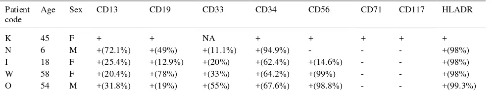
Table 2. The results of cell-surface antigen analysis of t(8;21)(q22;q22)
Patient code
Age Sex CD13 CD19 CD33 CD34 CD56 CD71 CD117 HLADR
K 45 F + + NA + + + + +
N 6 M +(72.1%) +(49%) +(11.1%) +(94.9%) - - - +(98%)
I 18 F +(25.4%) +(12.9%) +(20%) +(62.4%) +(14.6%) - - +(98%)
W 58 F +(20.4%) +(78%) +(33%) +(64.2%) +(99%) - - +(98%)
O 54 M +(31.8%) +(19%) +(55%) +(67.6%) +(98.8%) - - +(99.3%)
Figure 1. Specimens taken from t(8;21)- and t(8;21)+ cases Panel A: a specimen from a t(8;21)- case showed mature neutrophils with pale colored cytoplasm without any granules and myeloblasts type I, panel B: mature neutrophils from a t(8;21)+ case showing homogenous pink colored cytoplasm, panel C: the blast cells type III from a t(8;21)+ case, panel D: myelocytes with abundant granules from a t(8;21)+ case, panel E: eosinophils with abundant granules, from a t(8;21)+ case, and panel F: myeloperoxidase reaction from a t(8;21)+ case.
DISCUSSION
While two reports have described the morphologic features for AML-M2 with t(8;21)11,12 there have been no such description in the monographs or textbooks of hematology.13-16 Therefore, the reliability of the morphological characteristics to forecast t(8;21) is not known .
Jaffe et al reported several common morphological features in AML with t(8;21), but these features are not specific. In our attempt to diagnose AML-M2 patients with t(8;21) based on the morphological characteristics, we found pink colored cytoplasm in most of mature neutrophils, myelocytes with scarce granules, myelocytes with abundant granules, eosinophilia (eosinophilic myelocytes or metamyelocytes above 5%) very low percentage of type I blasts and a high percentage of type III blasts. These findings were not found in any of BM smears from AML-M2 patients without t(8;21)(q22;q22) (unpublished result). Among
these characteristics, myelocytes with abundant granules have not been reported yet, and supposed to be a new finding which could characterize AML-M2 with t(8;21) specifically.
Nakamura et al compared 30 AML-M2 with t(8;21) with 50 AML-M2 without t(8;21). It was disclosed that irregular nuclear shape, Auer bodies and homogeneous pink colored cytoplasm of mature neutrophils were observed in 90 to 100% of the AML with t(8;21). Pink colored cytoplasm were very characteristic. Conversely, pale-colored cytoplasm without any granules was observed in 84% of the AML-M2 without t(8;21)(q22;q22).12 Thus, most of our observations, except for “myelocytes with abundant
granules”, confirmed the findings of Nakamura et al.
CD13, CD19, CD33, CD34, and HLA-DR were expressed by the leukemia cells from all 5 tested patients, while CD56 was expressed in 4 of 5 patients. We found that the incidence of expression of CD34 and HLA-DR was quite high. This finding was in line with other findings which showed that the incidence of the expression of CD34 and HLA-DR in t(8;21) AML cells was significantly higher than those in AML-M2 without t(8;21).17,18 Borowitz et al (1989) have reported a high positivity of CD34 (45%) in the more immature leukemias,19 and Ferrara et al 1998 reported that t(8;21) showed significant high expressions of CD19,CD34, CD56, CD45RA and CD54.20
The CD19 antigen, which is usually a B-lineage specific antigen, has been known as very characteristic for AML with t(8;21).18 This observation seems to be associated with the absence of CD7 antigen expression.21 The high incidence of CD34 expression and the lineage promiscuity such as in those showing CD19 antigen expression suggest that this translocation occurs during early stage of hematopoietic differentiation.18
When t(8;21)(q22;q22) is suspected morphologically, the preservation of frozen samples or metaphase spread is strongly recommended to confirm the diagnosis. By using the frozen sample, the later reverse transcription (RT) PCR (RT-PCR) can indicate AML-1-ETO(MTG8) fusion gene. As long as suitable metaphase spreads are available, a later chromosomal analysis can confirm t(8;21)(q22;q22). The awareness of the various morphological characteristics for t(8;21) can keep hematologists being aware of the subclasses of AML, and can promote the use of the above laboratory procedures.
In conclusion, the characteristic of AML-M2 with t(8;21)(q22;q22) are pink colored cytoplasm in mature neutrophils, neutrophilic myelocytes or metamyelocytes without granules or with scarce granules, eosinophilia (eosinophilic myelocytes and metamyelocytes), myelocytes with abundant granules and low percentage of type I blasts.
REFERENCES
1. Bennett JM, Catovsky D, Daniel MT, Flandrin G, Galton DAG, Gralniick HR, et al. Proposal for revised criteria for the classification of acute myeloid leukemia. Ann Intern Med. 1985;103:620-5.
2. Zaccaria A, Valenti A, Toschi M, Salvucci M, Cipriani R, Ottaviani E, et al. Cryptic translocation of PML/RARA on 17q. A rare event in acute promyelocytic leukemia. Cancer Genetic Cytogenetic. 2002;138: 169-173.
3. Sainty D, Liso V, Cantu-Rajnoldi A, Head D, Mozziconacci MJ, Arnoulet C, et al. A new morphologic classification system for acute promyelocytic leukemia distinguishes cases with underlying PLZF/RARA gene rearrangements. Blood. 2000;96:1287-96.
4. Petti MC, Fazi F, Gentile M, Diverio D, Fabritiis PD, DePropris MS, et al. Complete remission through blast cell differentiation in PLZF/RARa-positive acute promyelocytic leukemia: in vitro and in vivo. Blood. 2002;100:1065-7. 5. Grimwade D, Biondi A, Mozziconacci MJ, Hagemeijer A,
Berger R, Neat M, et al. Characterization of acute promyelo-cytic leukemia cases lacking the classic t(15 ;17) : results of European Working Party. Blood. 2000;96:1297-308. 6. Foley R, Soamboonsrup P, Kouroukis T, Leber, B, Carter
RF, Sunisloe L, et al. PML/RARA APL with undifferentiated morphology and stem cell immunophenotype. Leukemia. 1998;12:1492-93.
7. Aventin A, Mateu R, Martino R, Colomer D, Bordes. A case of criptic acute promyelocytic leukemia. Leukemia. 1998;12:1490-1.
8. Jaffe SE, Harris NL, Stein H, Vardiman JW. World Health Organization classification of tumours : Pathology & genetics, tumours of haematopoietic and lymphoid tissues. Lyon: IARC; 2001.
9. Mrozek K, Heinonen K, Bloomfield, CD. Clinical importance of cytogenetics in acute myeloid leukaemia. Best Practice & Research Clinical Haematology. 2001; 14(1):19-47.
10. Tkachuk D, Hirschmann JV, McArthur JR. Atlas of clinical hematology. Seattle: Saunders; 2002.
11. Swirsky DM, Li YS, Matthews JG, Flemans RJ, Rees JKH, Hayhoe FGJ. 8;21 translocation in acute granulocytic leukaemia : cytological, cytochemical and clinical features. Br J Haematol. 1984;56:199-213.
12. Nakamura N, Kuriyama K, Sadamori N, Mine M, Itoyama T, Sasagawa J, et al. Morphological subtyping of acute myeloid leukemia with maturation (AML-M2): Homogeneous pink-colored cytoplasm of matute neutrophils is most characteristic of AML-M2 with t(8;21). Leukemia. 1997;11:651-5.
13. Bain BJ. Leukaemia Diagnosis. 3rd edition. Massachusetts: Blackwell; 2003.
14. Gross S, Roath S. Hematology A problem-oriented approach. 1st edition. Baltimore: Williams & Wilkins; 1996.
15. Hoffbrand AV, Pettit JE. Color atlas of clinical hematology. 3rd edition. London: Mosby; 2000.
16. Mc Kenzie SB. Textbook of hematology. 2nd edition. Baltimore: Williams & Wilkins; 1996.
17. Ghosh S, Shinde SC, Kumaran GS, Sapre RS, Dhond SR, Badrinath Y, et al. Haematologic and immunophenotypic profile of acute myeloid leukemia: An experience of Tata memorial hospital. Indian Cancer. 2003; 40(2):71-6. 18. Kita K, Nakase K, Miwa H, Masuya M, Nishii K,
Morita N, et al. Phenotypical characteristics of acute myelocytic leukemia associated with the t(8;21)(q22;q22) chromosomal abnormality: Frequent expression of immature B-cell antigen CD 19 together with stem cell antigen CD34. Blood. 1992; 80:470-7.
19. Borowitz MJ, Gockerman JP, Moore JO, Civin CI, Page SO, Robertson J, et al. Clinicophatologic and cytogenetic features of CD34 (My10) positivity acute nonlymphocitic leukemia. Am J Clin Pathol. 1989;91:265-70.
20. Ferrara F, Noto RD, Annunziata M, Copia C, LoPardo C, Boccini P, et al. Immunophenotype analysis enables the correct prediction of t(8;21) in acute myeloid leukemia. B J Haematol. 1998;102:444-8.
21. Tatsumi E, Yoneda N, Kawano S, Yamaguchi N, Yabe H, Nagai KI, et al. Expression of CD7 antigen precludes t(8;21)(q22;q22) Chromosome aberration in acute myeloblastic leukemia. Blood. 1992; 79: 3092-3.
22. Kahl C, Florshutz A, Leuner S, Arland M, Janssen JWG, Franke A, et al. CD7/CD56 double-positive acute myelogenous leukemia. Lab Hematol. 1999; 5:115-20. 23. Seymour JF, Pierce SA, Kantarjian HM, Keating MJ,
Esley EH, Anderson M. Neural cell adhesion molecule (CD56) is associated with FAB subtype, cytogenetic and skin infiltration in acute myelogenous leukemia (AML). Blood. 1993;82(suppl 1):126 a.
24. Byrd JC, Edenfield WJ, Sheilds DJ, Dawson NA. Extra medullary myeloid cell tumor in acute non lymphocytic leukemia : A clinical review. J Clin Oncol. 1995;13:1800-16.