Vol 6, No 4, October - December 1997
BiLateral Radical Neck Dissection 255
Simultaneous
Bilateral Radical
Neck
Dissection
Follo\ryed
by
Internal
Jugular Vein
Reconstruction
Hartono Abdoerrachman *, Bambang Hermani *, Murnizal Dahlan x*, purnaman Sardjono pandi x.
Abstract
performing simultaneous bilaterar radicar neck dissection and vein using savena magna vein. Six month follow up showed no
Abstrak
ah lama
d
ukan sebabaliran da
ditakuknn
Y:::;T:"
Keywords: Laryngectomy, Carcinoma of the larynx, Neck nodes metastases
Tumor of the head and neck region is often followed
by
neck nodes involvement.iu*o.
that has been spreading to the cervical lymph gland, needs a special considerationin
the treatment, depending on the stadiumof
thetumor,
the primary tumor and theIocation of the spread.
There are several modalities
in
treating the cervical spread and one ofthe treatment is radical neck dissec_ tion. Radical neck dissection still holds a prominent position and plays a vital role in surgical mÀagement of cancer of head and neck to contràl regional metas_ tatic disease.Usually neck dissection is performed unilaterally, on the involved side
of
the neck, while bilateral neck* Department of Otorhinolaryngology, Facuhy of Medicine,
U n iv e r s i ty of In do ne s ia,/ D r. C ip t o M an g un kus umo H o spit al, J akart a, Indo ne s ia
** Department of Surgery, Facuby of
Medicine, University of I ndo ne s ia,/D r. C ipt o M an g un kus umo H o s pital, J a kar ta, Indonesia
dissection
is
more uncommonin
regardsto
serious complications.Bilater
havins
1926,"
iË,î""'T"in3;:liïii;,iTî
ong time ago.Major complications of bilateral radical neck dissec_
been reported.
We reported a case with carcinoma of the larynx and bilateral cervical metastasis.
To
apprehend the in_ and other serious complications, a joint with a vascular surgeon was organized to simultaneous bilateral radical neck dissec_tion,
followed
by
laryngectomy andfinally
to256 Abdoenachman et aI.
METHOD
Case
On June 7987, a man, 65 years of age, was referred to
the Department of Otorhinolaryngology, Faculty of Medicine, University
of
Indonesia,with
the com-plaints of hoarse voice and stridor inspiratoir.Deform-ity of cervical contour was
clearly visible due toswelling on both sides of the neck.
CT-Scan
of the
larynx and
direct
laryngoscopyrevealed that the tumor involved the supraglottic and glottic region of the larynx. Neck nodes were palpable on both sides of the neck, and fixed to the surrounding
tissue. Histological
finding
of
the tumor showed squamouscell
carcinoma. Laboratory finding werewithin
normal
limits
and
no
metastasis was demonstrated on chest X-photo.In a joint cancer meeting, it was decided to conduct a
joint
operationwith
a vascular surgeon to performsimultaneous bilateral radical neck dissection to secure the radicalization, than followed
by
jugular veinreconstruction on one side to prevent the unexpected
complication, and total laryngectomy.
Surgical procedure
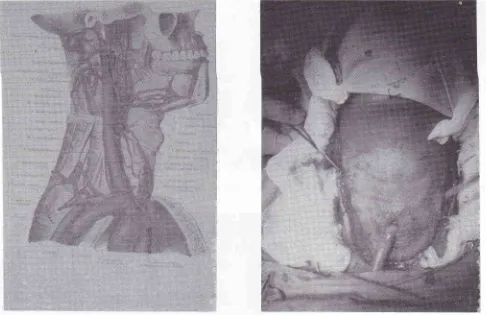
On June 1987 bilateral radical neck dissection by
removing both internal jugular veins was performed under general anesthesia followed by resection of the larynx, as shown
in
Figure 1-8. Figure 1 showed awide incision on the skin for extended access of the
tumor, followed by bilateral radical neck dissection and extirpation of the larynx, as shown in Figure 2. Figure 3 showed the surgical area afterwards. The
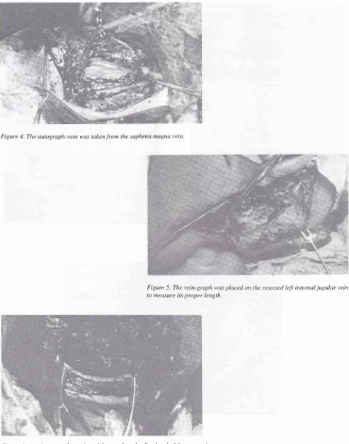
vascular surgeon resected the saphena magna vein
from the thigh
for
autograph(Figure
4).
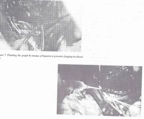
The autograph-vein was then placed at the siteof
the resected left internal jugular vein as shown in Figure5. After suturing the distal end of the graph (Figure 6), heparin flush was given to prevent clogging by blood clot (Figure 7). Overview of the excized larynx and the
reconstructed
left internal jugular vein
wasdemonstrated
in
Figure
8,
after closing
thehypopharynx. Finally, layer by layer of closing proce-dure of the wound was performed after the insertion of a haemovac-drain and nasogastric tube.
Med J Indones
Monitoring of complications
The patient was followed-up for 6 months post
opera-tively, to check for the presence
of
intracranial and local complications.RESULTS
One month later when the patient was discharged from
the hospital, no intracranial complications were noted. The patient was followed up intensively for 6 months post-operatively and no intracranial or local complica-tions were noted.
DISCUSSION
Our case had carcinoma
of
the larynx and bilateral cervical metastasis. The presence of bilateral cervical metastasis was regarded as a sign of inoperability and incurability by many surgeons. Some surgeonssug-gested
bilateral
neck dissection,but
anatomistdeclared that ligation of both internal jugular veins was very dangerous. Because of the serious complication and hesitancy in ligating both jugular veins, the
opera-tion did not become popular, until some experts, e.g.
Mc
Guirt and Razack reported their successful ex-periences.3'4 Since that time increasing number of the procedure have been performed, while others are reluc-tant to perform such a radical and simultaneousproce-dure for neck dissection due.to undesired serious
complications. However, sometimes they are
con-fronted
to
a
critical situation that needa
difficultdecision which sometimes controversial to an estab-lished and widely used procedure.
The venous return from cervical region is served
most-ly by both internal jugular veins and vertebral venous plexus. The studies of investigators documented
ade-quate collateral venous circulation after ligation of
both internal jugular veins. The main route of venous drainage
in the
absenceof
the jugular veinsis
thevertebral venous plexus, the emissary veins, and the veins of the posterior cervical region. Figure 9 shows the vertebral venous plexus.)
Several complications have been reported by the ex-perts. Therefore the surgeons should be aware of these
complications, and when they do occur, deal with them
Vol 6, No 4, October - December 1997
Figure
!.
Awide û-shape n,ide skin incisi.on on rhe neck_ ro provide wide accessibility to the operating.fie!J.BiLateral Radical Neck Dissection 251
Figure 2. BiLateral radicai neck
extiryation of the larynx. dissection and fol.lowed b1
Abdoerrachman et al.
Figure 4. The autograph-vein was taken from the saphena magna vein.
Med J Indones
Figure 5. The vein-graph was placed on the resected left internal jugular vein
[image:4.595.50.543.81.708.2]to measure its proper length.
Figure 6. Attachment and suturing ofthe graph to the distal end ofthe resected
Vol 6, No 4, October - December 1997
Figure 7. Flushing the graph by means ofheparin to prevent clogging by btood clot.
Bilateral Radical Neck Dissection 259
Figure-8. Overwew of the excisecl,operating field after closing of the
hypopharynx' The reconstructed
rjt i"t"-ti
i).gr1r, ,"in
,o,
demonstrated infront of the arrow.Figure 9 schematic ilru.çtration of the vertebrar venous pLexus in
260 Abdoerrachman et al.
Many experts suggested to perform bilateral neck
dis-section
in
two
stages, while othersdid
a modifiedsimultaneous bilateral neck dissection such as
func-tional neck dissection to prevent the undesired
com-plications.6 Razack meniioned
that
radical blockdissection of both sides of the neck in the same patient
had been performed as a two stage procedure for many
years, with the customary preservation of one internal
jugular vein due to the fear of fatal cerebro-vascular
complication. To his opinion, careful stripping and
preservation of the vein on one side is an acceptable
safety measure.a
Other writers said that there was no added risk in
removing both internal jugular vein. MartinT stated that
the indication for removing the second internal jugular
vein
in
abilateral neck dissection (either simultaneousor staged) was the same as for the removal of the first vein. His experiences in the clinic had shown that there
was no increase
in
post-operative mortalityor
dis-ability that could be ascribed to the removal
of
the second internal jugular vein. He believed that a com-plete neck clissgction was not possibieif
the vein wasnot resected.
In this case we dicl an extra ordinary procedure as an
alternative to overcome the disadvantages of wide and
extended dissection of the neck, by reconstructing one side of internal jugular vein using saphena magna vein.
Med J Indones
Our result showed that the reconstruction of internal
jugular vein could prevent intracranial complications.
CONCLUSION
In
wide
and extended dissectionof
the
neck,reconstruction of one side of internaljugular vein using
saphena magna vein can prevent intracranial
complica-tron.
REFERENCES
1
.
Crile G. Excision of cancer of the head and neck with speci a1reference to the plan of dissection based on I 32 patients.
JAMA 1906;47:1780-6.
2.
Bartlett EY, Callender CL. Neck Dissection' Surg Clin N Amer 1926;6:481-504.3.
Mc Guirt WF, Mc Cabe BF. Bilateral Radical Neck Dissec-tion. Arch Otolaryngol 1980l,106:427 -9.4.
Razack MS, Baffi R, Sako K. Bilateral Radical Neck Dissec-tion" Cancer 1 985'/7 :19i -9.-5. Spalteholz
W
Flandatias der anatomie des menschen. Amstsrdam Schslloma & Relkema, 19JQ,(:,. Abdoerrachman Ftr, Roesmarjono, Flermani B. Functionai
Neck Dissection. Kumpulan Naskah Ilmiah Konggres Nasional PERHATI VII; 1983, August 21-23; Surabaya. Surabaya: Perl.rati, 1 983 :846-5C.