12
The Ethanol Extract of
Physalis angulata
Linn Inhibits COX-2
Activity in MCF-7 Cell
In Vitro
EM Sutrisna1*, Indwianiastuti2, Haryadi2
1
Faculty of Pharmacy of Muhammadiyah of University of Surakarta, Surakarta 2
Faculty of Medicine of Gadjahmada University, Yogyakarta
* Corresponding author, email: [email protected]
Abstract
Some studies showed that ceplukan (Physalis angulata Linn) have cytotoxic effects toward HeLa (cervical cancer), KB (Nasopharyngeal), Colo 205 (colon), Calu (Lung) and MCF-7 cells in vitro. This plant has cytotoxic effects toward rat P388 lymphocytic leukemia
in vivo. One of the mechanisms of cytotoxicity is the inhibition of Cyclooxygenase-2 (COX-2) pathway. The purpose of this study was to determine if 70% ethanol extract of Physalis angulata Linn inhibited COX-2 activity in MCF-7 cell. The cytotoxic effect of ethanol extract
of Physalis angulata Linn toward MCF-7 cell was examined at concentration 20, 40, 80 and
160 µg/mL. Its estimated IC50 was used to test inhibition of COX-2 activity. After 24 hours of incubation, the inhibition of COX-2 activity was assayed by imunohistochemical staining with a monoclonal anti COX-2 antibody. COX-2 positive cells were counted using binocular microscope and IC50 for inhibition of COX-2 activity was calculated. The inhibition of COX-2 activity at 10, 20 and 40 µg/mL of the extract was 39.3%, 42.06% and 51.73%, respectively. The IC50 value of the ethanol extract of P. angulata Linn. for the inhibition of COX-2 activity was 37.57 ± 3.11 µg/mL, while the IC50 of celecoxib was 5.31 ± 0.27 µg/mL. The ethanol extract of Physalis angulata Linn. have an inhibitory effect on COX-2 activity in MCF-7 cell with IC50 37.57 ± 3.11 µg/mL
Key words:Physalis angulata Linn, cyclooxygenase-2 (COX-2), MCF-7 cell line
INTRODUCTION
In Indonesia, cancer is the sixth leading cause of death after infectious diseases, cardiovascular diseases, traffic accidents, malnutrition and congenital abnormalities (Tjindarbumi & Mangun-kusumo, 2002). According to WHO report in 1998, the most frequent type of cancer found in women are cervix (25.3%) and breast cancer (18.4%) (Anonim1, 2005).
Cancer is usually treated by surgery, radiotherapy, chemotherapy and immuno-therapy (Van de Velde et al., 1999). Those treatments cost highly and bring many side effects. Due to those reasons, many researchers hold studies to find new more effective and selective drugs.
Indonesia is a country that has great biodiversity. There are about 30.000 plant species found in Indonesia, and approxi-mately 1260 species can be used to cure diseases. Several plants are understood to have anti-cancer effect (Mangan, 2003).
One of them is ceplukan (Physalis
angulata Linn. and Physalis minima
vitro against H1477 (melanoma), Hep-2 (laryngeal) and 8401 (glioma) and they also showed antitumor effect against P388 lymphocytic leukemia in mice in vivo (Chiang² et al., 1992).
Other study conducted by Choi and Hwang (2003), showed P.angulata Linn. could inhibit inflammation in mice carrageenan (Choi & Hwang, 2003). According to Davies et al. (2002), the main mechanism of action of com-pounds/drugs that have anti-inflammatory effect is through inhibition of the cyclooxygenase (COX). Cyclooxygenase
also is known as prostaglandins
endoxyperoxide synthase. This enzyme is a
catalyst for the transformation of
prostaglandin endoxyperoxide (prostaglan-din H2) from arachidonic acid. Two
identified isoforms of prostaglandin
synthase are COX-1 and COX-2. COX-1 is expressed in most tissues and thought to be involved in the process of cellular homeostasis, while COX-2 is often not detected in normal tissue, but it will show up quickly in response to various stimuli including mitogen, hormones, cytokines, growth factors and factors. The excessive expression of COX-2 and the high concentrations of prostaglandins are often supposed to have correlation with chronic inflammatory diseases such as rheumatoid arthritis, and several human cancers including colon, lung, bladder, prostate, stomach and breast cancer.
Prostaglandins mediate tumor forma-tion through several mechanisms such as cell proliferation, apoptosis, modulation of the immune system and angiogenesis. In chronic inflammation, angiogenesis can help preventing the inflammatory process. (Davies et al. 2002). Angiogenesis is an important factor in tumor growth and metastasis (Costa et al., 2002). In experi-ment done on animals, inhibition of COX-2 expression by COX-COX-2 inhibitors can inhibit the angiogenesis process. COX-2 inhibitors may also prevent the deve-lopment of breast tumors in mice (Davies
et al., 2003). Non-steroidal
anti-inflam-matory drugs administration, for at least 3 times a week in at least a year, will reduce the risk of breast cancer (Haris et al., 1996). The administration of non-steroidal anti-inflammatory drugs for 13-36 months will reduce the risk of breast cancer (Langman, et al.,2000).
The present study was aimed at examining the inhibition of COX-2 activity of P angulata L. for MCF-7cells.
METHODS Tools
Glassware (Pyrex), analytical ba-lance, beaker glass (Pyrex), mixer, filter 0.2 um, micropipette, liquid-nitrogen tank, refrigerator, CO2 incubator (Nuaire),
lami-nar air flow cabinet (Nuaire), microplate 96 (Nuclone) wells, pipette ependorf,
inverted mycroscope, hemocytometer
(New Bouer), pH meter, glass objects, light microscopes, centrifuges, tissue cul-ture flasks (Olympus).
Materials
Test substance. ethanol extracts of
ceplukan (Physalis angulata Linn.), Posi-tive controle tamoxiphen (Tamoplex® Combiphar), and the positive control celecoxib (Celebrex® Pharmacia). MCF-7 cell line containing estrogen receptor alpha
and beta isoforms (Anonim2, 1998),
Medi-um RPMI 1640 (GIBCO), fetal bovine serum/FBS 10%, 3% penicillin-streptomy-cin, 1% fungison, gentamicin (Merck), aquabidest, 70% ethanol, 0.5% trypsin and thrypan blue, monoclonal antibody COX-2 (NovoCastra), PBS pH (7.4),
avidin-biotinylated antibody IgG secondary
biotinilated (Novo Castra), haematoxylin eosin (Dako), 3% H2O2 and methanol.
Extraction
14
MCF-7 cell propagation
MCF-7 cells taken from nitrogen tank were heated in 37° C immediately and then sprayed with 80% alcohol. The cells were transferred in a sterile centrifuge tube containing 10 ml RPMI 1640-serum me-dium. This suspension then was centri-fuged on 1200 rpm for 5 minutes. Super-natant liquids were removed and replaced with RPMI 1640 medium. After 20 mi-nutes, the cells were centrifuged at 1200 rpm for 5 minutes. Supernatant fluids were discarded, left 1 ml of suspension to be done again. Once homogenized, the cells were included in the tissue culture flask with medium containing 20% fetal bovine serum (FBS). The cells were observed by inverted microscope. After the cells grew confluent, cells were harvested and then centrifuged at 2000 rpm for 5 minutes. Supernatant fluids were removed and left for approximately 1 ml for re-suspension until homogeneous. The cells were added by medium containing 10% FBS after-wards (Bakhriansyah, 2004)
Cytotoxic test
Micro cultures with 96 wells were prepared. MCF-7 cells with density of 1.5 x 10⁴/mL were included in the 96 wells plate dissolved in 100 µl RPMI 1640 culture medium. The MCF-7 cells were incubated for 24 hours. Then 100 µl of ethanol extract of Physalis angulata Linn and tamoxiphen were added. Each con-centration was done three times. The micro cultures were incubated using CO2 incubator 37°C for 24 hours. The MCF-7 cells then were washed using 100 µl PBS 0.25% 2 times. To release the live cancer cells attached to the wells, samples were given with100 µl of 0.25% trypsin. 10 µl of cells were discarded and then were added with 10 µl tryphan blue on these wells. 10 µl of mixtures was taken, and placed on a haemocytometer chamber and the number of the living cells were counted. The percentage inhibition of the ethanol extract of Physalis angulata Linn, and tamoxiphen and negative control were
calculated by the following formula (Bakhriansyah, 2004):
Immunohistochemistry test
The 70% ethanol extract of P.
angulata Linn. at concentration 10, 20 and 40 µg/mL and celecoxib at 5, 10 and 20 µg/ml which had been incubated 24 hours were made on a glass object, then was soaked using 3% H2O2 in methanol for 20
DATA ANALYSIS
The inhibition of COX 2 activity was calculated by formula:
IC50 was determined based on the
inhibitory activity of COX-2 expression. Differences in IC50values were analyzed
by independent T-test(95%)
RESULTS
Immunohistochemistry test
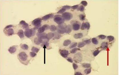
On visual observation, the cells that indicate COX-2 expression have dark brown -colored cytoplasm, while the cells that do not expressCOX-2 have light-colored cytoplasm. (figure 1).
Figure 1. MCF-7 cells that express COX-2 (red arrows) and did not express COX-2 (black arrows).
The calculation results from visual observation of cells with COX-2 expres-sion can be seen in table 1.
From Table 1, the percentage inhi-bition of ethanol extract of P angulata
Linn. can be calculated. The percentage inhibition of the ethanol extract of P angulata Linn on 10µg/mL, 20 µg/mL and 40 µg/mL were 39.3%, 42.06% and 51.73% respectively. From the results, IC50
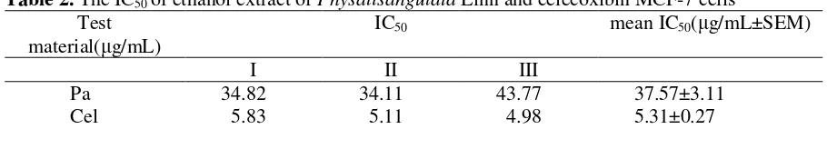
can be determined by linear regression. Table 2 shows that the IC50 value of
the ethanol extracts of P. angulata Linn.
on the inhibition of COX-2 activity 37.57 is ± 3.11 µg/mL, while the IC50 for
celecoxib 5.31 is ± 0.27 µg/mL. It means that although P. angulata Linn. is capable of inhibiting COX-2 activity but the potential of ethanol extracts P. angulata
Linn is lower than celexocib. The independent T-test result shows that there are significant differences on IC50 values
between P. angulata Linn. and celexocib p. 0.000 (95% CI)
DISCUSSION
Many studies claim that the
inhibition of COX-2 has effect on tumor formation. In an experiment using mice, excessive expression of COX-2 can cause mamae tumor formation, but COX-2 selective inhibitors can suppress the tumor mamae (Sivulaet al., 2005). To investigate whether the cytotoxic effect of ethanol
extract P. angulata Linn. through
inhibition of COX-2 activity, the cells are stained by antibody of COX-2. From table 2, it is known that ethanol extract of P. angulata Linn can deter COX-2 activity with IC50 37.57 ± 3.11 µg / mL. This
happens because celecoxib is COX-2 selective medicine.
Several studies on COX-2 states that COX-2 inhibitors affect cell proliferation. Celecoxib (selective COX-2 inhibitor) and the SC560 (COX-1 inhibitor) can induce cells to rest in G0/G1 phase in three different colon cancer cells regardless of whether these cells express COX-2 or not (Groschet al., 2001). A study conducted by Hu et al. (2003) showed that the adminis-tration of selective COX-2 inhibitors, Ns-398 in Hep G2 cells in humans will lead to inhibition of proliferation which correlates with the reduction of 5-bromo-2-deoxy-uridin (BrdU) obtaining. This causes the reduction of cell cycle progression in the G1-S transition phase.
16
Table 1. Quantitative results of visual observation of COX-2 expression cells
BU( g/mL) CI 1 %I probit C2 2 %I probit C3 3 %I probit PA 10 96 60 37,50 4,68 96 58 39,58 4,73 98 58 40,82 4,77 PA20 96 57 40,63 4,78 96 56 41,67 4,79 98 55 43,88 4,84 PA40 96 45 53,13 5,08 96 47 51,04 5 98 48 51,02 5 Cel 5 96 51 46,88 4,92 96 49 48,96 4,98 98 51 47,96 4,95 Cel10 96 38 60,42 5,26 96 39 59,38 5,23 98 36 63,27 5,34 Cel20 96 29 69,79 5,52 96 33 65,63 5,4 98 33 66,33 5,42
Notes: PA (P. angulata L.), Cel (Celexocib), % I: % inhibition, C1: control on replication1, I: number cells on replication 1
Table 2. The IC50 of ethanol extract of Physalisangulata Linn and celecoxibin MCF-7 cells
Test material( g/mL)
IC50 mean IC50( g/mL±SEM)
I II III
Pa 34.82 34.11 43.77 37.57±3.11
Cel 5.83 5.11 4.98 5.31±0.27
Notes : Pa: P. angulata Linn, Cel: celecoxib
decreases the proportion of cells in S phase and G2/M on the cell cycle (Li et al., 2003). COX-2 selective inhibitor, SC'236, given to mice suffering from sarcoma 6mg/bw inhibits tumor growth and neo-angiogenesis (Kishiet al., 2000). The provision of COX-2 inhibitors reduce cancer through the reduction of blood vessel formation (Davies et al., 2002). COX-2 expression inhibits angiogenesis and apoptosis in breast cancer (Costa, et al., 2002). The study by Chiang1 et al. (1992), suggests that the extract of P. angulata Linn. inhibits protein synthesis in leukemia cells. The ethanol extract of
Physalis peruviana (EEPP) triggers
apoptosis through the release of
cytochrome c, SMAC / DIABLO and Om/HtrA2 from mitochondria to cytosol that may lead caspase 3 activation (Wu, et al., 2004). Apoptosis occurs when cells are treated by 50µg / mL EEPP. After 48 hours of treatment with EEPP, apoptosis of Hep G2 cells is correlated with increased expression of p53, CD95 and CD95L. The administration of this EEPP also causes down-regulation of Bcl-2, Bcl-xl and XIAP and up-regulation of Bax. The
results also indicate that apoptosis
triggered by EEPP in Hep G2 cells is possibly through CD95 and CD95L system
and the mitochondrial signaling trans-duction pathway. Other study claimed that the EEPP has strong antihepatoma activity. Besides, it has apoptosis effects that are correlated with mitochondrial dysfunction (Wu et al., 2004)
CONCLUSION
The ethanol extract of Ceplukan (P. angulata Linn.) inhibits COX-2 activity in
MCF-7 cells with IC5037.57±3.11 µg / mL
SUGGESTION
Further investigation is needed to find out active compounds that have effect on inhibition of COX-2 expression.
REFERENCES
Anonim1, 2005. Disease Specific NCD
Morbidity and Mortality Profile,
Http://w3.whosea.org/linkfiles/NCD_i nforbase.disease-spesific.pdf, diakses,
April 2005
Anonim2,1998, Feasibility Demonstration
Project for HTPS, Http://www.
bdbiosciences.com/ clontech, diakses Juli 2005
Bakhriansyah, M., 2004. Pengaruh Ekstrak Etanol Biji Mahkota Dewa pada Sel
Kanker Payudara T47D: Kajian
dan Penghambatan Ekspresi Siklo-oksigenase-2, Thesis, Fakultas Pasca-sarjana UGM Jogjakarta.
Chiang¹,H.C., Jaw SM, Chen CF, Kan WS, 1992. Antitumor Agent, Physalin F from Physalisangulata L., Anticancer Res., 12(3): 837-43.
Choi, EM, Hwang JK, 2003. Investigations of Anti Inflammatory and Antinoci-ceptive Activities of Piper Cubeba, Physalis angulata and Rosa hibrida, J Ethnopharmacol, 89(1): 171-5.
Costa, C., Soares, R., Reis-Filho, J.S., Amendoeira, I and Schmitt, FC., 2002. Cyclooxygenase 2 Expression is Asso-ciated with Angiogenesis and Lym-phonode Metastasis in Human Breast Cancer, J. ClinPathol, 55: 429-34 Davies, G, Martin, L.A., Sacks, N.,
Dowsett, M., 2002. Cyclooxygenase-2 (COX-2), Aromatase and Breast cancer: A Possible Role in for COX-2 Inhibitors in Breast cancer Chemopre-vention, Ann Oncol. 13: 669-78. Davies, G., Salter, J., Hills, M,
Ann-Martin, L. Sacks, N., Dowsett, M., 2003. Correlation between Cyclooxy-genase-2 Expression and Angio-genesis in Human Breast cancer, Clin cancer Res. 9: 2651-2656
Grosch, S., Tegeder, I., Neiderberger, E., Brautigam, L., and Geislinger, G., 2001. COX-2 Independent Induction of Cycle Cells Arest and Apoptosis in Colon cancer Cells by The Selective
COX-2 Inhibitor Celecoxib, FASEB,
J., 10 (15): 2742-44.
McCracken, J.D., Hillebrand, D.J., and Hasan, M., 2003. Inhibited Prolife-ration of Cyclooxygenase-2
Express-ing Human Hepatoma Cells by NS-398, A Selective COX-2 Inhibitor. Int. J. Oncol. 22: 757-63.
Kishi, K., Petersen, S., Petersen, C., Hun-ter, N., Mason, K., Masferrer, J.L., Tofilon, P.J. and Milas, L., 2000. Preferential Enhancement of Tumor Radioresponse by a Cyclooxygenase-2 Inhibitor, Cancer Res. 60: 1326-31. Langman, M.J.S., Cheng, K.K., Gilman,
E.A. and Lankankershire, R.J., 2000. Effect of Antiinflamatory Drugs on Overall Risk of Common Cancer: Case Control Study in General Practice Research Database. Br Med J. 320: 1642- 46.
Li, J., Wang, X., Chen, F., Yu., J., and Luo, H., 2003. Nimesulide Inhibits Proliferation via Induction of Apop-tosis and Cell Cycle Arrest in Human Gastric Adenocarcinoma Cell Line.
World J. Gastroenterol, 9(5): 915-20. Mangan, Y., 2003. Cara Bijak
Menakluk-kan Kanker, Cetakan I, Agrome-diaPustaka, Jakarta, 28-44
MOH, 1986, Sedia Galenik, Jakarta
Sivula, A.,Talvensaarimatilla, A., Lundin, J., Joensuu, H., Haglund, C., Risti-maki, A., and Turpeenniemi-Hujanen, T., 2005. Association of Cyclooxy-genase-2 and Matrix Metal-loprotein-ase-2 Expression in Human Breast
2004. Antihepatoma Activity of
Physalis angulata and P. peruviana
Extracts and Their Effects on
Apoptosis in Human Hep G2 Cells,