DOI: 10.12928/TELKOMNIKA.v11i4.1273 783
Separability Filter for Localizing Abnormal Pupil:
Identification of Input Image
Retno Supriyanti*1, Elvin Pranata1, Yogi Ramadhani1, Tutik Ida Rosanti2
1
Electrical Engineering Dept, Jenderal Soedirman University, Purwokerto, Indonesia
2 Medical Science Dept, Jenderal Soedirman University, Purwokerto, Indonesia
*Corresponding author, e-mail: [email protected], [email protected], [email protected], [email protected]
Abstrak
Metode yang cukup handal untuk pendektesian pupil adalah metode separability filter, namun selama ini metode ini hanya diterapkan dalam mendeteksi pupil pada mata normal saja. Di lain pihak, pada mata abnormal seperti pada penderita katarak atau glukoma memiliki karakteristik yang berbeda dengan mata normal. Penelitian ini mencoba untuk menerapkan separability filter dalam mendeteksi pupil pada mata abnormal. Masalah yang dihadapi adalah perbedaan ukuran, bentuk dan warna pupil, sehingga perlu untuk mengimplementasikan transformasi hough, blob area, dan kecerahan pada citra masukan sebelum menggunakan separability filter. Hasil eksperimen menunjukkan bahwa penambahan pengolahan awal citra dapat meningkatkan unjuk kerja deteksi pupil pada mata abnormal hingga 95.65%.
Kata kunci: separability filter, hough transform, blob area, kecerahan, deteksi pupil
Abstract
Separability filter method is a reliable method for pupil detection. However, so far this method is implemented for detecting pupil of normal eye, while for abnormal eye such as cataract and glaucoma patients; they have different characteristics of pupil such as color, shape and radius size of pupil. In this paper we propose to use separability filter for detecting pupil of abnormal patients with different characteristics. We faced a problem about radius size, shape and color of pupil; therefore we implemented Hough Transform, Blob area and Brightness for identifying input images before applying separability filter. The experiment results show that we can increase performance of pupil detection for abnormal eye to be 95.65%.
Keywords: separability filter, hough transform, blob area, brightness, pupil detection
1. Introduction
Rapid developments of Information Technology especially in the field of Digital Image Processing give very big impact in many areas. Face recognition is a part of fields in digital image processing techniques in which iris and pupil detection as one of its parts is a topic widely discussed in various research areas such as in medical field area. In this field, usually pupil detection is implemented for early detection of eye diseases such as cataract [1-6].
robustly localizing the iris and pupil boundaries of a human eye in close-up images. Such an algorithm can be critical for iris identification, or for applications that must determine the subject’s gaze direction, e.g., human-computer interaction or driver attentiveness determination. Masek developed an open source iris recognition system in order to verify both the uniqueness of the human iris and also its performance as a biometric [15]. Fukui and Yamaguchi [16] proposed point feature extraction such as nostril and pupil using separability filter. Originally, separability filter was proposed as a method for edge extracting [17]. In this method an edge is defined as a boundary not as a point in which the intensity changed drastically. However, due to the separability filter using size of circle radius as a main parameter, then this method requires same size of pupil radius of input images relatively.
Almost all works above using normal eye as an object. While in fact, normal eye has different characteristics with abnormal eye especially in color, shape or radius size of pupil. In this paper we emphasize for applying separability filter to localize pupil area for abnormal eye such as cataract and glaucoma patient automatically. We study to consider for using Hough transform, blob area and brightness value to compare performance of using separability filter to localize pupil on abnormal eye. The problem will arise when using separability filter to localize pupil on abnormal pupil detection system are radius size, shape and color of pupil. Due to the fact that input images of abnormal pupil detection system is taken under uncontrolled illumination and also different pupil response between abnormal and normal eye cause to the difference radius size, shape and color of input images significantly.
According to this case, we need to sort input images in order to improve performance of pupil detection based separability filter. Images sorting are done by identifying characteristics of input images refer to the main parameter of separability filter. In this case we consider for using Hough transform for getting fix size of pupil radius, blob area and brightness for segmenting eye area.
2. Research Method
In this research we used primary data in which we take data independently. Data acquisition is done by taking photograph of patient using compact digital camera. A criterion of input image is focused on the whole of patient’s face without background as described in Figure 1.
Figure 1. An example of input image (source: private documentation)
The aim for using face area only is for reducing computation time. Also we have to consider about distance between camera and patient, therefore there is no blur in the input image especially in an eye area. The good level of light focuses also an important factor for getting an ideal input image. Figure 2 shows a flow chart of our research.
In the first step, input image will be compressed for saving computation time and facilitating segmentation process that will be done in the next step. Original size of input image is quite large; therefore we compress our input image to be n x 160 pixel size. n is image width that appropriate to aspect ratio of original image
2.1. Separability Filter
The main purpose of using separability filter is to determine the center of eye roundness by performing convolution of separability filter algorithm to ROI of input images in which the filter is expressed by Equation 1.
Figure 2. Flow chart of research
. . . . (1)
This filter also can be expressed by two vectors as described in Equation 2 and Equation 3.
. . . . (2)
Such that for each (I,j) ([0…(width -1)],[0……(height-1)])
∗ (3)
If the filter is separable, the convolution operation may be performed using only (width + height) multiplications for each output pixel. Applying the separability filter to Equation 2 becomes Equation 4.
Figure 3. Region of Interest
ROI
Input image Collecting characteristics
information
Processing and Analysis
Congruence identification
Classification of input images based on congruence level
Identification Process
(4)
This can be simplified to be Equation 5.
(5)
To apply the separable convolution, first apply Grow as though it were a width by filter.
Then apply Gcol as though it were a by height filter. In our research, the result of this step is
a separablity map image that contains the possible values of each pixel as the center of circle. The greater value of separability in each pixel will be the best candidate of the circle center. In the separability map image, values distribution in which the greatest separability value appears as a bright dots as described in Figure 4.
Figure 4. Image result of separability filter
2.2. Non-Maximum Suppression
According to section 2.1 by using separability filter we able to determine a center of eye. The center is a point that has greatest separability value. However the maximum separability value is distributed around eye center. In order to reduce distributed area to be maximum values only, therefore we implement non-maximum suppression. The result is described in Figure 5.
Figure 5. Non-maximum suppression image
2.3 Hough Transform
Hough transform method is used for classifying process according to the similarity to separability filter that usually using fix radius. Equation 6 described general equation for detecting a circle in an image.
r2 = (x-a)2 + (y-b)2 (6)
(a,b) is a circle center and r is radius.
The main purpose of using Hough Transform is to get eye shape, mark and find the center point of eye shape. Then we have to find correlation between eye circle from separability
separability map image
Bright dot Bright dot
After implementing non-maximum suppression
filter and eye circle from Hough Transform. If we get the same position of circle center between both methods, therefore we can judge that this is an appropriate input image.
3. Results and Analysis 3.1. Initial Performance
In this section, we use both of methods Separability Filter in order to get initial performance of this method when we use for localizing pupil in abnormal pupil. The results are shown in Table 1.
Table 1. Performance of Separability Filter
Image (*. jpg) Separability Filter
Success Failed
According to Table 1, when we implement Separability filter to localize abnormal pupil directly, the performance is 73.91%. In other hand, Hough Transform method is a famous method for detecting a shape. Therefore, we include this method as a factor in identifying the appropriate input image in the implementation of separability filter. We have to note here that we implement original algorithm which is developed by Fukui [16-17], but using abnormal pupil as an object. The result shows as in Table 1that we have to improve algorithm by modifying this method using color and brightness. Also we have to consider about Hough Transform as will described in the following subsection.
3.2. Comparison of Separability Value and Local Maximum
The main reason for comparing separability value and local maximum because there are some input images in which an end point is detected as a local maximum, in fact this is not desired eye point. This is caused by separability value of eye point less than surrounding area. Separability will be a seeker while local maximum will be an eliminator. On separability, sought the greatest value in an area, store this value and continue for finding separability value in other area until getting the greatest value of separability. While for non-maximum suppression as a method for finding local maximum, separability values of each region is comparing, therefore the greatest value will be assumed as an eye point candidate. According to the fact that between separability value and local maximum always sustainable and have the same value, therefore both methods could be used for identifying input image in the implementation of separability filter.
3.3. Colour
In this step, we emphasize our identification of colour element on the analysis of blob (binary large object). Blob is a collection of pixels that have a neighbor relationship. Blob calculation process can be done by analyzing neighboring pixels. Neighboring pixels at a pixel is determined as pixels within one of the original pixels.
On the Blob filter process, the filter is based on height and width of the Blob. Blob with height or width below the minimum value will be removed from the object map. Then blob was detected is labeled. The aim for using Blob is to get Blob on the eye area segmentation; therefore finally we can get eye position to be detected later. Figure 6 shows an example of Blob area segmentation.
Figure 6. Result of Blob area segmentation
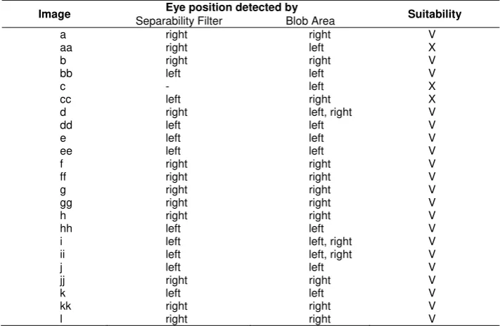
In this step, identification process adjusts eye position segmented by Blob area and eye position detected by separability filter. Table 2 shows results of identification process of this step.
Table 2. Result of eye position detected by separability filter and blob area
Image Eye position detected by Suitability
Separability Filter Blob Area
a right right V
According to Table 2, only 3 of 23 images have different eye position. Therefore Blob area has accuracy 86.95%.
3.4 Brightness
Citra separability map Citra separability map
separability filter as described in Table 1. Figure 7 describes an example of changing brightness value in an image.
Figure 7. An Example of changing brightness value
Refer to the result of this step, before we change brightness value, distribution of separability value tend more and look like noise, also local maximum value obtained is not eye point candidate. While after we change brightness value, distribution of separability value less visible and tend to gather into one edge and a point. Local maximum value obtained is an eye point candidate. Table 3 describes the result of pupil localizing performance after we change brightness value.
Table 3. Performance of Separability Filter after changing brightness value
Image
Brightness Changing Implementation of separability filter
Increase Decrease Before After Success Failed Success Failed c v v v v d v v v
ee v v v
ff v v v
i v v v
k v v v
Refer to the Table 2, by changing intensity, we can improve performance of separability filter to localize abnormal pupil from 73.91% to 95.65%.
4. Conclusion
According to our experiment results, we conclude that for abnormal pupil, separability filter method and Hough transform has similarities in use of radius value. Hough transform also can be use as sorting process for input images in order to detect pupil based on separability filter by giving accuracy about 95%. In this research we choose for using Hough transform as a sorting method because separability filter has characteristic for using fix radius value and also it has reliability to change image information. We also note here that brightness element influence to separability value on separability filter and bloob area segmentation also can be use to segment eye area in order to detect pupil based on separabililty filter.
Acknowledgment
This work is supported by research grant Hibah Bersaing Directorate General of Higher Education Fiscal Year 2013 under contract number 2740/UN23.10/PN/2013.
References
[1] R Supriyanti, Hitoshi Habe, Masatsugu Kidode, Satoru Nagata. A Simple and Robust Method to Screen Cataract using Specular Reflection Appearance. SPIE Medical Imaging Conference, San Diego, California. 2008.
[2] R Supriyanti, Hitoshi Habe, Masatsugu Kidode, Satoru Nagata. Cataract Screening by Specular Reflection and Texture Analysis, Communications of SIWN, 2009. 6: 59-64.
[3] R Supriyanti, Hitoshi Habe, Masatsugu Kidode and Satoru Nagata. Extracting Appearance Information inside the Pupil for Cataract Screening. IAPR Conference on Machine Vision Application. Tokyo, Japan. 2009: 342-345.
[4] R Supriyanti, Y Ramadhani, Hitoshi Habe, Masatsugu Kidode. Performance of Various Digital Cameras for Cataract Screening Techniques based on Digital Images. International Seminar of Electrical Power, Electronics, Communications, Control and Informatics (EECCIS), Malang, East Java, Indonesia, December, 2010.
[5] R Supriyanti, Y Ramadhani. The Achievement of Various Shapes of Specular Reflections for Cataract Screening System Based on Digital Images. International Conference on Biomedical Engineering and Technology (ICBET). Kualalumpur, Malaysia. 2011:
[6] R Supriyanti, B Setiawan, HB Widodo, E Murdyantoro. Detecting Pupil and Iris under Uncontrolled Illumination using Fixed-Hough Circle Transform. International Journal of Signal Processing, Image Processing and Pattern Recognition (IJSIP). 2012; 5(4): 175-188.
[7] F Arnia, N Pramita. Enhancement of Iris Recognition System Based on Phase Phase Only Correlation. TELKOMNIKA Telecommunication Computing Electronics and Control. 2011: 9(2): 387-394.
[8] IKG Darma Putra, A Cahyawan, Y Perdana. Low-Cost Based Eye Tracking and Eye Gaze Estimation. TELKOMNIKA Telecommunication Computing Electronics and Control. 2011; 9(2): 377-386.
[9] Michal Ciesla, Przemyslaw. Eye Pupil Location Using Webcam. Jagiellonian University. Poland. 2012. [10] Samira Kooshkestani, Mohammad Pooyan, Hamed Sadjedi. A New Method for Iris Recognition
Systems Based on Fast Pupil Localization. Lecture Notes in Computer Science. 2008. 5072: 555-564. [11] Haider Ali, Ahmad Ali2, Riaz Ul Husnain, Roman Khan, Mohsin Khan, Ihsan Ullah Khan. Automatic
Localization of Iris Using Region Properties. International Journal of Computer Science and Network Security. 2011: 11(7): 93-97.
[12] Zhaofeng He, Tieniu Tan, Zhenan Sun. Iris Localization via Pulling and Pushing. The 18th International Conference on Pattern Recognition. Hongkong. 2006: 366-369.
[13] Peng Yang, Bo Du, Shiguang Shan, Wen Gao. A Novel Pupil Localization Method Based on Gaboreye Model and Radial Symmetry Operator. International Conference on Image Processing (IClP). Singapore. 2004: 67-70.
[14] TA Camus, R Wildes. Reliable and Fast Eye Finding in Close-up Images. The 16th International Conference on Pattern Recognition (ICPR). Canada. 2002: 389-394.
[15] Libor Masek. Recognition of Human Iris Patterns for Biometric Identification. PhD thesis, The University of Western Australia, 2003.
[16] K Fukui. Edge Extraction Method Based on Separability of Image Features. IEICE Transactions on Information and Systems. 1995; E78(12): 1533-1538.