24
Susanto et al. Med J IndonesDiagnosis of
Malaria
by
the Rapid
Manual
Test
Lisawati
Susanto,Wita
Pribadi,
Hendri Astuty
Abstrak
Diagnosis penyakit
nalaria
ditegakkan dengan nenenukan parasit dalan darah penderita. Hingga saat ini diagnosisnalaria
dilnkukan dengan cara konvensional dengan nenbuat sediaan darah tebal atau tipis yang dipulas dengan pewarnaan Giensa dan diperiksa dengan nùkroskop cahaya. Dalant penelitian ini dikcnukakan suaru cara baru wttuk mendiagnosis nalariafalsiparwn, yaitu dengan "Rapid Manual Test" ("RM test"). Cara ini lebih mudah dilakukan karena tidak nenerlukan pulasan warna dan petneriksaan nûkroskop. Tes ini dapat nrendeteksi antigen PJalciparunt terlarut y(t,19 berasal dari stadiun trofozoit, yaitu histidine-rich protein-Il (HRP-II). RM tes ini ircrupakan suatu "dipstick test" yang nengandung antibodi nonoklonal terhadap HRP-
IL Pada
penelitian ini dilakukanuji
coba "RM tes" ini yang dilakuknn pada pengunjung Runnh Sakit International Tinùer Corporation Indonesia (ITCI), Kenangan, Balikpapan, Kalinantan Tinur yang nerupakan daerah endeniknalaria.
Tujuan penelitianini
ialah untuk mengetahui sensitivitasdan
spesifisitas tes tersebut terhadap infeksi P.falciparwn dan menbandingkarurya dengan netode diagnostik yang konvensional. Berdasarkan I 17 sediaan darah yang diperiksa, 33,3% sediaan nenunjukkan positif malaria falsiparutn dan 53,0% sediaan negaûf, baik dengan "RM test" naupun dengan cara konvensional, sedangkan sisanya 13,7% sediaan nenunjukkan hasil yang berbeda, yaitu 93,870 sediaan positifpada "RM test", tetapi negatif dengan cara konvensional dan 6,2% sediaan negaîifpada "RM testn tetapi positif dengan cora konvensional. Ternyata sensitivitas pada 'RM test" adalah 97.5% dan spesifisitasnya 80,5% dibandingkan dengan cara konvensional. Gejala klinis pada 6O penderita yang diperiksa yang terbanyak adalah sakit kepala (42,9%) disusul dengan tnenggigil (42,6%), denandi
atas 37,50C (37,5%), tnual ataununtah (48,8%) dan "RM test" posirif 44,2% padasalahsaru gejalaklinis atau lebih. Penderita dengan splenonegali sebanyak 50,0% dan dengan konjungtiva pucat 45,5%. Sebagai kesimpulan dapat dikntakan bahwa " RM test" cukup sersitifsehingga dapat digunakan untuk nenggantikan cara konvensional dalan nenegakkan diagnosis penyakit nalaria falsiparum secara cepat dan tidak nenerlukan nikroskLtp. Caraini
dapat digunakandi
runah sakit4erifer
yang tidak uenpunyaifasilitas tersebut dan tidak uenerlu.knn tenaga nikroskopis khusus untuk nnlaria yang nanang sangat kurang.Abstract
The diagnosis of
nalaria
is based on the finding of malaria parasites in blood. Until trow the diagnosis is carried out by ,neans of the conventional ntethod usitrg Gietnsa stained of a blood smear (thin or thick) and then exanined by ordinary light nicroscope. In this study a Rapid Manual Test (RM test)for
diagnositrgnalaria
was introduced. The test was sinpler than the conventional nethod and did not need staining as well as nicroscope. The test could detect soluble Plaxnodiwn falciparun antigen of the trophozoite stage which consisted nainly of histidine-rich-protein-Il (HRP-ll). This test is a "dipstick test" which contains nonoclonal antibody against HRP-ILIn
this study blood samples were collectedfron
patients who were treated aî the International Tinber Corporation Indonesia (ITCI) Hospital at Kenangan, Balikltapan, East Kalinnntan, an endeuic areafor nalaria. The aim of this study was to know the sensitivily and specificity of the RM test in detecting P.falciparwn infectiott and conparing to the conventional diagnostic ,nethod. Based on I 17 blood snrears etauined, 33.3% were positiv'efor nalariafalciparwtt whereas 53.0% were rtegative by both iltethods. Howeve4 l3-7% blood snears showed different results: 93.8Vo were positive wilh the RM test but were negative with the conventional ,nethod, whereas 6.2% were negative with the RM test but positive with the conventional ilrcthod. By statistical analysisit
was shown that the sensitivity and specificity of the RM testwere 97.5% and 80.5% respectively. The major clinical synptorns of the 60 patients exaninedwere headache (42.9%)followed by chills (42.6%),fever above37.f
C G7.5Vo), nausea or voniting (45.8%) and rhe RM tesr positive rate was 44.2%. Fifty percent of the patients showed splenontegaly whereas 45.5% showed a nentic cottjuttctivae.It
was concluded that the RM test could replace the conventional nethod. It did not need a nicroscope and was quite sensitivefor diagnosing nalariafalciparutn. Furthennore it could be applied in small peripheral hospitals where nicroscopes and nalaria nûcroscopist are not available.Reywords : Conventional nethod, plasuodiutnfalciparun, histidine-rich protein-Il (HRP-ll), dipstick test, tilonoclornl antibody
Vol 4, No
l,
January-March, 1995INTRODUCTION
Until
now malaria isstill a
public health problem in somecountries
including
Indonesia.The
mortality
andmor-bidity
causedby
malaria
is
still
high, particularly in
infants
and
children
underfives
i,e
6
Væ.and
ll
Voo.respectively.l
H".r""
early diagnosis
is
required to
reduce the
mortality
and
morbidity
rates.Usually
thediagnosis
of
malaria
is
carried out
by
means
of
theconventional
method,using
Giemsa stainedof
ablood
smear and then examined unde^r the ordinarylight
micro-scope asintroduced
by
Rosszin
1903.However,
this method needs an experiencedmicroscopist
and the ex-aminationitself
istime-consuming
(abo^ut one hour).To
solve this problemWardlaw
andLeviner in
1983 intro-duced theQuantitativeBuffy
Coat technique (QBCtech-nique)
for
diagnosing malaria.This
method is based on staining ofDNA
andRNA
of
the parasitewith
acridine-orange;the nucleus
will
show
green fluorescence and thecytoplasm
will appear
orange under theultra-violet
light. This
method
is
more
simple
andrapid
than
theconventional
method and anyone can master thetechni-que
in
(do
it
accurately)
less than one day.aThe
sen-sitivity of theQBC
technique has been provedby
someinvestigators.al
Nevertheless, theQBC
technique is notpractical to be
usedunder
field
conditions
becauseit
needs electrical power for acentrifuge
and anultraviolet
(UV)
fluorescence microscope.
In 1993,
Taylor
and Vol lero introduced anEnzyme-linked immunosorbent
assay
@LISA)
for
detecting
Plasmodiumfalciparunt
antigen.This
assay wasquite
sensitivebut
it
was time-consuming also and it needed many laboratoryfacilities.
A
new
method, the Rapid-Manual
test
(RM
test)
wasintroduced by
Shiff
et al.r0in 1993.
This
test was morerapid
and easyto perform
andit
did
not
needspecific
laboratory equipments such as thecentrifugeand
micro-scope sofhat this
test was morepractical to
be usedin
the field.12'13The Rapid Manuaf test which
wasintro-duced
by
Becton Dickinson
Company
was
aimed to
detect
P.falciparum
antigen
of
the
trophozoite
stagewhich
consisted mainly
of
histidine-rich-protein-Il
(HRP-II)
orPfHRP-II.
This
test is adipstick
testwhich
used amonoclonal
antibody againstHRP-II.
The aimof
this study was to compare thesensitivity
andspecificity
of
the Rapid Manual test against the
conventional
methodfor
diagnosin gP.falciparum infection.
MATERIALS
AND
METIIODS
Methodology
In this
study, the
single blind
method
wasused ;
oneinvestigator
carried out theRM
testwhile
the other oneperformed
theconventional method
separately.Blood
Rapid Manual
Test
25samples were
collected from
patientswith
fever,chills,
headache
and anemic conjunctivae who visited
theITCI
Hospital
at
Kenangan,
Balikpapan,
East
Kali-mantan. The samples wereexamined
with
theRM
test and stainedwith
Giemsafor
conventional blood
smearexamination.
Materials
The test
sticks
( 7 cmlong
and 3 | 4 cmwide
) were madeof
nitrocellulose fibre
consisting
of
2
parts :
part A
which
contained monoclonal antibody
against
his-tidine-rich-protein-Il
(HRP-II)
andpart
B which
con-tained
a dessicant(Figure
1).The test
stick holder
was madeof
cardboardwith
circled
wells
onit
(Figure
2).Methods
a.
TheRapid
Manual
test methodExactly 50
ul
blood
was drawn
with
a
micro-hematocrit
tube
directly
from a finger
puncture,
was pouredinto
adispensing
tubewhich contained
3 dropsof lysing
reagent andthen mixed
until
it
becamehomogenous
by
tapping the tube
with
the indexfinger.
Subsequently, one dropof
lysedblood
was dropped onto onecircle of
the teststick holder,
then partA of
the teststick
was placed onthe circle
until all
blood
was absorbed. Onedrop
of
detector
reagent was added to the samecircle
and afterit
wascompletely
absorbed,2
drops
of
washing
reagentwere
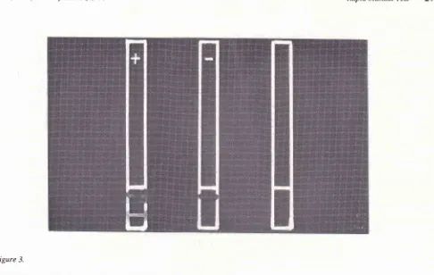
added.The results could
be read atpart
A
of
the test
stick. Positive
results were shownby
apink
line,
that
appearedon
the test
stick, i.e
the blood
sample tested contained PJalciparum antigen
which
reacted
with
the monoclonal antibody.
A
pink
dot could
also be seen as anindicator
that the test was conductedproperly,
hence anegative
testresult
showedonly
apink
dot. The testcould
not beinterpreted
if
thepink
dot
did
not
appear andsub-sequently
the testshould
be repeated.s(Figure
3;b.
Theconventional method
For this method,
thick
andthin
blood
smear were prepared. Eachblood
sample was thenstained
with
8%
Giemsa solution (pH7.2).
For
athick
blood
smear,
the blood
sample
was
stained
for 20
minutes,
whereasfor
athin
blood
smear, theblood
Susanto et al- Med J Indones
Figure 1.
Vol 4, No
I,
January-March, 1995Figure 3.
Test sticks show positive (lefi), negativ'e (nriddle) and not be interpretated (right) reactiot'ts.
Rapid Manual Test 27
immersion
oil.
The result
was negative
when
malaria
parasites were notfound in
theblood
smearafter examining
200
fields,
under the
light
micro-scope
(5x100).
In
positive blood
smears,
the
nucleus
of
the
parasite appeared
red
and
the
cytoplasm
violet-blue
while
the pigment
was
brown-black.9
RESULTS
Based
on
117blood
samplescollected
(113 adults aged15-43
years
andonly 4 children aged2-lO
years),
39samples
(33.3%) were
positive
for
P.falciparunt
whereas 62 samples (53.O%)
were negative
by
both
methods. However,
13.7%
samplesshowed
different
results. Statistical
analysis showedthat
thesensitivity
andspecificity of
theRM
test
were73.3%
and 82.5%respectively;
thepositive predictive
value was
87.27o and the negativepredictive
value was 7 4.6%(Table
l).
Out
of
the 33 samples
wlth P.falciparum positive
by
the
conventional
method, 32 showed
positive
resultswith the
RM
test. 5 out
of
20
samples
with
P-vivax
infection
showedpositive
resultswith
theRM
test, andall
of the 7 sampleswith
mixed infection
(P.falciparunt
andP.vivax)
showed
positive
results
by the RM
test(Table
2).The major
clinical
symptoms
of
the 60
patients examined were shownin Table
3.Table
l.
Comparison of resultsof
the Rapid Manual test and theconventional nrcthod on
l17
paticnts with P.falciparuminf'ection lrom ITCI Hospital, Kenangan, Balikpapan, East
Kalinrantan.
Rapid Manual lcst Conventional mcthod
positive
ncgativepositive
negative
44
t6
l0
47
54 63
Total
Scrsitivity = 4416O x l0o% ='73.3%
Spcciticity = 47 157 x IOO% = 82.57o
Positive Pre<lictive Value = 44154 x tOO% = 81.2%
Negative Predictive Value = 47163 x IOO% ='14.6%
Table
2.
Comparison of positive results of conventional nrethod andRapid Manual test according to spccies.
Species Conventional
mclhod
Rapid Manual Testpositive
positive
negative57
P. falciparunt
P. vivau
Mixed infection
(P.fulciparum
+
P.vilar)
33 20 7
32 5 7
I
l5
[image:4.595.50.536.63.371.2] [image:4.595.306.538.456.622.2]28
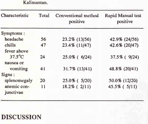
Susanto et al.Table
3.
Results of both test according to symptoms and sign on 60 patients on ITCI Hospilal, Kenangan, Balikpapan, EastKalimantan.
Characteristic
Total
Convenlional method positiveRapid Manual lest positive
Symptoms:
headache
56chills
47fever above
37.50C
24naùsea of
vomiting
4l
Signs :
splenomegaly 20
anemic
con-
I I junctivae23.2% (13156) 23.4% (tU47)
2s.o% ( 6124)
3t.716
(t3l4t)
25.016
(
sl2o) t8.2%( 2ltt)
42.9% (241s6) 42.6% (20147)
37.s%
(
9124\48.8% (2ol4t)
5o.o% (t2l2o)
4s.5.h (
5ltt)
DISCUSSION
In
1993,Taylor
andVoller6
introduced
anELISA
testto
detect
P.falciparuta
antigen.
This
assaywas quite
sensilive, however
it
was time-consuming and
re-quired laboratory
supportfacilities.
In this study, anew
method, the Rapid
Manual
test
was
used. Compared to theELISA
test,the RM
test hascertain
advantages;it
doesnot require special equipments
andthe
resultscould
beobtained
morerapidly.
The resultsof
theRM
test
differed from
the conventional method.
Based on117 samples
examined, only
33.3% sampleswereposi-tive by
both
methods
(Table
l),
whereas I7.5%
samples
which were
negative
with
the
conventional
method
were positive
with the
RM
test,
It
might
be possible that thenumkr
of
the parasites was toosmall
(submicroscopic
level)
so thatonly
theRM
testcould
detect.
According
to
Shiff
er al.l0'12PfHRP-II
antigen
persisted longer in the
blood
than the parasiteitself;
the antigen canstill
be detecteduntil
2 weeks. In thisstudy,
samples
were
collected from
patients
with clinical
symptoms
(acute phase) andPfiRP-II
antigen could
be
found
in
large amounts,
that
was
during
schizont
rupture. However, not
all
malaria infections
are
ac-companied
by specific clinical
symptoms
like
fever,
chills
and headache.
The
results showed
only
9(37.5%)
out
of
the patients
with
fever
who
had
apositive
RM
test.
A
possible explanation was
thatprobably
the patients had been
living in
an endemic
malaria area and
were infected continuously
so
theybecame
semi-immune
andasymptomatic
with
nofever
on
physical
examination, although they
had ahistory
of
fever or
of
taking analgetic-antipyretic
drugs.
By
statistical
analysisit
was shown that thesensitivity
andspecificity
of
the
RM
test were
73.370 and
825%,
which
was
different
from
the results
of
Shiff
et al.l0
who found that the sensitivity and specificity
wereMed J Indones
88.97o
and
87.57o
respectively.
In
mixed infection
(P.falciparu,?r
and P.vivax)
both
tests gave
almost
equal results. On the
contrary in
infection with P.vivar
as asingle
species theRM
testshould give
anegative
result,
but 5of
20P.vivcx
slides werepositive
with
theRM
test,which
could
be dueto
the presenceof
mixed
infection with P.falciparum;
not
detectedby
the
con-ventional method
because
of
the
low
parasitemia
(Table
2).
To
detect
parasites
in
the blood
by
theconventional method,
thenumber
of
counts should
beminimally 50
parasites
per
pl
of
blood,
whereasac-cording
to
Beadle
et
al.ll
an
inexperienced
micros-copist often had
difficulties
in detecting parasitesif
thecount was
lessthan
60 parasitesper
pl
of blood.
Ac-cording to
Shiff
et al. l0 àpositive
iesult of
theRM
testcould be
reached
if
the
parasite
count
was about
40 parasites per prlblood;
*h"."u,
Beadle etal.l
Iclaimed
that
the testwas
965%-lOO%
sensitive
if
the parasitecount
wasmore than 60
parasitesper
pl
blood,TO%-81% sensitive
for
11-60 parasites
per
pl
blood
andll%-67%
sensitive
for
l0
parasites
9r
less
per
Fl
blood.
Until now
theconventional method
is assumedas
a
reliable
gold
standard
for
detecting malaria
parasites.
In
this
study the
conventional
method
was conductedby
an experiencedmicroscopist.
CONCLUSION
The
Rapid Manual
test is anew diagnostic
methodfor
detecting
P.falciparutn
antigen.
As
adipstick
testthis
method
is
very
simple and could be perfomed
by
anyone.
The results can
be
obtained
within
ap-proximately
10minutes
andit
doesnot
need amicro-scope.
Therefore
it
is
applicable
in
primary
health
centers and
peripheral hospitals. The
RM
testis quite
sensitive
andspecific,
and severemalaria
can bediag-nosed
quickly
so thattreatment could
begiven
imme-diately.
In
Indonesia,
P.falciparum
andP.vivax
are
the mostprevalent
species, hence acombined
RM
testfor
both P.
falciparum
andP.vivax in
one teststrip should
be
developed
and speciesspecific
diagnosis could
bemade so that proper treatment
could
begiven instantly.
Acknowledgements
We thank the
manager
of
PT
International Timber
Corporation
Indonesiafor
allowing
us to use thehospi-tal
facilities, Also
toDr.
Tekky
Jokom,
Director
of PT
ITCI
Hospital
andhis
staff for their
assistancein
per-forming
this
study,
we would
like to
extend our
gratitude. The
RM
test
kits
were
provided by
Becton
[image:5.595.47.288.106.307.2]Vol 4,.No 1, January-March, 1995
REFERENCES
l.
World
Health
Organization.Weekly
Epidemiologicalrecord. Geneva, 1991.
2. Pomsilapatip
P,
Namsiripongpun V, Wilde H, HanvanichM,
ChutivongseS.
Detectionof
plasmodiain
acridineorange stained
capillary
tubes(
The QBC
System ).Southeast Asian
I
Trop Med Pub Hlth 1990;2l
: 3 - 15.3. Wardlaw SC, Levine RA. Quantitative
buffy coat
analysis:
a
new laboiatory tool functioning as a screening completeblood cell count.
IAMA
1983;249 : 617 - 20.4. Riclcnan LS, Long GW, Oberst
R
et aL Rapid diagnosisof
malaria by acridine orange staining ofcentrifuged parasites.
Lancet 1989; 68 -71.
5. Parzy D, Raphenon B, Martet G, et al. Quantitative buffy
coat test ( QBC te,st) monofluo kit falciparum. Comparative
value in the rapid diagnosis of malaria. Medicine Tropicale
1990;
50 :97 -l.
6. Taylor DW, Voller A. The development and validation of a
simple antigen detection ELIS Afor Plasnodi um falci parunr
malaria. Trans R Soc Trop Med Hyg, 1993; 87 :29 - 31.
Rapid Manual
Test
297. Pata ME, Evans CB, Taylor
DW.
Identification ofPlas-nrcdiutn falciparulr histidine-rich-protein-2 in the plasma
of
humans with malaria.
I
Clin Microbiol 1991; 29 t 1629-34.8. Rapid test for P.falciparun malaria. Becton Dickinson and
Company. Iakarta, L992
9. World Health Organization. Manual
for
processing andexamination
of
blood slides
in
malaria
eradicationprogrammes. Geneva, 1961.
10. Shiff CI, Premji Z and Minjas I. The rapid manual
ParaSight-^-
F test. A new diagnostic tool for P/as nodiumfalciparuminfection. Trans R Soc Trop Med Hyg 1993; 87 : 646 - 8.
11. Beadle
C,
Long GW, Weiss WR, McElroy PD, Maret S\,I,Oloo
AI,
HoffmanSL.
Diagnosis of malaria by detectionof
Plasnodiutnfalciparun
HRP-2 antigenwith
a rapiddipstick antigen- capture assây. lâncet 1994; 343 : 564 - 8.
12. Shiff Ct, Minjas J, Premji Z. The ParaSightR-F test: a simple
rapid manual dipstick test to detect Plasmodiumfalciparum
infection. Parasitology Today 1994; 10(12)
: 494
- 5.13. Premji Z, Minjas I, Shiff CL Laboratory diagnosis of malaria
by
villaç.e,