F2α-isoprostane, Na
+-K
+ATPase and membrane luidity of placental
syncytiotrophoblast cell in preeclamptic women with vitamin E
supplementation
Rusdi,1 Oentoeng Soeradi,2 Sri B. Subakir,3 Franciscus D. Suyatna4
1 Department of Biology, Faculty of Mathematics and Natural Sciences, State University of Jakarta, Jakarta, Indonesia 2 Department of Biology, Faculty of Medicine, Universitas Indonesia, Jakarta, Indonesia
3 Department of Physiology, Faculty of Medicine, Universitas Indonesia, Jakarta, Indonesia 4 Department of Pharmacology, Faculty of Medicine, Universitas Indonesia, Jakarta, Indonesia
Abstrak
Latar belakang: Penelitian ini bertujuan untuk menganalisis kadar F2α-isoprostan, aktivitas enzim Na+-K+ ATPase dan
luiditas membran sel sinsitiotrofoblas plasenta penderita pre-eklampsia yang diberi vitamin E.
Metode: Penelitian dilakukan pada bulan September 2003 – Februari 2005 di Rumah Sakit Bersalin Budi Kemuliaan, Jakarta Pusat. Sampel penelitian adalah 6 wanita pre-eklampsia yang mendapatkan vitamin E, 6 wanita pre-eklampsia yang tidak mendapat vitamin E dan 6 wanita hamil normal. F2α-isoprostan diukur dengan ELISA Reader pada λ = 450 nm. Fluiditas diukur dengan membandingkan rasio molar kolesterol total dan kadar fosfolipid membran sel. Kolesterol diukur menggunakan Modular C800 dengan reagen Roch. Fosfolipid diukur menggunakan spektroluorometer Shimadzu RF5301PC dengan ilter eksitasi 267 nm dan emisi 307 nm. Aktivitas Na+-K+ ATPase dihambat dengan ouabain. Produksi
Pi diukur dengan metode Fiske dan Subbarow menggunakan spektrofotometer pada λ = 660 nm. Data dianalisis menggunakan uji F melalui ANOVA 1 arah.
Hasil: Pemberian vitamin E pada penderita pre-eklampsia menurunkan stres oksidatif dengan indikasi turunnya F2α -isoprostan secara bermakna (26,72 ± 11,21 vs 41,85 ± 7,09 ng/mL, p = 0.017). Vitamin E mampu menangkal radikal
bebas sehingga peroksidasi fosfolipid dapat dihambat dan luiditas membran sel dapat dipertahankan pada 0,39 ± 0,08
dibandingkan tanpa pemberian vitamin E yaitu 0,53 ± 0,14 (p = 0,024). Aktivitas enzim Na+-K+ ATPase membran sel
sinsitiotrofoblas tidak dipengaruhi oleh vitamin E (p = 0,915).
Kesimpulan: Suplementasi vitamin E pada wanita pre-eklampsia menurunkan kadar F2α-isoprostan, mempertahankan luiditas membran sel, namun tidak meningkatkan aktivitas enzim Na+-K+ ATPase sel
sinsitiotrofoblas. (Med J Indones. 2012;21:225-9) Abstract
Background: The aim of our study was to analyze F2α-isoprostane level, Na+-K+ ATPase activity and placental syncytiotrophoblast cell membrane luidity in preeclamptic women who received vitamin E supplementation.
Methods: The study was conducted between September 2003 and February 2005 at Budi Kemuliaan Maternity Hospital,
Central Jakarta. Samples were 6 preeclamptic women with vitamin E supplementation, 6 preeclamptic women without vitamin E supplementation and 6 normal pregnant women. The dose of vitamin E was 200 mg daily. F2α-isoprostane was
measured with ELISA reader at λ of 450 nm. Cell membrane luidity was measured by comparing the molar ratio of total cholesterol and cell membrane phospholipid concentration. The cholesterol was measured by Modular C800 using Roche reagent. Phospholipid was measured by Shimadzu RF5301PC spectroluorometer (excitation 267 nm, emission 307 nm).
Na+-K+ ATPase activity was inhibited by ouabain. Pi production was measured with Fiske and Subbarow method using spectrophotometer at λ of 660 nm. Data was analyzed using F test with one-way ANOVA.
Results: Vitamin E supplementation in preeclamptic women decreased the oxidative stress, indicated by signiicantly lower
level of F2α-isoprostane compared to those without vitamin E (26.72 ± 11.21 vs 41.85 ± 7.09 ng/mL, respectively, p =
0.017). Membrane luidity in syncytiotrophoblast cell of preeclampsia with vitamin E group was maintained at 0.39 ± 0.08 while in those without vitamin E was 0.53 ± 0.14 (p = 0.04). Na+-K+ ATPase activity in syncytiotrophoblast cell membrane was not affected by vitamin E (p = 0.915).
Conclusion: Vitamin E supplementation in preeclamptic women decreases F2α-isoprostane level and maintains cell
membrane luidity of syncytiotrophoblast cells; however, it does not increase Na+-K+ ATPase enzyme activity. (Med
J Indones. 2012;21:225-9)
Keywords: F2α-isoprostane, membrane luidity, Na+-K+ ATPase, preeclampsia, vitamin E
Correspondence email to: rusdibioma@yahoo.com
the theories describing the etiology of preeclampsia is oxidative stress including increased free radicals and reduced antioxidant. Free radicals can disrupt
endothelial cell and prevent trophoblastic cell invasion
into spiral artery in decidua.3
Preeclampsia is a disorder in pregnancy which marked
by increased blood pressure and proteinuria, decreased uterus-placental blood perfusion, increase of trophoblast
Low oxygen partial pressure as a result of spiral artery vasoconstriction in decidua has negative effect on trophoblast development in placental villi. Hypoxic condition also increases the formation of radical oxygen species through hypoxanthine reoxygenation pathway with the aid of xanthine oxidase enzyme.4
Free radicals exert a destructive effect to the cells. The
destructive effects can be observed from the products of bio-macromolecular reaction, such as F2α-isoprostane.5
The reaction between nitric oxide (NO) and oxygen radicals (O2) results in peroxynitrite, which causes the
oxidation of arachidonic acid product, F2α-isoprostane. 5
Increased level of F2α-isoprostane is a characteristic of
oxidative stress that disrupts cell membrane.6
Increased F2α-isoprostane level is assumed to induce
vasoconstriction of placental spiral arteries. This will cause entrapment of syncytiotrophoblast cell growing in decidua due to vasoconstriction.7 This condition may
cause syncytiotrophoblast cell destruction and necrosis
in fetal artery and increased apoptosis of trophoblast.8
Destruction of placental syncytiotrophoblast cells may also bring negative effects on fetal development, because it is the epithelial cell responsible for exchange of nutrients and substances between mother and fetus.
Free radicals can attack cell membrane protein such as Na+-K+ ATPase enzyme, which is a Na+ and K+ ion
pump. Free radicals can also attack cell membrane
phospholipid, which can disrupt cell membrane
luidity. If Na+-K+ ATPase enzyme activity and luidity in syncytiotrophoblast cell membrane were disrupted,
it will destruct the cell and causes apoptosis.
One of modalities to overcome the effects of free radicals is by vitamin E supplementation such as alpha tocopherol. Vitamin E supplementation in 36 weeks of pregnancy will decrease plasma lipid peroxide, thus negative effect of free radicals to the cells can be prevented.9
This study was aimed to recognize the protecting effect of vitamin E on syncytiotrophoblast cell in preeclamptic women by measuring F2α-isoprostane level, Na+-K+ ATPase enzyme activity, and fetal
placental syncytiotrophoblast cell membrane luidity.
METHODS
The study was conducted between September 2003 and February 2005 at Budi Kemuliaan Maternity Hospital,
Central Jakarta. Ethical clearance from Ethical Research Committee of Medical Faculty Universitas Indonesia had been obtained. It is an experimental study with
total-random sampling of 6 preeclamptic women given 200 mg/day of vitamin E from 30 weeks of pregnancy until delivery on top of routine medication, six preeclamptic women without vitamin E supplementation, and 6 normal pregnant women.10
Isolation of syncytiotrophoblast cell was performed based on modiied methods developed by Smith et al,11
Rand et al,12 and Subakir.13 Immediately after delivery,
placenta was cut ± 10 g, and submerged into buffer
solution of 0.05 Mol/L Tris-HCl, 0.1 Mol/L NaCl, 1 mMol/L CaCl2, pH 7.4 (Tris-buffer saline solution/
TSS) with 4oC temperature.
Chorionic plate and the surface of decidua were removed, washed with TSS solution to remove maternal
blood. Tissue was crushed with TSS solution and iltered with gauze. Then, tissue pellet was centrifuged with 2000 rpm for 10 minutes in 4oC temperature.
Erythrocyte-lysing solution was added; incubated for 10 minutes in 37oC temperature. Afterward, it was
centrifuged and syncytiotrophoblasts were isolated. Erythrocyte-lysing solution contained 2.0727 g of
NH4Cl, 0.25 g of NaHCO3, 0.009275 g EDTA and 250
mL of 131.3329 osmolality distilled water.13
Cytosol and syncytiotrophoblast cell membrane preparation
Syncytiotrophoblast cells were weighted 500 mg, and then crushed with Wheaton voter. Subsequently, the cells were centrifuged at 3,000 G for 15 minutes in 4oC.
Supernatant was added and centrifuged with 22,000 G for another 15 minutes in 4oC. Pellet were discarded
and supernatant was centrifuged with 100,000 G for 30 minutes. If after this centrifugation no more pellet were found, then the whole part were taken and 20 mL TSS
was added as cytosol and cell membrane sample and
kept at -80oC until measurement.
Parameter measurement
F2α-isoprostane was isolated with chromatography and measured with F2α-isoprostane kit from Oxford
Biomedical Research using ELISA Reader at 450 nm wavelength.
Na+-K+ ATPase enzyme activity was inhibited using
ouabain and inorganic phosphate (Pi) production was measured with Fiske and Subbarow methods using spectrophotometer at 660 nm wavelength.14 Protein
level was measured at 280 nm wavelength.
Cell membrane luidity was measured by comparing
phospholipid. Cholesterol was measured by C800 Modular with Roch reagent and Phospholipid was measured with RF5301PC Shimadzu spectroluometer (excitation: 267 nm, emission 307 nm) in 37oC.
1,6-diphenyl-1,3,5-hexatrin (DPH) probe were dissolved in tetrahydrofuran until 0.6 µg/mL concentration.
Data analysis
Data normality was analyzed with Kolmogorov-Smirnov, and one-way ANOVA was used for comparisons of mean values of each parameter. P value of < 0.05 was taken as the limit of statistical signiicance.
RESULTS
Data of mean blood pressure, proteinuria, maternal age, weight and length of the baby were shown in Table 1.
Data of mean F2α-isoprostane, Na
+-K+ ATPase
enzyme activity and placental syncytiotrophoblast cell
membrane luidity were shown in Table 2.
DISCUSSION
In this study, vitamin E supplementation has no effect on the weight and length of the newborn. Mean
Parameters Normal PE PE + Vit E
Sample size (n) 6 6 6
Systolic blood pressure (mmHg) Baseline 120 ± 0.00 146 ± 8.16 150 ± 7.53
End 110 ± 5.77 144 ± 12.25 150 ± 14.14
Diastolic blood pressure (mmHg) Baseline 80 ± 0.00 96.67 ± 8.16 98.30 ± 7.53
End 73.33 ± 4.71 100 ± 10.95 96.67 ± 8.16
Proteinuria Baseline - ++/+ ++/+
End - +/-
+/-Gestational age (weeks) 39 ± 0.82 37.33 ± 0.52 37.50 ± 0.84
Newborn weight (g) 2.937 ± 221.11 2.775 ± 216.02 2.760± 216.02
Newborn length (cm) 45.50 ± 1.67 47.00 ± 0.63 45.00 ± 1.26
Maternal age (years) 30 ± 1.63 32 ± 1.90 33 ± 1.67
Table 1. Mean blood pressure, proteinuria, maternal age, newborn weight and length
PE = preeclampsia
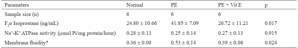
Parameters Normal PE PE + Vit E p
Sample size (n) 6 6 6
F2α Isoprostane (ng/mL) 24.80 ± 10.66 41.85 ± 7.09 26.72 ± 11.21 0.017
Na+-K+ ATPase activity (µmol Pi/mg protein/hour) 0.28 ± 0.13 0.25 ± 0.14 0.27 ± 0.13 0.915
Membrane luidity* 0.36 ± 0.08 0.53 ± 0.14 0.39 ± 0.08 0.024
Table 2.Mean F2α-isoprostane level, Na+-K+ ATPase enzyme activity, and syncytiotrophoblast cell membrane luidity
Membrane luidity = total cholesterol/phospholipid ratio
diastolic pressure in preeclamptic women with vitamin
E supplementation were decreased, but still in higher range, as also with the proteinuria. However, diastolic pressures of preeclamptic women with vitamin E
supplementation at the end of the study were lower,
although it was still higher than normal pregnant woman. Similar result has also been reported by Bastani et al who found that supplementation of 400 IU vitamin E started from 14 weeks of pregnancy has no effects on fetal weight and preeclampsia incidence.15
Wangkhemayum also reported that 400 IU vitamin E
supplementation daily is not positively correlated with
blood pressure.16
In preeclampsia, there is oxidative stress marked with
increase of F2α-isoprostane or 8-isoprostane.17 In
our study, 200 mg daily vitamin E supplementation started from 30 weeks of pregnancy in preeclamptic women has signiicantly lowered F2α-isoprostane level of syncytiotrophoblast cell than those preeclamptic
women without vitamin E (p = 0.017). Lower F2α-
isoprostane indicates that vitamin E supplementation can reduce oxidative stress since it functions as a chain breaking antioxidant which neutralizes free radicals effects.7,18 Vitamin E is hydrophobic and lipid soluble,16
thus enables it to enter cell membrane and deposited
protect cell membrane fatty acid against free radicals attack, by inhibiting oxidation of arachidonic acid
into F2α-isoprostane. This result is consistent with the
study of Poston et al who found that vitamin C and E
supplementation could decrease the concentration of
8-epi and F2α-isoprostane.
19
In preeclampsia, trophoblast cells fail to invade spiral artery.8 This can be shown by an increase
of mannose-binding lectin (MBL) which inhibits trophoblastic and endothelial cell interaction.8 Failure
of syncytiotrophoblast invasion into maternal spiral arteries leads to inability of the spiral arteries to
transform, hence caused reduced oxygen transportation and hypoxia. This condition will induce oxidative stress. Vitamin E supplementation reduces oxidative
stress thus decrease the producion of F2α-isoprostane.
Therefore, it is expected to induce syncytiotrophoblast growth and invasion to spiral artery. Transformation of spiral arteries to cone-shaped can reduced hypoxia
effect, and formation of free F2α-isoprostane through
hypoxanthine pathway can be reduced.
By decreasing oxidative stress in placenta, it is expected that syncytiotrophoblast cell destruction,
necrosis, and apoptosis in fetal placenta trophoblast can
be prevented. It means that vitamin E supplementation in preeclamptic women will prevent ischemic damage in syncytiotrophoblast cell which may bring negative effects on the fetus. It is important because placental
syncytiotrophoblast is the epithelial cell responsible for
substances transportation between mother and fetus.
Vitamin E supplementation in preeclamptic women has
shown no effect on Na+-K+ ATPase enzyme activity
in syncytiotrophoblast cell membrane. Total enzyme
activity was measured with and without ouabain to cease the Na+-K+ ATPase activity (sensitive to ouabain).
Data analysis showed that vitamin E did not increase
Na+-K+ ATPase enzyme activity (p = 0.915).
In oxidative stress, free radical can destruct cells through many pathways, such as oxidation of enzyme protein. One of the enzymes in cell membrane is
Na+-K+ ATPase, which pump 3Na+ out and 2K+ into
the cell.20 This pump maintains ion charges balance in
syncytiotrophoblast cell. Decreased activity of Na+-K+ ATPase enzyme will disrupt Na+ and K+ ion transport.
It could be fatal as cell membrane might have a malfunction with transmembrane potential leading
to accumulation of intracellular Na+ and it may cause
apoptosis.20
It has been assumed that increased oxidative stress may
disrupt Na+-K+ ATPase, thus decreasing its activity. In
our study, vitamin E supplementation did not increase
Na+-K+ ATPase enzyme activity in syncytiotrophoblast
cell membrane. Presumably, there is a speciic mechanism to regulate Na+-K+ ATPase, i.e. subunit α of the enzyme, which function is controlled directly
by subunit β.7 Subunit α and β protein are peripheral
membrane cell protein and hydrophilic in nature.20
Vitamin E is lipid soluble and hydrophobic, therefore it is less likely to protect the hydrophilic subunit α and β protein. Therefore, Na+-K+ ATPase enzyme activity is
not affected by vitamin E supplementation.
Meanwhile, vitamin E supplementation in preeclamptic women is capable to maintain luidity of syncytiotrophoblast cell membrane signiicantly. Membrane luidity is a ratio of cholesterol and phospholipid concentration. Membrane is more lexible with higher phospholipid, which shown by low ratio of cholesterol and phospholipid. Free radicals might cause oxidation of cell membrane phospholipid, thus decreasing phospholipid level, and make the cell membrane more rigid. Fiore and Capasso also suggested that vitamin E and C supplementation alone
or in combination can protect the cell from reactive
oxygen species (ROS) effects.3
Peroxidation of lipid cell membrane by free radicals affects membrane luidity and permeability, which leads to cell membrane destruction.21,22 In preeclampsia,
increased cholesterol and phospholipid ratio indicates
that membrane luidity is decreased.
In preeclamptic women who received vitamin E
supplementation, cholesterol and phospholipid ratio
were lower. It indicates that vitamin E supplementation is able to maintain cell membrane luidity. Vitamin E maintain membrane luidity by: (1) insertion of vitamin E between phospholipid preventing free radicals to attack phospholipid, (2) vitamin E as an antioxidant can neutralize free radicals effect against phospholipid oxidation.23 Therefore, vitamin E is able
to protect phospholipid, hence maintaining the luidity of cell membrane. Cell membrane luidity is important to maintain membrane integrity, thus preventing membrane destruction.
In conclusion, vitamin E supplementation of 200 mg daily is beneicial to provide protection against oxidative
stress as indicated by reduced level of F2α-isoprostane.
Vitamin E can overcome free radicals effect and inhibit phospholipid peroxidation, thus maintain cell membrane luidity. However, Na+-K+ ATPase enzyme activity of fetal placental syncytiotrophoblast cell in preeclampsia
regulated by peripheral protein, subunit α and β, which are hydrophilic in nature. Further studies regarding
Na+-K+ ATPase enzyme physiology through subunit α
and β protein, with water soluble antioxidant such as vitamin C are needed.
Acknowledgments
The authors thank Prof. Dr. dr. Sri Bekti Subakir, MS (deceased) of her support in Graduate Research Grant Funding Medical Faculty Universitas Indonesia, from 2003 to 2005, Director of Budi Kemuliaan Maternity Hospital, Staff of Cardiovascular Laboratory Physiology Department, Biochemistry laboratory, Cytogenetic laboratory, Faculty of Medicine, Universitas Indonesia, Food and Drug Regulatory Agency (Badan POM), and Prodia laboratory.
REFERENCES
1. Vatten LJ, Skjaerven R. Is pre-eclampsia more than one disease? Br J Obstet Gynaecol. 2004;111:298-302. 2. Padmini E, Lavanya S, Uthra V. Preeclamptic placental
stress and over expression of mitochondrial HSP70. Clin Chem Lab Med. 2009;47(9):1073-80.
3. Fiore G, Capasso A. Effects of vitamin E and C on placental oxidative stress: an in vitro evidence for the potential therapeutic or prophylactic treatment of preeclampsia. Med Chem. 2008;4:526-30.
4. Ali S, Diwakar G, Pawa S, Siddiqui MR, Abdin MZ, Ahmad FJ, et al. Xanthine oxidase-derived reactive oxygen metabolites contribute to liver necrosis: protection by 4-hydroperoypyrazolo(3,4-d) pyrimidine. Biochim Biophys Acta. 2001;1536:21-30.
5. Monthuschi P, Barnes P, Roberts LJ 2nd. Insights into oxidative stress: the isoprostanes. Curr Med Chem. 2007;14(6):703-17.
6. Yen HC. Detection of F2-isoprostanes and F4-neuroprostanes
in clinical studies. J Biomed Lab Sci. 2010;22(1):1-10. 7. Habib A, Badr KF. Molecular pharmacology of
isoprostanes in vascular smooth muscle. Chem Phys Lipids. 2004;128:69-73.
8. Agostinis C, Bossi F, Masat E, Radillo O, Tonon M, De Seta F, et al. MBL interferes with endovascular trophoblast invasion in pre-eclampsia. Clinical and Developmental Immunology. 2012;2012:1-7.
9. Chappell LC, Seed PT, Kelly FJ, Briley A, Hunt BJ, Charnock-Jones DS, et al. Vitamin C and E supplementation in women at risk of pre-eclampsia is associated with changes
in indices of oxidative stress and placental function. Am J Obstet Gynecol. 2002;187:777-84.
10. Lemeshow S, Hosmer DW, Klar J, Lwanga SK. Adequacy of sample size in health studies. New York: John Wiley and Sons; 1993. p. 36-40.
11. Smith CH, Nelson DM, King BF, Donohue TM, Ruzycki S, Kelley LK. Characterization of a microvillous membrane preparation from human placental syncytiotrophoblast: a morphologic, biochemical, and physiologic study. Am J Obstet Gynecol. 1977;128:190-6.
12. Rand JH, Wu XX, Guller S, Scher J, Andree HA, Lockwood CJ. Antiphosfolipid immunoglobulin G antibodies reduce annexin-V level on syncytiotrophoblast apical membranes and in culture media of placental villi. Am J Obstet Gynecol. 1997;177:918-23.
13. Subakir SB. Kadar tromboksan B2dan NO pada kultur jaringan endotel penderita pre-eklampsia dan wanita hamil normal. Laporan Penelitian. Fakultas Kedokteran Universitas Indonesia; 2004. Indonesian.
14. Ridwan R. Aktivitas spesiik enzim Na+-K+ ATPase membrane eritrosit normal dan penderita thalasemiaβ [Thesis]. Ilmu Biomedik Universitas Indonesia; 1998. Indonesian.
15. Bastani P, Hamdi K, Abasalizadeh F, Navali N. Effect of vitamin E supplemetation on some pregnancy health indices: a randomized clinical trial. Int J Gen Med. 2011;4:461-4. 16. Wangkheimayum S, Kumar S, Suri V. Effect of vitamin
E on sP-selectin levels in pre-eclampsia. Indian J Clin Biochem. 2011;26(2):169-71.
17. Pramono BA, Kristanto H. Serum 8-isoprostane increased in pre-eclampsia. Universa Medicina. 2012;31(1):43-51. 18. Janicka M, Kot-Wasik A, Kot J, Namieśnik J.
Isoprostanes-biomarkers of lipid peroxidation: Their utility in evaluating oxidative stress and analysis. Int J Mol Sci. 2010;11:4631-59.
19. Poston L, Igosheva N, Mistry HD, Seed PT, Shennan AH, Rana S, et al. Role of oxidative stress and antioxidant suplementation in pregnancy disorders. Am J Clin Nutr. 2011;94:1980S-5S.
20. Suhail M. Na+,K+-ATPase: ubiquitous multifunctional
transmembrane protein and its relevance to various
pathophysiological conditions. J Clin Med Res. 2010;2(1):1-17.
21. Adele AA. Oxidative stress and disease: an updated review. Res J Immunol. 2010;3(2):129-45.
22. Gohil JT, Patel PK, Pryanka G. Evaluation of oxidative stress and antioxidant defence in subjects of preeclampsia. The Journal of Obstetrics and Gynaecology of India. 2011;61(6):638-40.