78 Ranelan Med J Indones
Down
Syndrome
: Report of
a
Cytogenetic
Study
and a New
Proposed
Practical
Classification
Wahyuning Ramelan
Abstrak
Sindron Down nerupakan kelainan genetik yang paling sering dijumpai, yang dkebabkan oleh kelninan jwnlah krontosom atau aberasi numerilç yaitutrisoni-21. Secarasitogenetik, kelainan ini dapat nrenanpilkanberbagai aberasi kromosont. Klasifiknsi sindrom
ini
yang ada sekarang didasarkan pada keadaan krontosontnya, terasa lauang pralais, terutanta untuk kanseling, sehingga dapat dikatakan tidak berguna. Makalah ini nenyantpaikan hasil penteriksaan sitogenetik pada 280 penderita sindron Down. Berdasarlcan hasil peuerilcsaan itu, diusulknn klasifikasi baru yang hanya terdiri dnri 4 kelonpolç yang lebih praktis sehingga lebih bernanfaat, yaitu: Sindrotn Down reguler ataupriileL
sindron Down translolusi atau sekunder dan sindron Down mosaik atau tersier serta sindron Down lain atau kuarterner- Enpat kelonrpok sindron Down r,rcnurut Hasifikasi ini, hanpir sejalan dengan 4 lælas sindrom Down nenurut International Classification of Diseases yang terbaru dari WHO (WHO-ICD IO).Abstract
Down syndrourc is the no$ frequent genetic abnormality, with nunerical chrouosone aberration (nanely trisonry-2l) æ the aetiology. Cytogenetically, this disease has nany q,pes of chrontosonre aberrations. The existing classification based on the chronrosone picture, is not very practical, especially
for
counseling, and hence not very beneficial, This paper reports 280 cases of Down syndrone which had been exanined cytogeneilcally. In conelation withthe
results, this paper proposes a new, ntore beneficial and practical classification of Down syndronrc. This proposed classification consrsts only of 4 groups:
Regular or primary Down syndrone, trarclocation or secondary Down syndrotne, uosaic or tertiary Down syndronte, and other types or quarternary Down Syndrone. The eûstence of 4 groups of Down syndronein
this new proposed classification fits the newest International Classificationof
Diseasesof
the WHO (WHO-ICD 10) quite well.Keywords : Down syndrone, c\togenetic stud1,, cytogenetic classification.
INTRODUCTION
Down syndrome (DS)
is the most frequent
genetic
abnormality with numerical
chromosome aberration
(namely
trisomy
2l)
asthe
aetiology.t Th"
syndrome
was
named
after
Dr. Langdon Down,
who
in
1866reported
asyndrome
of
mental deficiency. The facial
appearanceof the patient,
resembles
the
face
of
thepeople
of
the
Mongol
race.The
likeness
is
undepen-dent
to
the
raceof
the patient. Once
you
have
seen apatient
with
DSyou
will
alwaysrecognize
other cases, evenin
patients of other
races.
Becauseof
theabnor-mal mongoloid-like 'facies',
he n-amedthe syndrome
mongolism or mongoloid idiocy.2
Becausethe
namesounded very racial,
it
was
then
named Down
syndrome.l Nearly
acentury went by before its
naturecould be
concealed,
although
Waardenburg
in
L932
and
Penrosein
1939 had suggestedthe
possibility
of
chromosome abnormalities
iihe
aetiolùy.
t't
Mittwoch
in
1952 studied meiosis
of
male
patientswith
DS,
andfound
24 bodies/chromosomes.2Her
conclusion
of
human
meiotic
chromosome
num-ber
as24 and made thediploid
numberof
48 in human, madeit
farther
from
thenature
of
DS.
It
wasLejeune
in
1959
who
showed
the actual
existence
of
chromosome aberration
in
DS which later was
alsoconfirmed
by
others.2Down
syndrome
canoccur
in
themale
aswell
asin
thefemale,
it
can befound all
over
theworld
andit
is not
restricted
to
any
raceor
any
season.In
general-Vol 4, No 2, April-June, 1995
the incidence
is
1out of 700
-
800newborns,
or
I
in
900-
l0OOof
the generalpopulation.l'3
Epidemiologi-cally,
about 957o
of
the
cases, areinfluenced
by
the maternal age atconception. Recently,
a higher percent-ageof
dependancyto mother
age wasalso confirmed
by
Antonarakis
et al.a Using
DNA
polymorphisms,
they got
188DS patients with
anextra
chromosome-21 from the mother, compared
to only 9 DS with
anextra chromosome-2l which
was
paternally derived.
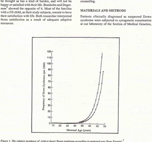
Therelativerisk
of amotherto
givebirth
to a babywith
DS
increasesslowly
until
the
ageof
30
years,rather
fast after the ageof
30 andit
becomes much fasterafter
35 yearof
age, asshown in Figure
1.From logical
thinking, family with
a DSchild
canbe thought
ashas a kind
of
burden, and
will
not
be happJ orsatisfied with their life. Branholm
andDeger-man'
showed the opposite
of it.
Most of the families
with
a DSchild,
astheir
study subjects, remain to havetheir satisfaction
with life. Both
researcherinterpreted
those satisfaction as
a
result
of
adequate adaptive
resources.Down Syndrotne: Cytogenetic and
Classification
79Now
we know that
there are many types
of
chromosome aberrations
in
DS. Cytogenetically
DSor
trisomy-21 was classified
in
several groups. The
classification
is notpractical,
especially
for
counsell-ing. In correlation
with
the newestInternational
Clas-sification of
Diseasesof
theWHO (WHO ICD l0),
theexisting classifications
are not well
matched.A more
practical
DSclassification should
be made and used.The purpose
of
this
study
is
to
compare
thecytogenetic
condition found
in
the
DS
patients in
Jakarta to DS patients
in
generalpopulation
(litera-ture), and to put forward
a new classification
of
DSmacthing with
theWHO-ICD
10,which
is moreprac-tical
and hencemore beneficial especially
for genetic
counseling.
MATERIALS
AND
METIIODS
Patients
clinically
diagnosed
as suspected Down
syndrome were subjected
to cytogenetic examination
atour laboratory
of the Section
of
Medical
Genetics,120
110
100
90
80
70
60
50
40
30 20
10
^
oo
o o.q)
E !
>. </,
É o
A
ox
o É oa d o tr.
25 30 35
40 [image:2.595.55.549.258.719.2]Maternal Age (years) 45
Ranelnn
Department of Medical
Biology Faculty
of Medicine,
University
of
Iâdonesia. There were 283
patients
suspectedof
Down
syndrome, examined
cytogeneti-cally over
aperiod of
6 years(1989
-
1994).The cytogenetic examination performed
waschromosome analysis. The
method used was the
modified
method
of Moorhead.
The cell preparation
was stained and thenexamined morphologically.
RESULTS
Three
from
the 283 patients showednormal 46,XY or
46,XX karyotype.
The remaining 280 patients,
con-sisting
of I25
females and 155 males, werecytogeneti-cally confirmed
astrisomy 21. As predicted, most of
them were 47,XY
or 47,XY
with an additional
free
chromosome-2l (229
patients
or
8I.79%). Four
patients (1.43%)
hadRobertsonian translocation
to
Gchromosome
(3
patients) and to D chromosome
(onepatient). The
rest
(47
patients
or
16.78%)
had
mosaicism
or mixoploid (46,XX/47,XX,+ll
sy 46,XYl47,){Y,+21)
(table
l).
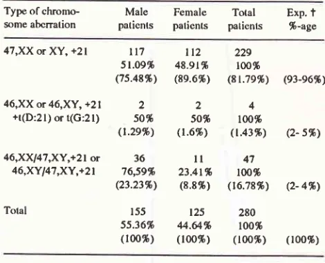
Table l. Nunrbcr and perccntage* of the Down syndronre according to
sex and type of chromosome aberration found in the study
Type of
chromo-
Male
Female Total
Exp.t
some
abcrralion
paticnts paticnts paticnts
%-ageMed J Indones
As known very well,
DS canoccur in
the male aswell
asin
thefemale. This
means that theincidence
of
thisabnormality
is the samein
male andin
female.The
number
of DS in this study is very different
in
male and female patients (155 and 125respectively),
but theratio of male to female patients
with
DS found in our
report (1.24 :
1.0) isnot
toodifferent from Chili6
with
ratio of (1.18
:
1.0).In our study rough calculation
of
theX'
for
thedifference between male
andfemale is
>
3.This
can beinterpreted
assignificantly
different.
This can
be the result of
an actual
difference of
DSincidence
in the general
population, or a lower
com-patibility
tosurvive
in the female compared to male DSpatients. Another possible explanation
for
the
dif-ferencein this study might
be thedifferent
attitude of
our society toward male
andfemale DS patients. Our
society cares and hopesfor
betterment in the male more thanthey
dofor
thefemale
DSpatients. Further study
should
be done to
ensure
which one
of
those
pos-sibilities
is
theright
one.Table
I
shows that
the percentageof
eachgroup
of DS is rather
different from
the expectedin
generalpopulation. Epstein in his review had
93-96%regular
or
free,2-5% translocation and2-47o
mosaictrisomy
21.' Verma et al from New Delhi
have rather similar
percentageswith
theexpected
in
generalpopulation,
that was 93%
fre,e,4.1% translocation,
2.6%
mosaic
trisomy
2l
and
0.3% trisomy
21 with other
con-comitant
aberration.o
Cortes et al from Chili
found
rather different
percentages
:
89,9%
classic
or free
3,9%
trarslo
aic trisomy
2l
and3.47otrisomy
21
other aberration.6
The
mosaic trisomy
2l
found in this study is much higher
(16.787o) than expectedin conected
percentageof
the generalpopulation (2
- 47o). Correlationally,
the per-centageof free trisomy
2l
is lower in this study,
thanthe corrected percentage
in
the general population.
Thosedifferences
can beinterpreted
aslaboratory
er-rors due to the processing
of the slide preparation
of
cells.It
has been shown before thatlaboratory
process-ing
can cause thelost of
anindividual
chromosomeof
the
metaphase,
although more
of
the
small
chromosome'. The other possibility that
can explain
thedifference
ofthe
percentagefound in this study
isthat it is
due toracial or genetic
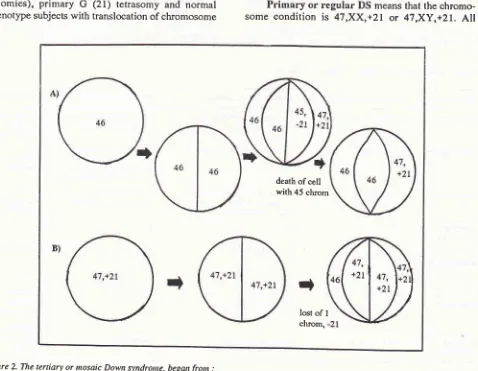
reason.From
thetheoretical point of view,
mosaicism can arisein
themitotic cell
division, post-zygotic
non-dis-junction
or
chromosome
lost.
In
vitro
study
of
chromosome
lost frorn normal subjects has
beenshown in our
lab.eMosaicism
of
DS
can begin from
a normal
46,XX
or 46,XY
zygote.During
theearly
cleavage acell divides and yields to daughter cells with trisomy
2L
and monosomy
21,
followed
by death
of
the 47,XX or XY,+21
Il7
51.og.fr (75.48%)
46,XX or 46,XY,
+21
2+t(D:21) or
t(G:21)
50* (t.2e%)46,XX47,XY,+21
or
3646,XYl41,XY,+2t
76,59% (23.23%)Tolal
t5555.36.h (100%)
tt2
22948.9t%
1001b(8e.6%)
(8t.'19%) (93-e6%)24
50%
ro0*(r.6%)
(1.43%)
(2- s%',)ll
4723.41%
tOO%(8.8%) (16.78%)
(2- 4%'r25
28044.&%
100%(r00%)
(100%)
(100%)* pcrcentages without brackets are pcrcentage of ûc total in each type
of aberration and percenlages in brackcls are pcrccntage of thc tolal fcmale/male group.
texpccted- perccnlage
in
a gcneral population (comectcd), from Epstcin.'DISCUSSION
[image:3.595.49.284.398.588.2]Vol 4, No 2, April-June, 1995
monosomy
21 cells. The blastocyst thenwill
consisted [image:4.595.69.547.339.710.2]of normal 46,XX or
XY
cells
and trisomy
2l
cells.
Another
possibility
is that a mosaic DS can arisefrom
a trisomy
2l
zygote.
Lost
of
one
of
the
free
chromosome
21 during mitotic division
in the early
period
of
embryo
yield
to daughter
cells, one with
trisomy
andthe other
onewith
normal
chromosomes(Figure 2). Which
oneof those 2 possibilities results
in mosaic DS, should be investigated. The result
of this
study, a higher number of mosaicism in the male, needs
explanation.
An explanation
for this maybe that
non-disjunction (for
zygotewith
46 chromosome) or lostof
chromosome
during rnitotic
division
(for
47,+21
zygote)
happens more
easily
in
zygote
with
y
chromosome.Cytogenetic classification of Down syndrome
Hamerton2
hasproposed
acytogenetic classification
of
DS : primary, secondary, mixoploid or
mosaictrisomy 21, double trisomy
(21 with
other
type
of
trisomies),
primary
G
(21) tetrasomy and normal
phenotype
subjectswith translocation of chromosome
Figure 2. The tertiary or nosaic Down syndronte, beganfron : A) a nornal 46,XX or 46,Xy zygote and
B) an already
tisony
21 zygote, (XXor
Xy)Down Syndrotne: Cytogenetic and
Classifcation 8l
21 to either
D
[t(D;21)] or G
chromosome
tt(2|;G)l
(Iable
2).Primary
Gtetrasomy
and doubletrisomy
arevery
rare. Theinclusion of normal
phenotype subjectswith
t(D;21)
and
r(21;c)
in
rhe DS classification
provoked many
questions.De Grouchy and Turleauio also classified
(al-though
not
explicitly)
the DS cytogenetically,
the groupsbeing
:
free,
translocation,
mosaic,partial
andassociation
trisomy 21 (Table
2).
All
kinds
of double
trisomies belonged
to the last group. Partial and
as-sociation
trisomy
2l
are also very rare.
So a
more
simple and
practical cytogenetic classifcation of
DSshould
be proposed and used.A new
proposed cytogenetic classification of Down
Syndrome
As seen
in table
1, mostof
the subjectswith
DSbelong
to
free additional chromosome 21, so primary
oi
regular
DS still
can be usedefficiently. According
to
Epstein, at:rlul95%
of DS patients belong to this class.TPrimary
or regular DS
meansthat
thechromo-some
condition
is
4'l,XX,+21
or
47,XY,+21.
All
death of cell with 45 chrom
t
82
Ranelanpatients
with
regular orprimary
DS
have thesimilarity
that
their
parentswere normal cytogenetically. Still
further, the
recurrence
risk
of
this
abnormality
is
almost
stable,
doesn't
increase
drastically. The risk
does increase but
still
correlatewith
the increasing ageof
the
mother,
which conform
to
general population.
The
clinical picture of
thesepatients
aretypical,
clas-sical or
standards.Secondary or translocation
DS
alsostill
can beused,
although
a
litlle bit
rare. Only
2
- 5% of
the
patients
with
DS
can
be
categorized
in this
class.?Although
the nature
of
thetranslocations is
different,
there
is
still
asimilarity in
that one
of
theparent
hasthe
same
chromosomal translocation
as
the
DSpatient
has,but
the parent
hasno
additional
chromo-some (balancedtranslocation). The
recurrencerisk
of
this type of DS is
increaseddrastically.
Therisk could
bevery
high up
to
IOO%in
2l-21
translocation
in
themother. Patients
belonging to this
category have
the sameclinical
picture
as theregular
orprimary
DS.Tertiary or
mosaic
DS can be proposed and usedfor
DS patientswith
mosaicism ormixoploid condition
of their
chromosome number.
Although
this group
of
DS is also rather rare (almost the same as the secondary
Tablc 2. A new proposcd classil'ication of Down syndrome (DS)
Med J Indones
DS),7
they
still
can be grouped
into
one.
They
havethese
similarities
:
their
parents
are
cytogenetically
normal
andthey
still
havecells
with
normal,
disomic
chromosome number (a6). The
recurrencerisk of
this
condition
is
very low, may be much lower
than
theprimary DS. Of
all
the patients
with DS,
mosaic
DS has a broad rangeof clinical
picture. They
can appearclinically
normal, but they
canalso
appear asprimary
DS. The proportion and
bodily
distribution
of
thetrisomic cells play an important role
in
this clinical
picture.
Quarternary
DS is
anew category
for
any
kind
of
cytogenetic conditon
other than
those
already
categorized above.
This kind
of
DS
is clearly
below
I %in
frequency
andmight
beunder 5%o.
As
shown
in
this study,
from
280patients
with
confirmed
DS nopatient can be
categorized
in this
group.
The
chromosome aberration
of
a
patient
in
this
group
should be statedclearly.
For instance, a doubletrisomy
of
Klinefelter
syndrome
andDS,
should be
followed
by its cytogenetic nature i.e.
48,XXY,+21.
All
thosecharacteristics of
the DSfor
eachgroup
of
the new proposed classification,
which
already
ilustrated,
arebriefly
summarized
in
table
2.DS classific Regular or Primary DS Translocation or Secondary DS Mosaic or Tcrtiary DS Quartemary DS
Chromosome 4'l ,XX,+21 or 47 ,XY,+21
Clinical
picture
spccific, typicalRecurrence risk low, depcnd on
mothcr's age
Depcndancy
yeson mothcr's age
Chronrosome
nobcncfitanalysis in parent
Frcquency
*
92-96%46,XX or 46,XY, with
transloca-ton, e.g. 46,XY,-l 3,+t(l 3;2
l)
specific, same as the regular DS
high, can be 100% [as in t(2 I ;2
l)
of thc mothcrl, dcpend on the
nature of the translocation no
ycs; vcry inrportant
2-5%
Secondary or
Translocalions DS
Translocation trisomy 2l
has 2 or more ccll line
e.g.46,XN47,XX,+21
variable appearence, can be normal as a non Down
syndronre and can also be
the same as the rcgular one
might be very low,
lowcr tlran the rcgular DS
no
might be bcncficial
2-4%
Mixoploid DS
olher type, does
not bclong to other groups variable,
same as the nrosaic DS
very low,
lower than the
tertiary DS
no
might be bcncficial
<t%
- Primary G tctrasomy
- Double trisomy
- Partial trisonry
- Asociation
trisomy Ilamerton's
Classific.
t
de
Grouchy-Turleau's Classific.#
Primary DS
Frce trisomy 2l Mosaic DS
*
from Epsteiil ^t
from Hamerlon'Vol 4, No 2, April-June, 1995
This new
proposed
classification
of
the Down
syndrome
fit
the newestInternational Classification
of
Diseases
of
theWHO (WHO-ICD
10),nearly
IOO%.rlDown
syndrome
in this ICD
aregrouped
in
4
classesthat are Q90.0
for trisomy
21,meiotic non-disjunction,
Q90.1
for trisomy
21, mosaic
or mitotic
non-disjunc-tion, Q90.2 trisomy 21,
translocation and
Q90.9 for
unspecified
Down
syndrome. The
first
and thefourth
class
inthe ICD fit
into
theprimary
and theguarternary
DSof
the newclassification.
While
the second and thethird
class
in
the
ICD
fit
into
the tertiary and
the secondary DS of this newclassification.
The secondaryDS
in
this classification
are the
same asHamerton2
andalso
by
de Grouchy
andThere were no explanation
why
the
WHO
madetheir
classification
in
such away. Lack
of
amedical
geneticist
in
the ICD-10
team
especially
in
the
sub-team
for
congenital malformation, and they didn't
make
any
corsultation
to amedical
geneticist, might
be the
explanation.
As
already
discussed, theprimary
and secondary DS
in this
newclassification,
havevery
similar clinical picture, while the tertiary DS
has
avariable spectrum
of clinical
picture.
CONCLUSION
Two
hundred and
eighty
patients
from
283 (98.94%)
clinically suspected
as
Down
syndrom
(DS)
were
proven cytogenetically as having trisomy 21. Only
l.06%
was
misdiagnosed
as Down syndrome.
The
primary or regular DS
was
found
in
229 patients
or
81.79%,
which
was
lower
than expected
(93
-
9670),The
secondary
or
translocation DS was found
in
4patients
(1.437o), and wasslightly
below
the expected(2-5%). The tertiary
or
mosaic
DS
was higher
(47patients
or
l6.78Vo) than expected (2 -4%). This result
Down Syndrotne: Cytogenetic and
Classification
83need
to be studied further,
and
combined
to
reports
from
other
centresto
get
ahigher number
of
patients
with
DS.This
new
proposedclassification
has the same 4 classesof
DS
as the
WHO-ICD
10
and
fits
it
quite
well.
Furthermore,
it
is
more
practical
and beneficial
for
counseling.
REFERENCES
l.
VogelF,
MotulskyAG.
Human Genetics. Problems and approaches. Berlin : Springer Verlag, 1986,2. Hamerton
JL.
Human Cytogenetics. Clinical Cytogenetics. NewYork:
Academic Press, 1971.3. Gelehrter TD, Collins FS. Principals of Medical Genetics.
Baltimore : Williams-Wilkins, 1990.
4. Antonarakis SE, Petersen MB, MCInnis MG, Adelsberger
PA, Schinzel AA, Binkert F, et al. The meiotic stage of non-disjunction
in
trisomy 21:
determinationby
using DNApolymorphisms. Am
I
Hum Genet L992;50;544.5. Branholm IB, Degerman EA. Life satisfaction and activity
preferences in parents of Down's syndrome children. Scand
I
Soc Med L992;20:37 (abstr.).6, Cortes F, Alliende
M,
CurottoB.
Cytogenetic finding inpatient with Down syndrome. Rev Chil Pediatr 1990; 6 : 313 (abstr.).
7. Epstein CJ. Down syndrome (frisomy 21). In : Scriver CR,
Beaudet
A\
Sly WS, Valle D, editors. The Metabolic Basisof krherited Disease. New York: McGraw-Hill, 1989.
8. Verma
IC,
Mathew S, ElangoR,
ShuklaA.
Cytogenetic studies in Down syndrome. Indian Pediatr 1991; 28 : 991(abstr.).
9. Adiwinata
J.
Kromosom yang hilang pada pemrosesanmenurut modifikasi cara van Hemmel [Magister Thesis]. Jakarta
:
Facultyof
Post-graduate Studies Universityof
Indonesia, 1987.