EISSN: 2086-4094
Myoglobin Expression in
Chelonia mydas
Brain, Heart and Liver Tissues
RINI PUSPITANINGRUM1,2, SEPTELIA INAWATI WANANDI3, RONDANG ROEMIATI SOEGIANTO3, MOHAMAD SADIKIN3, DARYL ROBERT WILLIAMS4, ANDREW ROBERT COSSINS4∗∗∗∗∗
1Department of Biology, Faculty of Mathematics and Natural Sciences, Universitas Negeri Jakarta, Jalan Pemuda 10 Rawamangun, Jakarta 13220, Indonesia
2Doctoral Programme in Biomedical Sciences, Faculty of Medicine, Universitas Indonesia,
Jalan Salemba 6, Jakarta 10430, Indonesia
3Department of Biochemistry and Molecular Biology, Faculty of Medicine, University Indonesia,
Jalan Salemba 6, Jakarta 10430, Indonesia
4School of Biological Sciences, University of Liverpool, Crown Street, Liverpool L69 7ZB, UK Received September 14, 2009/Accepted August 30, 2010
An understanding of the underpinning physiology and biochemistry of animals is essential to properly understand the impact of anthropogenic changes and natural catastrophes upon the conservation of endangered species. An observation on the tissue location of the key respiratory protein, myoglobin, now opens up new opportunities for understanding how hypoxia tolerance impacts on diving lifestyle in turtles. The respiratory protein, myoglobin has functions other than oxygen binding which are involved in hypoxia tolerance, including metabolism of reactive oxygen species and of the vascular function by metabolism of nitric oxide. Our work aims to determine whether myoglobin expression in the green turtle exists in multiple non muscle tissues and to confirm the hypothesis that reptiles also have a distributed myoglobin expression which is linked to the hypoxia-tolerant trait. This initial work in turtle hatch Chelonia mydas confirms the presence of myoglobin transcriptin brain, heart and liver tissues. Furthermore, it will serve as a tool for completing the sequence and generating an in situ hybridization probe for verifying of cell location in expressing tissues.
Key words: hypoxia tolerance, endangered species, diving lifestyle, non muscle tissues
___________________________________________________________________________
DOI: 10.4308/hjb.17.3.110
_________________
∗ ∗ ∗ ∗
∗Corresponding author. Phone: +44-01517954510,
Fax: +44-0151795, E-mail: [email protected]
INTRODUCTION
Global climate change has profound significance for the maintenance of biodiversity. This particularly includes the green turtle, a species which is critically endangered and listed as scarce in CITES Appendix 1. Tsunami events in recent last years have placed local populations under even greater pressure. Thus, none of the turtles inhabiting the tsunami affected coastline generated hatchlings for one year after the 2004 Tsunami event. This suggests that the reinforcement of these local populations by supplementing them with farmed and hatchery-reared progeny, as already achieved in some areas of Indonesia including Pangumbahan Beach-Sukabumi, West Java and Bali Island. Hatchling success would increase by enhancing the husbandary quality in turtle farms. This requires an ability to assess quality from physiological and molecular analysis.
Hatchling turtles called tukik in Indonesian have a critical, susceptible juvenile phase, in which the hatching moves from the nest to the coast in order to grow into the mature turtle. The proportion of survival of this event and growth to adults is low at approximately 1% and the survival of this phase will determine its contribution to
the next generation. In juvenile phase, turtle needs energy to power its movement from environmental stress such as the lack of oxygen (at the first time after hatching in the sand to reach the surface), the heat of the sun and the present of predators. A critically important feature of the
tukik phase is the rapid development of the physiology and adaptive features necessary to support the diving lifestyle. Thus it is important to understand which are the important features that limit the survival and need to be optimized in hatchery protocols.
Currently, release of hatchlings at farms is an arbitrary practice; in some locations they are released on hatching and on others they are hold until strong enough to be released. A proper understanding of hatchling quality would have important implications for the best conservation strategy in “Indonesian Turtle Farming”. However, maintaining the hatchlings in captivity is expensive and also exposes the animals to potential diseases. It is thus important to determine these trade-offs in order to better predict the optimal size or physiological condition for maximising hatchling success, allowing its natural resilience to disease and to the diving lifestyle.
myoglobin research has been significantly affected by the discovery in the hypoxia - tolerant common carp,
Cyprinus carpio that myoglobin is expressed in many tissues other than oxidative muscle fibres (Fraser et al.
2006). Moreover, myoglobin is now known to participate in the metabolism of reactive oxygen species and nitric oxide (Lowenstein et al. 1994; Brunori 2001; Wittenberg & Wittenberg 2003) a well-known physiological signal (Rassaf et al. 2007), so myoglobin may underpin a new kind of physiological response. This new understanding may address the questions posed by the lack of a phenotype in the knockout mouse that was deficient in myoglobin expression. Because myoglobin expression is up-regulated by hypoxic treatment of carp, it might well participate in or even endow hypoxia tolerance to tissues. New findings indicate that myoglobin is expressed in vascular endothelial cells lining tissue capillaries, which if expressed in other vertebrates would have major physiological implications.
Moreover, increasing the quality of these hatchlings through by optimizing farming practices and farm conditions will also have beneficial in maximizing survival of released hatchlings. This requires an ability to assess quality from physiological and molecular analysis. Therefore, the aim of this research is to analyse the presence of myoglobin transcripts and protein in multiple non-muscle tissues of C. mydas hatchlings. We also want to determine the extent to which myoglobin expression increases during the period of tukik maturation in hatchlings. This would give new information the state of hypoxia-tolerance of hatchling turtles and how this changes over time, and whether this corresponds to changes in myoglobin expression.
So far, we have confirmed the presence of PCR products generated from myoglobin transcripts in a range of non-muscle tissues. We have cloned and sequenced a 114 bp fragment of C. mydas myoglobin from which we propose to isolate the full-length transcript. Obtaining further sequence data of green turtle myoglobin will allow the production of an in situ hybridization probe for analysis of cell location in expressing tissues.
MATERIALS AND METHODS
Tissue Preparation. Liver, heart, and brain tissues were taken from C. mydas hatchlings which were obtained from Pangumbahan Beach-Sukabumi, West Java.This animal has CITES certificate from Department of Forestry of Indonesia: SK.136/IV-SET/2008 and CITES Bristol United Kingdom in Certificate No. 315631/02/2008. All of this exploratory research was conducted with the approval by the institutional animal care and use committee-Department of Health of Indonesia 2008.
Design of Myoturtle Primers. Myoglobin specific PCR primers (synthesized by Sigma, Poole, UK) were the following: myoturtle F2 5’AAGAAGCATGGAACTAC TGTC3’; myoturtle R1 5’GATTTTATGCTTGGTGGCAT GGC3’ and myoturtle R2 5’GTATTTG-CTGGCCATG TCATTCCTG3’. Since no turtle myoglobin cDNA
sequence was available, oligonucleotides were designed based on Iguana iguana myoglobin cDNA obtained from NCBI (Acc Num EF061937.1) (Xi et al. 2007). Iguana iguana myoglobin showed highest homology to turtle myoglobin at amino acid level. Primers were designed to areas where amino acid sequence were identical between turtle and iguana with the assumption they coded for similar codons.
Total RNA and Synthesis of cDNA.Total RNA from
C. mydas multiple tissues were extracted using the TRIzol reagent (Invitrogen, Paisley, UK) according to the manufactur’s protocol. Furthermore, to obtain nucleotide
tukik myoglobin fragments were isolated using the SMART PCR technique. As a template, cDNA was synthesized from total RNA brain, heart and liver tukik
using Super Script II Reverse Transcriptase (Invitrogen). To synthesize cDNA 1, sfi IA oligonucleotide 5’ AAGCAGTGGTATCAACGCAGAGTGGCCATT-ACGG ccrgrgrg3’ (Sigma) was combined with 3’ CDS primer-sfi IB 5’AAGCAGTGGTA-TCAACGCAGAGTGGCCGAGGC GGCCdT20-3’ (Sigma), whereas to synthesize cDNA 2, CDS primer – sfi IB 5’ AAGCAGTGGTATCAACGCAGAGTGG CCGAGGCGGCCdT20-3’ (Sigma) was combined with sfi IA oligo 2 5’AGCCGTACCAGTAAGGCTATGCC GGCCATTACGGCCGGG3’ (Sigma).
PCR. Isolation of PCR fragment performed using the cDNA 1 template and PCR primer 2 5' AGCCGTACC AGTAAGGCTATGCC 3‘. Isolation of PCR fragment 2 performed using the cDNA 2 template with PCR primer 1 5’ AAGCAGTGGTATCAACGCAGAGT 3‘. Amplification was done by following the PCR program in the temperature
of denaturation at 95 oC for 7s followed by 34 cycles
consisting of temperature of 65 oC for 20s and 72 oC during
3 minutes. Each PCR reaction was performed using 1 uM of PCR primer 1 or 2, 1 µl of cDNA 1 or 2 and a PCR 2x Red Biomix ready mix (Bioline). Reaction volumes typically 30 µl and were carried out in a tetrad thermocycler (MJ Research). Myoglobin tukik 114 bp fragment generated from PCR fragment amplified as template respectively using primers myoturtle F2 and R1. Each PCR reaction was performed using primers myoturtle 1 µM of each, 1 µl of PCR 1 or 2 template and a 2x Biomix Red PCR Ready mix using the following conditions of myo PCR 1 program: 2 min 95 oC; 34 cycles of 15s, 95 oC; 30s, 55 oC, 1 min , 72 oC with a final elongation step for 7 min at 72 oC. Reaction volumes typically 20 µl and were carried out in a tetrad thermocycler. Myoglobin PCR product were subsequently sequenced by using the same primer as used in the amplification process. Furthermore, we submitted to Genbank (www.ncbi.nlm.nih.gov) the myoglobin DNA and its putative amino acid sequence.
with myoglobin PCR primers. Bacterial colonies containing the expected size insert were grown on in LB - ampicillin and submitted for sequencing by GATC, Germany. Based on the resulting sequence primers designed specifically to turtle myoglobin cDNA were designed: Sirnagalih F2-SGF2: 5’GGATCCTGAAGCAGAAAAACAATC3’ and Sirnagalih R1-SGR1: 5’CAGCTC-CTGTTCATGATT GTTTTTC). These primers were subsequently used to perform 3’ Rapid amplification of cDNA ends (RACE) to allow isolation of additional C. mydas myoglobin sequence. Furthermore, it will form the basis for completing the C. mydas myoglobin sequence.
Western Blot Analysis.Protein extracts were prepared by homogenising tissue samples in 10 mmol–1 Tris buffer (pH 8.0) containing Complete Mini Protease Inhibitor Cocktail Tablets (1 tablet/20 ml buffer; Roche Diagnostics, Burgess Hill, UK), followed by centrifugation at 10,000 xg
for 10 min at 4 oC. Supernatants were decanted and stored frozen at -20 oC. Protein concentration was determined (Bradford 1976) using bovine serum albumin as standard. For all tissues a sample of 50 µg of supernatant protein was electrophoresed on polyacrylamide gels. All supernatants were heated at 70 oC for 10 min in Laemmli buffer (Sigma, Poole, UK) before electrophoresed alongside PageRuler TM Prestained Protein Ladder (Fermentas, York, UK) used as marker. Electrophoresis and blotting were performed using precast NuPAGE® MES gels in Laemmli buffer employing the X Cell Sure LockTM Mini-Cell apparatus (Invitrogen, Paisley, UK), following the manufacturer instructions. The corresponding blotting module for this unit was also used to transfer the protein onto the nitrocellulose membrane. The membrane was blocked overnight in 5% (w/v) nonfatmilk (NFM) in Tris-buffered saline (TBS, pH 7.4). A commercial polyclonal anti-mouse myoglobin (abcam, cambs, UK) was incubated at 1:2000 dilution in 0.5% (w/v) NFM in TBS for 2 h and, after 5 x 5 min washes in TBS, the secondary antibody sheep anti-rabbit horseradish peroxidase (GE Healthcare, Amersham, UK) was incubated at 1:2000 dilution in 2% (w/v) NFM in 1 x TBS for 2 h. The membrane was washed for 5 x 5min in 1 x TBS before application of ECL reagent (GE Healthcare) and exposured of the membrane to autoradiography film.
RESULTS
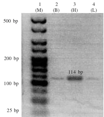
This study revealed the first result of the hatchling turtle Chelonia mydas cDNA fragment from the brain, heart and liver tissues. By using myoturtle F2R2 and F2R1 primer, we obtained 64 and 114 bp of hatchling’s tukik
myoglobin fragments, respectively (Figure 1). Hence, only F2 R1 primers were used for the subsequent process. This study also found that C. mydas myoglobin cDNA were expressed in the brain, heart and liver tissue of the hatchlings (Figure 2, lane 2-4).
Based on SMART PCR techniques we obtained PCR products more than 500 bp (Figure 3) that contained myoglobin cardiac 114 bp of tukik. This fragment was cloned into a suitable vector and positive clone were detected by using universal SP6 and T7 primers and were
500 bp
1 2 3 4 5 6 7 (M) (B) (B) (H) (H) (L) (L)
Figure 3. SMART PCR cDNA fragment (> 500 bp) of C. mydas; lane 1 = DNA marker Hyperladder V (Bioline); lane 2-7 = 114 bp of myoglobin sequences of turtle hatchlings from brain (B), heart (H), and liver (L) tissues.
used for DNA sequencing (Figure 4, lane 2). The myoglobin cDNA (Figure 5) and amino acid sequences (Figure 6) of C. mydas were submitted to Genbank under Acc Num HQ148882. This submission was the first of C. mydas cDNA in Genbank database. The amino acids resulted from this study showed 100% homology to the complete sequence of C. mydas’ myoglobin amino acids from Acc Num P02202. We observed that the C. mydas
myoglobin amino acids obtained from this study were at the amino acids number 63 to 100 of C. mydas myoglobin under Acc Num P02202 (Figure 6).
To detect myoglobin protein expression in heart, brain, and liver tissues of hatchling, the protein was analyzed
500 bp 1 2 3 4 5
(H) (H) (L) (L) (M)
200 bp
100 bp
25 bp
Figure 1. Partial cDNA fragment of C. mydas myoglobin from
tukik heart (H) and liver (L); lane 1, 3= 64 bp amplified using F2 and R2 primer; lane 2 = 114 bp amplified using F2 and R1 primers.
114 bp 64 bp
1 2 3 4 (M) (B) (H) (L)
500 bp
25 bp 100 bp 200 bp
Figure 2. Partial cDNA fragment (114 bp) of C. mydas myoglobin from tukik brain (B), heart (H), liver (L) by using F2 and R1 primers; lane 1 = DNA marker Hyperladder V-Bioline (M).
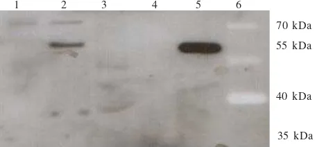
by using polyclonal anti-mouse myoglobin in western blot technique. Blot assay was conducted by using hatchling’s protein of brain, heart, and liver homogenate and as a standard the homogenate of mouse’s cardiac protein was used. The results can be seen in Figure 7. The positive presence of myoglobin protein band was showed by hatchling’s cardiac (Figure 7 lane 2), but not brain and liver tissues (lane 1 & 3). Figure 7 also showed that mouse heart myoglobin protein band displayed high intensity (lane 5).
DISCUSSION
We have demonstrated the presence of a 114 bp myoglobinfragment in cDNA from multiple tissues of turtle hatchlings (Acc Num HQ148882). The PCR products were cloned into E.coli and then sequenced to provided an open reading frame corresponding to 38 amino acids from 63-100 of the published C. mydas putative amino acid sequence (Acc Num HQ148882 – Figure 5 & 6). As cited by Ordway and Gary (2004) myoglobin protein is widely known to be expressed only in oxidative muscle fibres particularly in cardiac myocytes (Hochachka & Somero 2002; Wittenberg & Wittenberg 2003; Flogel et al. 2004). The identification of gene product corresponding to myoglobin confirms that this gene is also express in turtle cardiac tissues as well as liver and brain.
A second gene product of 64 bp (using myoturtle F2 and R2) was also successfully generated from cDNA of both brain and liver tissue of turtle hatchlings consistent
with the expression of the myoglobin gene in these tissues. Fraser et al. (2006) have previously shown that myoglobin gene expression occurs in non muscular tissue in carp, and this was linked to expression of the corresponding protein in the liver. These authors also showed that chronic exposure to environmental hypoxia increases the level of both transcript and protein expression. As it has known that turtle hatch is the hardest atrocious phase for turtle. It is likely that in this hypoxic state it may be require myoglobin to be expressed in multiple non muscle tissues in order to survive. It is required further laboratory research to answer these questions.
To determine the presence turtle hatch C. mydas
myoglobin protein - approx 17 kDa (Sadikin et al. 2010 – in press) in multiple non muscle tissues we employed a western blotting technique. We could only detect myoglobin protein in cardiac tissue of turtle hatch C. mydas
(Figure 7). As explained by Ordway and Garry (2004) that overwhelm of myoglobin protein can be found in heart muscle tissues (Hochachka & Somero 2002; Wittenberg & Wittenberg 2003; Flogel et al. 2004). Under these conditions we could not detect myoglobin protein in brain and liver tissues of turtle hatch C. mydas. There are some reason than could be use to explain this fact.
1 2 3 4 5 6
70 kDa 55 kDa
40 kDa
35 kDa Figure 7. Western blot analysis of tukikChelonia mydas myoglobin homogenate protein from brain, heart and liver tissue using antibody anti-myoglobin mouse. lane 1 = homogenate protein from brain tissue; lane 2 = homogenate protein from cardiac tissue; lanes 3 = homogenate protein from liver tissue; lanes 4 = no sample; lanes 5 = homogenate protein from mouse cardiac tissue; lanes 6 = protein marker (Fermentas– SM0671).
1 2 3 4 5 6 7 HF2-1 HF2-2 HF2-3 HF2-4 HF2-5 HF2-6
500 bp
200 bp 100 bp 25 bp
Figure 4. E. coli positive colonies contained the 114 bp cDNA fragment (lane 2) of tukik cardiac myoglobin using general primer SP6 and T7; lane 1 = DNA marker Hyperladder V (Bioline); lane 3-7 = negative E. coli colonies.
Figure 6. Complete amino acid sequence of Chelonia mydas myoglobin (Acc Num P02202); underlined amino acid were the homology position of 38 putative amino acids of Chelonia mydas cardiac myoglobin resulted from this study (Acc Num HQ148882). Figure 5. Partial myoglobin DNA and amino acid sequences of C. mydas resulted from this study (Acc Num HQ 148882); lower letter
First, the mouse’s antibody as the primary antibody is not sensitive to detect myoglobin in non-muscle tissue of
C. mydas. This is shown in Figure 7 that the commercial antibody can recognize mouse’s heart myoglobin much better than the turtle’s. Hence, to verify the model of myoglobin expression on various non-muscle tissues of
C. mydas, a protein analysis technique and turtle-specific myoglobin antibody would be needed.
The second reason is the highly cell-specific pattern of expression means that, although gross tissue or organ concentrations might be low, the cellular concentration of myoglobin in expressing cells might not be different from those of the heart, where 100% of the cardiac myocite contain at concentrations of 100-400 µmol l-1 (Cossins et
al. 2009). This fact may be explain the absence of myoglobin in brain and liver tissue of hatchlings C. mydas; it was not sensitive when was detected using mouse’s anti-myoglobin on western-blot protein analysis technique. Therefore, more sensitive technique is required in related to protein analysis. Roesner et al. (2008) used different experimental technique from Fraser et al. (2006) and proved that myoglobin expression occurred in many non-muscular tissues.
The data gained from this research is the basic information of myoglobin expression development at the turtle hatch phase. This information is useful for the basis to determine exact turtle hatch treatment technique of farm and hatchery-rear progeny in the field. We aim to improve hatchling survival and help development into strong and healthy adults.
ACKNOWLEDGEMENT
We thank Lisa Olohan, James Wilson, and Gregor Govan from School of Biological Sciences, University of Liverpool, UK. This research was supported by Directorate Generale of Higher Education (DGHE) Ministry of Education, Indonesia through Research Grant Batch IV year 2009, Sandwich Programme Indonesia 2008-2009 and the collaboration of University of Liverpool-UK with universitas Negeri Jakarta.
REFERENCES
Bradford MM. 1976. A rapid and sensitive method for quantitation of microgram quantities of protein utilizing the principle of protein-dye-binding. Anal Biochem 72:248-254.
Brunori M. 2001. Nitric oxide, cytochrome-c oxidase and myoglobin. Trends Biochem Sci 26:21-23.
Cossins A, Berenbrink M. 2008. Physiology: Myoglobin’s new clothes. Nature 454:416-417.
Cossins AR, Williams DR, Foulkes NS, Berenbrink M, Kipar A. 2009. Diverse cell-specific expression of myoglobinisoforms in brain, kidney, gill and liver of the hypoxia-tolerant carp and zebrafish. J Exp Biol 212:627-638.
Flogel U, Godecke A, Oliver Klotz L, Schrader J. 2004. Role of myoglobin in the antioxidant defense of the heart. FASEB J
8:1156-1158.
Fraser J, De Mello VL, Ward D, Rees HR, Williams DR, Fang Y, Brass A, Gracey AY, Cossins AR. 2006. Hypoxia-inducible myoglobin expression in nonmuscle tissues. PNAS 103:2977-2981.
Hochachka P, Somero G. 2002. Biochemical Adaptation: Mechanism and process in physiological evolution. Oxford: Univ Pr.
Lowenstein CJ, Dinerman JL, Snyder SH. 1994 Nitric Oxide: A physiologic Messenger. Ann Intern Med 120:227-237. Ordway GA, Garry DJ. 2004. Myoglobin: an essential hemoprotein
in striated muscle. J Exp Biol 207:3441-3446.
Rassaf T, Flogel U, Drexhage C, Hendgen-Cotta U, Kelm M, Schrader J. 2007. Nitrite reductase function of deoxymyoglobin: oxygen sensor and regulator of heart energetics and function. Circulation Research 100:1749-1754. Roesner A, Hankeln T, Burmester T. 2006. Hypoxia induces a complex response of globin expression in zebrafish (Daniorerio). J Exp Biol 209:2129-2137.
Roesner A, Mitz SA, Hankeln TA, Burmester T. 2008. Globins and hypoxia in the goldfish, Crassius auratus. FEBS J
275:3633-3643.
Sadikin M, Puspitaningrum R, Wanandi SI, Soegianto RR. 2010. Isolation and purification of turtle hatch Chelonia mydas
myoglobin. J Makara (In press).
Wittenberg BA. 2009. Both hypoxia and work are required to enhance expression in myoglobin in skeletal muscle. Am J Physiol Cell Physiol 296:C390-C392.
Wittenberg JB, Witternberg BA. 2003. Myoglobin function reassessed. J Exp Biol 206:2011-2020.