http://mji.ui.ac.id
Primary malignant giant cell tumor of the patella: report of a rare case
Achmad F. Kamal,1 Errol U. Hutagalung,1 Saukani Gumay,2 Yogi Prabowo,1 Yanuarso3
1 Department of Orthopaedic and Traumatology, Faculty of Medicine, Universitas Indonesia, Cipto Mangunkusumo Hospital, Jakarta, Indonesia
2 Department of Anatomical Pathology, Faculty of Medicine, Universitas Indonesia, Cipto Mangunkusumo Hospital, Jakarta, Indonesia 3 Department of Orthopaedic and Traumatology, Gatot Subroto Central Army Hospital, Jakarta, Indonesia
Abstrak
Primary malignant giant cell tumor di patela merupakan tumor tulang yang sangat jarang. Tumor tersebut merupakan tumor derajat tinggi yang ditandai adanya komponen sarkoma dan gambaran giant cell tumor (GCT) konvensional pada pemeriksaan histopatologi. Pembedahan limb sparing merupakan prosedur pembedahan yang dilakukan untuk menyelamatkan ekstremitas pada keganasan dengan mempertahankan fungsi yang optimal. Berikut kami laporkan satu kasus tentang prosedur diagnosis sampai dengan prinsip teknik pembedahan limb sparing pada primary malignant GCT di patela. Evaluasi 15 bulan pasca-pembedahan, pasien memiliki fungsi ekstremitas yang baik tanpa ditandai dengan rekurensi lokal dan metastasis jauh. (Med J Indones. 2013;22:238-42. doi: 10.13181/mji.v22i4.607)
Abstract
Primary malignant giant cell tumor (GCT) at the patella bone is a very rare case. It is a high-grade sarcoma characterized by a lesion in which there are areas of synchronous high-grade sarcomatous next to areas of benign GCT. A limb sparing approach is applicable to virtually any bone lesion, whether low grade or high grade. We report a case with primary malignant GCT at the patella, from diagnostic to detailed limb sparing surgery procedure. Fifteen months after surgery, the patient had a good functional outcome without local recurrence and distant metastasis. (Med J Indones. 2013;22:238-42. doi: 10.13181/mji.v22i4.607)
Keywords: Limb sparing surgery, primary malignant GCT, total patellectomy
Correspondence email to: [email protected]
goal, which is to obtain functional union and avoid arthrodesis.
CASE PRESENTATION
A 31 year-old man presented to Orthopaedic Clinic Cipto Mangunkusumo Hospital, with chief complaint of ulcer on the lump at his left knee. Four years before admission, patient underwent trauma at the left knee and there was swelling. He went to bonesetter several times and had massages. The lump got worsen and it was getting bigger. Ten months before admission, the lump became bigger (15 x 12 x 10 cm3), more painful,
ulcerated, and produced sticky brown-yellowish and foul smelling exudate (Figure 1). Patient came to oncology division of surgery department, and had open biopsy with suspicion of malignant GCT. Patient then referred to orthopaedic oncology.
On physical examination, general condition was good and no abnormality was found in other organ. At the left knee, we found a big tumor at the patellar bone accompanied by necrotic tissue and a large ulcer at lower part of the tumor. No swelling or oedema on distal part of the tumor. The knee movement decreased with range of motion 0-60°.
The laboratory indings showed haemoglobin level 6.4 g/dL, white blood cells 8,100 cells/μL, an increase of erythrocyte sedimentation rate 40 mm/h (ref. < 20), Giant cell tumor (GCT) of bone was irst described by
Sir Astley Cooper in 1818. Historically, the lesion has been referred by numerous terms, including myeloid sarcoma, tumor of myeloplaxus, osteoblastoclastoma, and osteoclastoma. GCT is a relatively common skeletal
tumor, accounting for 4%-9.5% of all primary osseous
neoplasms and 18%-23% of benign bone neoplasms.1
It is characterized by the presence of multinucleated giant cells. Of these, the majority of cases occur in the epiphysis of long bones.2,3
Malignant GCTs, whether representing primary or secondary changes from an originally benign tumor, are extremely rare. It is a high grade sarcoma arising in GCT as a primary or at the site of previously documented GCT (secondary). The incidence of malignant GCT is less
than 0.5% of the total group of malignancy and 5.8% of
all GCT. In Mayo Clinic, there were 35 malignant GCT documented from 11,087 tumor cases, and of these,
there were only ive cases of primary malignant GCT.4
There is a slight female predominance and patients are generally about a decade older than patients with GCT.5
We report a rare case of primary malignant GCT arising at the left patella focusing on diagnostic procedure
and surgical management. This was the irst case
http://mji.ui.ac.id
slightly increase of serum alkaline phosphatase 194 µ/L (ref. 35-187), and normal lactic dehydrogenase 97µ/L (ref. 100-190).
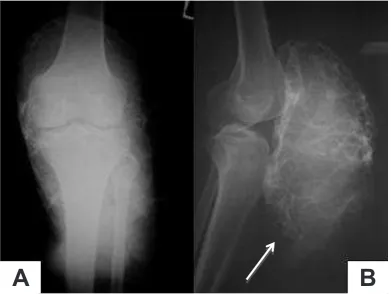
Radiographic examination of left knee anteroposterior and lateral projections showed expansile lytic lesion
at the patella with well-deined margin and cortical
thinning, multiple septation (soap bubble appearance), and destruction of the lowerpool of patella (Figure 2).
Figure 1. Ulcerated tumor at left knee, anteroposterior (A) and lateral (B) view. Big tumor at the patellar bone accompanied with necrotic tissue in large ulcer at lower part of the tumor, no swelling or edema in distal part of the extremity (compared with contralateral side)
A
B
Figure 2. Radiographic pictures. Anteroposterior (A) and lateral (B) projections showed an expanding area of lytic lesion, multiple septations (soap bubble appearances), and destruction of the lower pool of patella (white arrow)
Radiographic indings might suggest benign aggresive
tumor (GCT or aneurysmal bone cyst) or malignancy,
but was not enough to conirm whether it was malignant
or not. Chest radiography and bone scintigraphy showed no evidence of metastasis on the lung and the other bones.
Patient underwent limb sparing surgery with total patellectomy and reconstruction of the extensor
http://mji.ui.ac.id
mechanism using medial gastrocnemius muscle lap.
The incision was designed from the proximal part of the mass in the distal femur and extended to shaft of the tibia. Wide excision with total patellectomy was done.
Soft tissue attached to the tumor were sacriiced, thus
it produced extensor mechanism defect. Then rotational
lap was performed from medial gastrocnemius
muscle to close the defect and to enhance the extensor mechanism. The distal incision was extended on the medial side of the leg to proximal ankle. After identifying medial gastrocnemius muscle, we detached that muscle from its aponeurosis and divided it from lateral part of gastrocnemius toward its proximal pedicle at the medial margin of sural nerve. After that, we rotated the muscle as
a lap to close the defect and sutured it to the quadriceps.
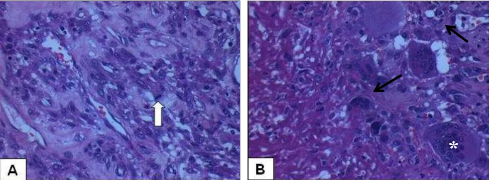
Histopathological indings showed features of GCT with
malignant component. Macroscopically, The resected tumor was yellowish, separated by bony tissue, and
had central necrosis. Microscopic indings showed the
characteristics of typical benign GCT, with ovoid-shaped tumor cells, mitosis, nuclear atypia, and multinucleated giant cells. Osteoid sarcomatous component was also found with its atypical, pleomorphic, bizarre cells, abnormal mitoses, and lacy pattern of osteoid (Figure 3). The margin of excision was free from the tumor.
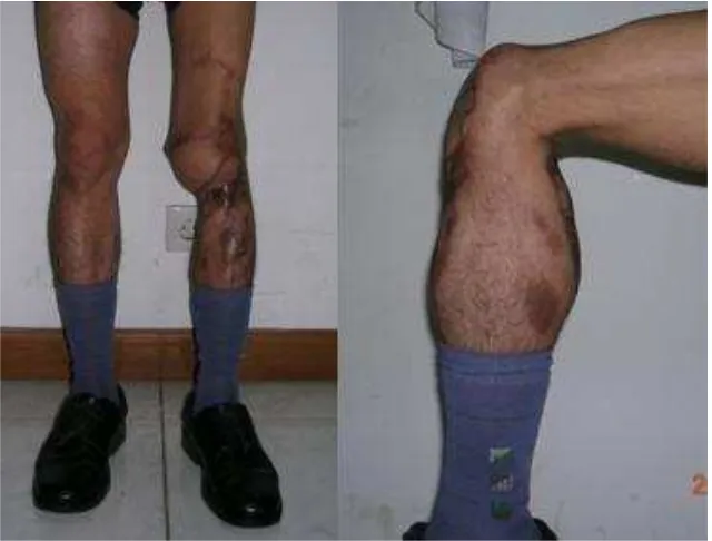
After surgery, patient routinely came to the outpatient clinic. We performed evaluation by clinical and radiological examinations. Fifteen months after surgery, there was no local recurrence and distant metastasis. Patient had good functional outcome, he could walk and stand up without being assisted, ride a motorcycle, and work as a courier at an expedition company. There was improvement of knee motion,
range of motion improved from 0-60° to 0-90°.
According to Musculoskeletal Tumor Society scoring system (MSTS), functional outcome after surgery was
29 out of 30 (Figure 4).
DISCUSSION
GCT of the bone is a benign, locally aggressive neoplasm that is composed of sheets of neoplastic ovoid mononuclear cells interspersed with uniformly distributed large, osteoclast like giant cell. It represents
around 4-5% of all bone tumors, and approximately
20% of all benign primary bone tumors.1,3 Although
GCT affects all races, there is high prevalence in China and Southern India (State of Andhra Pradesh).1,6,7
The vast majority of GCTs affect skeletally mature
patients, with approximately 80% occurring in patients
between 20 and 50 years of age.2,3,8,9 The location of GCT is one of the most important features suggesting
the diagnosis because approximately 84-99% of lesions
extend within 1 cm of subarticular bone. The exact site of origin of GCT has been a controversy, commonly these lesions arise on the metaphyseal side of the epiphyseal plate. GCT usually occurs in the epiphysis of long bones, involving the lower end of the femur, the upper end of the tibia, and the lower end of the radius, in descending order.1-3,5 A small portion of GCT also
occurs in patella, vertebrae, and skull.10
Primary malignant GCT shows a slight female predominance, they are generally about a decade older than patients with GCT.5 Primary malignant GCT is
histopathologically different from GCT. It is a lesion in which there are areas of synchronous high-grade
Figure 3. Photomicrographs of the primary malignant GCT. (A) Low magniication image showed mitosis, atypic, pleomorphic, and bi
-zarre cells (white arrow). (B) Low magniication image showed atypic, pleomorphic, bi-zarre cells, and a lacy pattern of osteoid
– representing sarcomatous component (black arrow) – and giant cells (asteriks). (Haematoxylin & eosin, x40)
∆
*
∆
http://mji.ui.ac.id Figure 4. There was no local recurrence and range of movement of the knee was 0-90°
at ifteen month follow-up
sarcomatous growth next to areas of benign GCT.3,5,9,10 It is very rare and considered as a representation of dedifferentiation in GCT,4 approximately 5 to 10% of GCT cases. 2,10
The patient have had a growing lump at the patella since ten years before, without other organ disturbance. Slow tumor growth in this case conforms the benign tumor characteristics. Tumor growth acceleration was thought due to physical manipulation (massage) on the lump. Large ulcerated tumor with wide necrotic tissue depicted existent or probable character shift from benign to malignant lesion. Radiographic picture of expanding geographic lytic lesion with soap
bubble appearance further conirmed the existence
of aggressive benign tumor.1,11 From initial clinical
and radiological examination, GCT or an aneurysmal bone cyst were the possible diagnoses. Since the available examinations were not adequate to work up the diagnosis, histopathological examination was then performed.
The histological diagnosis of malignant GCT is established only when the stromal cells are truly sarcomatous, as indicated by pleomorphism, atypic, and high mitotic activity. In addition, the areas of typical benign GCT should be in the tumor at the time of diagnosis, or in previous tissue taken from the same lesion. The histological features of the reported
sarcomas include ibrosarcoma, osteosarcoma, malignant ibrous histiocytoma, and undifferentiated
sarcoma.4,5,10
In this patient, the resected tumor was yellowish, showed central necrosis, and separated by bony
tissue. Microscopic indings revealed the presence of
ovoid-shaped tumor cells, mitosis, and atypic nuclear with multinucleated giant cells as typical benign GCT components and also sarcomatous component. The sarcomatous component that we found was osteosarcoma. Because of no history of tumor reported
at the same site, therefore this inding was consistent
and highly suggestive with primary malignant GCT.
The role of chemotherapy is unclear in malignant GCT,6 so the choice of treatment for the tumor is a
complete surgical excision.3,8-10 In this case, the tumor
had been expanding out of patellar bone and involving the surrounding soft tissue, but it did not involve the femoral and tibial bones. The patient had wide excision with total patellectomy and then the defect on the extensor mechanism was reconstructed in order to enable him to walk. The lateral or medial gastrocnemius
lap is useful for defects on the proximal third of the leg. We chose medial gastrocnemius lap because it is
more easily done and have less complication than the
lateral lap.12
Reports on primary malignant GCT affecting other site such as distal femur or proximal tibia were found4,13 but no previous publication has ever reported primary malignant GCT of patella.
In conclusion, primary malignant GCT is an extremely
http://mji.ui.ac.id malignant GCT that was found and reported in Cipto
Mangunkusumo Hospital and probably in Indonesia. The diagnosis of primary malignant GCT was
established based on histopathological indings such as
sarcomatous component and the areas of typical benign GCT. Complete surgical excision and reconstruction
with medial gastrocnemius lap is the treatment of
choice of this primary malignant GCT at the patella.
REFERENCES
1. Murphey MD, Nomikos GC, Flemming DJ, Gannon FH, Temple HT, Kransdorf MJ. From the archives of AFIP. Imaging of giant cell tumor and giant cell reparative granuloma of bone: radiologic-pathologic correlation.
Radiographics. 2001;21(5):1283-309.
2. Unni KK. Giant cell tumor (osteoclastoma). In: Dahlin’s bone tumors - general aspects and data on 11,087 cases. 5th edition. Philadelphia: Lippincott Williams & Wilkins;
1996.p.263-82.
3. Unni KK, Inwards CY, Bridge JA, Kindblom L-G, Wold
LE. AFIP atlas of tumour pathology series 4: tumours of the
bones and joints. Washington: AFIP-ARP; 2005.
4. Unni KK. Malignancy in giant cell tumor of bone. In:
Dahlin’s bone tumors - general aspects and data on 11,087 cases. 5th edition. Philadelphia: Lippincott Williams &
Wilkins; 1996.p.285-9.
5. Bullough PG, Bansal M. Malignancy in giant cell tumour. In: Fletcher CDM, Unni KK, Mertens F, editors. World
health organization classiication of tumours, pathology,
and genetics of tumours of soft tissue and bone. Geneva: IARC Press; 2002.p.313.
6. Sung HW, Kuo DP, Shu WP, Chai YB, Liu CC, Li SM. Giant-cell tumor of bone: analysis of two hundred and eight cases in Chinese patients. J Bone Joint Surg Am.
1982;64(5):755-61.
7. Reddy CRRM, Rao PS, Rajakumari K. Giant-cell tumors of
bone in south India. J Bone Joint Surg Am. 1974;56(3):617-9.
8. Manaster BJ, Doyle AJ. Giant cell tumors of bone. Radiol
Clin North Am. 1993;31(2):299-323.
9. Larsson SE, Lorentzon R, Boquist L. Giant-cell tumor
of bone. A demographic, clinical, and histopathological study of all cases recorded in the Swedish Cancer Registry
for the years 1958 through 1968. J Bone Joint Surg Am. 1975;57(2):167-73.
10. Hong SW, Choi HY. Malignant giant cell tumor of the skull.
J Korean Neurosurg Soc. 2004;36(4):324-7.
11. Greenspan A. Orthopedic imaging: a practical approach.
4th ed. Philadelphia: Lippincott Williams & Wilkins;
2004.p.136-54.
12. Armen KK, Nolan SK. Lower extremity reconstruction. In: Charles HT, editor. Grabb and Smith’s plastic surgery. 6th ed.
Washington: Lippincott Williams & Wilkins; 2007.p.1031-49.
13. Domovitov SV, Healey JH. Primary malignant giant-cell tumor of bone has high survival rate. Ann Surg Oncol.