Hepatitis
B.
A
Preliminary
Report.
Purnomo
SoeharsoAbstrak
Lintfusit T mengenal antigen nelalui reseptor di pernukaan sel (TCR) setelnh berinteraksi dengan nolekul MHC sendiri ("setf
MHC').
Untuk mengenal antigen yang sangat besar junilahnya" TCR mentpunyai struktur molekul yang sangat bemariasi, dihasilkandari rearrangement gen TCR pada maturasi limfusit seperti yang terjadi pada gen inunoglobulin. Meskipun telah diketahui bahwa sel Tberperan pada proses serokonversi penderitahepatitis B khronikatau karier, aknn tetapi mekanismenyabelumjelas. (Jntukntengetahui rearranlgentent gen TCR pada penderita hepatitis B khronik, dilakukan transfer DNA yang diisolasi dari lekosit darah perifer (PBMC) ,rtenurut cara Southern, dan dihibridisasi dengan pelacak gen TCR yang spesifik Rearrangentent dapat dideteksi pada sekurang-kurangnya 57o dari populasi PBMC. Rearrangenent lebih sering ditenrukan pada penderita dengan antigen e positip dan lebih jarang pada penderitayang antigen e negatip. Patron rearrangenent 5 kb EcoRI dan 5,5 kb Hind
III
nterupakan penandayang sering dijwnpai pada karier hepatitis B dengan antigen e positip. Penreriksaan berkala pada 6 penderita hepatitis B khronik ntemperlihatknn adanya penanda rearrangement sebelun serokonversi dan dapat terus terdeteksi selann 6 bulan sesudah serokonversi. Dengan detnikian dapat diduga bahwa munculnya klon sel T dengan patron rearrangement tertentu menjad.i penanda terjadinya serokonversi. Identifikrxi kLon tersebut akan diteliti lebih lanjut.Abstract
T cells recogttize speciftc antigens through the T celL receptor (TCR) and self M HC nolecules. Structural variation in the TCR to recognise the enonnous diversity, of antigens
is
effected b), naturation rearrange,nentof
TCR genesin a
tilaruter analogous to inrnunoglobulin genes. Although it is believed that T cells play auajor
role in the seroconversiotr of e Ag positive hepatitis Bcaliers,
the tnechanisnts are still unclear. To etanine the role of TCR rearrangetne,lts in chronic hepatitis B we have carried ouI Southertl transfers of DNA aliqu.otsfrom peripheral blood tttononuclear celk (PBMC) usitrg TCR chain specific probes. Rearranget,rcn$ affecting as few as 5 % of PBMC could. be detected. Rearrangentents were iltote cornnnn in e Ag positive than negative. Of several rearrangenrcnt patterns a 5 kb EcoRl and 5.5 kb HindIII
were ùtost coilmrcn. Longitudinal studies of 6 patients suggested the appearence of the rearrat'tgement band preceded seroconversion and could be detected in PBMCfor 6 nonths after seroconversion.It
is postulated that duritrg seroconversion the appearance of a clone of T cells with a particular rearrangernentis
associated with seroconversion and characterisation ofthese clones is currently being undertaken.Keywords
:
T cell receptor, Gene rearrrangement, Chronic hepatitis BINTRODUCTION
The
diseasecourse
of chronic
hepatitis
B is
variable,
with patients
fluctuating between
remission
andactive
disease, asmonitored
by
markers
of liver
destruction
and
virus replication.
It
is important
to
understandT
cell activity during
the course
of chronic
hepatitis B,
as
it
may
provide
a meansof monitoring
the disease,and possibly
will
be
of
benefit
for
immunological
intervention
in care
or treatment
strategies.From
several studies,
it
appearedthat different
phases
of
disease progression
can
be
recognized
over the
yÇa^rsduring the long
course
of
chronic
hepatitis
B."'''
These phasesreflect variations in
host7
128
Soeharsoimmune
responseto HBV during different
occasionsof
chronic
infection.
Application of
TCR
gene rearrangement analysisduring
the courseof
chronic
HBV infection would
be usefulin defining
the disease progress, asit
mayreflect
theimmunological
statein different phases of
chronic
disease.The observation
of TCR
gene rearrangement bandsmay thus
belinked
to
the featuresmost
predic-tive of outcome in chronic hepatitis B. As
discussedin
severalpublications,
these are serum aminotransferaselevel
indicating
liver
destruction,
and
serologic
markers
of
virus replication
indicated
by
HBeAg
presentin
theserum.*
The possibility that
there are
changesof T
cell
clonality during
the
courseof chronic HBV infection
and the use of serum aminotransferase level and avirus
replication
marker
as theindicators
of disease
chroni-city,
prompted
theapplication of TCR
generearrange-ment analysis
in
association
with
serum transaminaselevel and the
presence
of
HBeAg
in
the serum
of
patients
from whom blood
had been takenon
several occasions.This examination may provide
a meansfor
the
evaluation
of
host
immunity
in
virus-induced
chronic hepatitis
B,
that may
be advantageousfor
thestrategy
of
chronic hepatitis B
treatment.SUBJECTS AND
METIIODS
Subjects
Subjects
were
6patients
with
chronic hepatitis
B
who
had been testedserologically
for
HBeAg
andanti-HBe
as
virus replication
markers;
the
test was performed
usingcommercial
ELISA kit (Abbott laboratories,IL).
Subjects were also testedfor
serum alanine transferase(ALT)
level
as amarker
for liver
destruction, using
astandard
method. The
tests
were carried out
by
thePathology Department, Royal Brisbane Hospital, over
a
period ranging
from 15
to 37 months. Theblood
was taken at least oncein
2or 3
months.Methods
Genomic
DNA
was isolatedfrom
1Oml of heparinized
blood.
10
ug
of
genomic
DNA
was then
digestedcompletely with EcoRl or Hind
III
in
anappropriate
buffer.concentration
overnight. DNA
fragments
weresize fractionated
by
electrophoresis
in
0.8% agarose
and the
separatedDNA
fragments were transfered
toHybond
Nylon
membrane(Amersham).
Thefilter
con-taining DNA
fragments
wasthen
hybridized with
the"P-labelled
TCR
BcDNA
probeovernight.
At
the endMedJ Univ Indon
of
the hybridization thefilter was
washed severaltime-swith
2 xdown
to 0.1 x SSCcontaining 0.1%
SDS, andfinally
thefilter
was autoradiographed.RESULTS
To
determine
whether the detection
of
TCR
B generearrangements
might be correlated
with
the
patho-genesisof
chronic hepatitis
B,
the appearanceof
rear-rangement bandsduring
the disease courseof
these sixpatients was associated
with
the
disease progress
which
was assessed by regular testing ofALT
level and serumHBeAg.
The pattern
of TCR B
gene rearrange-ment,ALT
level
and serumHBeAg
wereplotted
in onechart,
and the results areshown
in Figure
l.
As
shown
in
case
I
(MY),
the
presenceof
rear-rangement bandsof
5.0 kbEcoRl and
/
or 5.5 kbHind
III
DNA
fragmentscoincided
with
high
ALT
level
andthe
presenceof
HBeAg
in
the serum, detected
in
theperiod
of
5/88 to
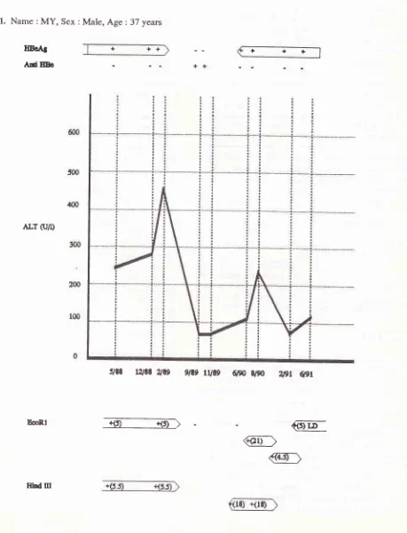
2189.As
thepatient underwent
sero-conversion and the ALT dropped to moderate level, the rearrangement bands become undetectable as shownin
9/89. Nine
months
later (6/90) the
serum was
againHBeAg positive, and the
ALT
level
increased
to
ahigher level, although
not
as
high as
in
the
period
beforeseroconversion;
rearrangement bands appearedagain,
but the size was different
from
those before
seroconversion.In
case 2 (MF), the 5.0 kbEcoRl
and 5.5 kbHind
III
rearrangement bands were also detectable andcoin-cided
with
high
ALT
level
andHBeAg
positive
serum asindicated
in
theperiod
of
8/88
to
1/89.At
thetime
of
seroconversion between
6/89 and 1l/89,
the
ALT
dropped
to normal level.
At
this time
the presenceof
rearrangement bands was
not
assessed.Four
monthslater (3/90) the
ALT
increased
ro
a
high
level
and reached amaximum
in5l9O;the
serum becomeHBeAg
positive
again, the 5.0
kb EcoRl
and 5.5 kb
Hind
III
rearrangement bands also
reappeared.
The
5,0
kb
EcoRl
band was undetectablein
thebegining
of
ALT
elevation
in
3/90,probably
because the amountof
theT
cell
clone
of
interest at that
time
was
lessthan
thethreshold
of
detection
sensitivity. Four
months later
(9/90)
the
patient
underwent seroconversion and
theALT
dropped tonormal level;
this stage persisteduntil
9/91,
during which
rearrangement bands
of
TCR
B gene were undetectable.In
case3
(BW),
there were no
detectableEcoRl
rearrangement bandsduring
23months of chronic
dis-easecourse.
the
appearanceof
the 5.5
kb
Hind
III
rearrangement band
coincided
with
serum
being
HBeAg positive
andwith a high
ALT
level
as in 3/89and 9/89.
As
patient
underwent seroconversion
anded so
through2lgl.
In
case4 (DD), the patient
had
not
cleared
thevirus
completely
following
20
months
of
diseasecourse.
The
5.0kb
EcoRl
and 5.5kb Hind
III
rearran-gement bandswere
detectable,coinciding with
serum beingHBeAg positive
andwith high
ALT
level
as seenin
theperiod between
10/88and 3/88.
Theserearran-gement bands
disappeared
as the
ALT
dropped
tonormal level
in
8/89, whenHBeAg
wasstill
presentin
trace amount
in
the serum (borderline).
This
stagepersisted
until
6/90.In
case 5(DHD),
theHBeAg
was alwaysnegative
during the
course
of chronic
disease.ALT level
in-creased
slightly
in
thefirst
5 monthperiod
and reached amaximum in
6/89,
during which
5.0
kb EcoRl
and5.5 kb
Hind
III
rearrangement bandswere
detectable.When
ALT
dropped to
normal
level
in
8i89 the
rear-rangement bands were
undetectable.
This
situation
proceed
until
3/90,
when
ALT
level
increased
to moderatelevel
and the 5.0kb
EcoRl
and 5.5kb
Hind
III
rearrangement bands reappeared.Six months later,
the rearrangement bands disappeared again,
coincident
with
the decreasedALT
level.
Case
6 (KC)
is unique
in
that
the rearrangementof TCR
B genein
peripheral blood
T
cells
wasdetec-table simultaneously
with
the rising
ALT
level
and become undetectable asALT
dropped tonormal level.
However,
theHBeAg
wasalways negative
during
the courseof
the disease.The results
of monitoring
TCR
B generearrange-ment
during
the
diseasecourse
on
these 6patients
issummarized
in table
1 andthe general feature
of
therelationship
of TCR
B gene rearrangement,ALT
level
and serumHBeAg
can beillustrated in
Figure
2.DISCUSSION
The
appearanceof TCR
gene rearrangement bandsin
peripheral blood
DNA,
is
associatedwith clonal
ex-pansionof
aT cell clone
to aproportion of
at least 5 %of total
mononuclear cells, as has
been confirmed
previously.s
Clonal
expansion
of T cells would
occur
if
there were
a
specific stimulation that
induced
specific
T
cells to become sensitized and
undergo
proliferation.
Some
previous
experiments
have
reported
thatperipheral blood lymphocytes
of
chronic
HBeAg
positive patients
are sensitizedto
HBcAg.6'7
Furthermore, some experiments suggest
that
the
peripheral
blood
mononuclear
cell
commpartments
in
patients
with
chronic active hepatitis
B
(CAH,B)
con-tain cytotoxic
T
cells specific
for
HBcAg.8,e
HB"Ag
induces
important immune
reactions tocytotoxicclass
tion of HBV-infected
hepatotytes.T'I0
In
addition
to
cellular cytotoxicity, lymphokine
secretion
is
anotherimportant
consequence of Tcell activation, which
con-tribute to
the outcome
of viral
disease. II
On"
of
theimportant lymphokines
is gamma-interferon, which
has been
well known for its antiviral effect, but
may
alsocontribute
tocytotoxic
eventsby upregulating
the expressionof HLA
molecules on the susceptible targetcells."
This
finding
is likely to
be
relevant to
theresult of this study.
The 5.0 kbEcoRl
and 5.5 kbHind
III
rearrangement
bands may correlate
with
clonal
expansion
of
cytotoxic
T
cells specific
to HBcAg
or
regulatory
T
cells that
mediate
immune
responseby
releasing lymphokines.
This
study examined the hypothesis that
TCR
Bgene rearrangement
in
peripheral blood
T
cells
is
amanifestation
of
immune
progression
in
chronic
hepatitis
B.
The experimental
approach was based on theconsideration
thatclonal
proliferation
of T cells
in
peripheral
blood
correlate
with
cell
mediated
im-munity
to
virus infection,
andlinked
to
theactivity
of
T
cells
in
thesite
of inflammation in
theliver.
It
wasfound in this
study that
only few
rearran-gement bands
of TCR
B genes were observed in
peripheral blood of chronic hepatitis B,
indicating few
clones
of T cells
toproliferate
and detectableby direct
examination
of
TCR
Bgene rearrangement
of
white
blood cells.
Dominant clones carrying
TCR
genedetectable
as 5.0
kb EcoRl
and 5.5 Hind
III
DNA
fragments appear in most patients examined.
This
rear-rangement
bands changedynamically during
the
dis-ease course
concurrent
with
immune
attack toinfected
liver
in
the processof
viral
clearance.A previous study
by Moebius et all3 found that liver
infiltrating
lym-phocytes
distribute
polyclonally
as detectedby TCR
Bgene rearrangement
analysis on
T cell
clones
from
liver
of chronic active hepatitis B
(CAH)
patients.It
islikely
that sensitized Tcell
clones expand inperipheral
blood
are part of those present in the site ofinflamation
in
the liver.
There are
only small proportion
of
sen-sitized
T
cells
in
peripheral blood
as mostof
them aresequestered
into
the
liver
for
clearance
of virus
in-fected
cells,
and
only cells with
considerable
expan-sion
would
be detectableby
the present method.sThe
activity
of T cells, asindicated
byTCR
l
generearrangement detectable
in
peripheral
blood
was ex-aminedfor
associationwith liver
destruction
andvirus
clearanceduring
diseaseprogression.
As
shown
infigure
1,presenting
6 cases, theTCR
B generearrangement bands
of
5.0 kb EcoRl
and/or
5.5 kb Hind
III
DNA
fragments were
usually
coinci-130 Soeharso Med.J Univ Indon
Figure
l.
Tine course of the appearance of narkers virus replication (HBeAg) and seroconversion (anti-HBe) in the seruttr, Iiver datnage (indicated by seru ALT level) and TCR0 gene rearrange,,rcnts detectable when peripheral blood ntononuclear cett DNA was digested with EcoRl or HindIII,
in 6 patients with chronic hepatitis B. The presence of HBeAg or anti-HBe in the serunr is indicated by pàsitive(+) or ,legative (-) sigtts. Changes ofALT level are indicated by graph lines- Dates on the abscissa indicate date
ofblood
coliections. Detectable reaftangement bands are indicated by a positive(+)
sign along with the size of rearrangement band (in ttrackets). A negative() sign
indicates that rearrange,nent fund bands were not detectable (caset,
2, 3, 4, 5 and 6).CASE
1.
Name
:MY,
Sex :Male,
Age:
37 yearsEoAS
AdEEo
ALT (r/l)
EcoRl
+
++
+++
@
@
@]
slæ
t2tEt
2139g/te
rUtg
6po
s,90
Wr
6pr
EAI
oûllDo
6m
5{D
/(x)
ALr (U/r)
:n
2m
tq)
0
rrrr
t/19
$e3ne
@@
rure 'l9l
qgr 9,9r3/90
9i90-@
86llrhd m
dn
50
4m
lqr
2û
tm
EoRl
I32
SoeharsoCASE
4.Name
:DD,
Sex :Male, Age
:51
yearsllBÈtt AdHBc
@
50
{(D
ALrCJrr)
m
tn
t(D
0
MedJ Univ Indon
B€Rl IltdItr
l. r-.
-;rr
qs'-<3t---<Jt) æ-er'---î!i-9>
l/le lqq
qe
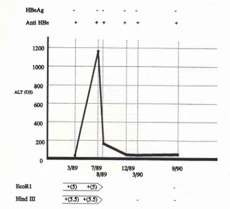
CASE
5.Name
:DHD,
Sex :Male, Age :
19 yearsfIBcAt
Ard
HBoALT (U/r)
++
EcoRl
Inrd Itr
HDoAg
Ad
HBe
+
++
+
+
1200
1000
t00
ALT
G'D
600
12189
3N
9/90
BcoR.l
IThd
Itr
+(s.t
+(5.t
Table
l.
TCR R gene rearrangement analysis during the disease course of six patients, monitored in association withserum
ALT
level and the presence of HBeAg in the serum.slEe
TEe
E/8e
+(5)
ALT
level reached
Average
Period e'le- Case6
E/H
Case 5
E/H
Case Case
Caset23
E/H
E/H
E/HCase
4
EIH
Prior to peak
At peak After peak Trough Prior to peak
At peak After peak Trough
5 mth
<lmth
5 mth
>7mth
5 mth
<lmth
5 mth>7mth
+ +
+
+ + +
5/s.5 5.5.5
-t-y'I8
2U18
4.sl-
-t-
4.sl-5/5.s
sl--t--ls.s 5/5.5
-t-J5.5 -/5.s
-t--t
5/5.5 5/5.5
-t-5/5.5 -t-sls.s
-t-5/5.5 5/s.5
-t-Note:
E = EcoRl rearrangementbandH = Hind
III
rearrangement bandmth = month
[image:7.595.67.523.105.518.2]t34
SoeharsoEcoRl
clÔ)
---F\
dent with the
increase
of ALT
level.
In
some
casesrearrangement bands
of different
sizewere
detectablesix months after soroconfersion
(as
in
case 1). The
results
indicate
thatin
somechronic
HBV
carriers,
the expansion ofperipheral blood
Tcell
cloneswith
5.0kb
EcoRl
and 5.5
kb Hind
III
TCR
B genefragments is
associated
with cytotoxic
clearance
of virus
infected
hepatocytes
and the loss
of HBeAg from circulation.
The
clonal
expansion
of T cells
detectableby TCR
Bgene rearrangement
apparently
represents theprogres-sive
development
of
immunity
to virus infection in
chronic
HBV
carriers. The
expandedT
cell
clones aremost
likely linked to
the
activity of cytotoxic T
cells
with
thepotential
to eliminate substantial
numbersof
virus
infected
liver
cells. During this elimination
phase, thedestruction
of liver
tissueis marked by
theelevation
of ALT level in
theserum. The
elimination
of virus
infected
hepatocytesby
T cells
is theeffector
mechanism
of
virus eradication, that
consequently
prevents
viral replication and
therefore reduced
the releaseof virions to
peripheral
blood. This
reduction
of
virus replication
is
characterized
by
the
disap-pearance
of HBeAg
andin
many patients by
serocon-version
from
being HBeAg positive
tobeing
anti-HBe
MedJ Univ Indon
antibody positive. When there
is
no sign
of
viral
replication
and thepatient
becomesanti-HBe positive,
liver cell lysis is
decreased,indicated by normal or
slightly
elevated
ALT
level. The specific
T
cells
responsible
for
liver cell
lysis would be
reduced
in
number, as antigenic stimulation leading
to
clonal
proliferation is over.
Consequently
the TCR
B gene rearrangement become undetectable, since thenumber
of
such
T
cells would be reduced
to
less
than
thethreshold
of
detection
sensitivity
for TCR
gene rear-rangement.Case
5 (DHD)
and 6
(KC)
areunique
in
that
the rearrangementof
the
TCR
B genein
peripheral blood
was detectable
simultaneously
with rising ALT
level,
however the
HBeAg
was always negative
during
thecourse
of
the disease.
As
there was evidence
of
cytotoxic
liver
cell
lysis
(high
ALT
level),
virus
replication must have occured
previously,
since
production
of viral
antigen on theliver
cells drives
thecytotoxic
reaction. The
absenceof HBeAg during
thereplication
phase
of
the
diseasecould be
associatedwith
the
failure
of
the
virus to prduce
and release eantigen to the
circulation. This situation
may occur dueto
mutation
in
the
virus
genes
responsible
for the
EIE!oAg
+++++
ALT
Ellnd
rrr
oô)
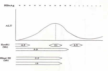
Figure 2. General pattern ofthe relationship
ofTCRf
gene rearrangenrent in peripheral blood, ALT level and HBeAg in the serunt [image:8.595.96.549.90.389.2]sid precore-core
geneproduct; when
thefull
precore-core
gene
is
transcribed,
the
transcript
will
beprocessed and the
resulting
mRNA
istranslated into
asecretory
protein. Transcription
of
the nucleocapsid
genes
starts
at the
second initiation codon, yielding
HBc protein which is not
secretedfrom cells
due to
lack
of
precore encoded peptide which functions
asleader directing
this
protein
to
the
cell
membrane,ultimately to be
secretedfrom liver cells.la Mutation
in
theprecore region
would
leadto failure of
produc-tion
of
leader peptide necessary
for
secretion
of
HBeAg.
Patients
infected
with
such mutant virus
would therefore
never have detectableHBeAg in their
serum, despite the
presenceof
HBV-DNA
and DNA
polymerase
simultaneously.
This
is
presumably
thecase
in patients
5 and 6.This study describes the relationship
of
TCR
Bgene
rearrangement
with
the mechanism
of
immune
mediated
liver injury
and virus clearance
in
chronic
hepatitis
B. It might
be suggestedthat specific
TCR
Bgene
rearrangement
of
5.0 kb EcoRl
and/
or 5.5 kb
Hind
III
DNA
fragments
in
peripheral
blood
aredetected
in
chronic hepatitis
B
patients just prior
to
seroconversion.
The21 kb EcoRl DNA fragment
wasfound
at the peakof
ALT
level,
if
the 5.0kb EcoRl or
5.5 kb Hind
III
DNA
fragments were
not
detectable.This band may
representclonal expansion
of T
cells
with
theactivity similar
to those expressing the 5.0kb
EcoRl or
5.5kb Hind
III
DNA fragment. However,
asthe presence
of
this
band
is
not
common,
its
sig-nificance is unclear.
As seroconversion
is completed,
the
5.0
kb EcoRl
and
/
or
5.5
kb
Hind
III
DNA
fragments usually
become undetectable
and
this
is
accompanied by
dropping of ALT
tonormal
level.This
general pattern
of
the.relationship
of
TCR
B
generearrangement
in peripheral blood with
ALT
level
andserum
HBeAg during
the courseof chronic hepatitis B
infection
was summarized
asin table
I
andis shown
diagrammatically
in
Figure 2.
In
general, the TCR
Bgene rearrangement band
usually
of
5.0kb EcoRl
and/ or
5.5 kb Hind
III
DNA fragments or
sometimes 4.5kb,
21 kb EcoRl
and
18kb Hind
III
DNA fragments
are detectable during
the
viral
clearance phase
of
chronic
diseasecourse. This is indicated
by the flare
up of
ALT
level and continue toresolution
phasewhere
the
ALT
level
decreasing tonormal level.
Therearran-gement band
dissappearswhen
ALT
dropped to
nor-mal level
andpatient
undergoesseroconversion,
whenthe marker
of
virus replication is undetectable in
theperipheral blood.
In conclusion, the
appearanceof specific
TCR
Bgene rearrangement
in
peripheral blood
T
cells
of
progression
of
host
immunity to clear the virus. The
TCR
B generearrangement may reflect clonal
expan-sion
of
T
cells responsible
for
elimination
of
virus
infected
hepatocytesin
the processof viral
clearance,bringing
thepatient to seroconversion.
REFERENCBS
l.
Lee PI, Chang MH, Lee CJ et al. Changes of serum hepatitis B virus DNA and aminotransferase levels during the courseof
chronic hepatitisB
infectionin
children. Hepatologyl99O:- 12 :657 - 60.
2. Desmet VJ. Liver lesions in hepatitis B viral infection. Yale.
J Biol
Med1988;2:61
- 83.3. Paronetto F, Colucci G, Colombo M. Lymphocytes in liver
diseases. vol
VIII.
Grune and Stratton. pp. l9l-2Ù1,1986.4. Hoofnagle IH. Chronic hepatitis B. New Engl J Med 1990;
323:33'l
-9.
5. Soeharso P. Definition of TCR gene rearrangement detection sensitivity in peripheral blood population. in T cell receptor
gene polymorphisms
and
rearrangementsin
chronichepatitis B, Ph.D thesis University of Queensland. pp.108 -9,1992.
6. Vento S, Hegarti JE, Alberti A et al. T lymphocyte
sensitiza-tion to
HBcAg and T-cell mediated unresponsiveness toHBsAg
in
hepatitisB
virus related chronic liver disease.Hepatol 1985;5 :192
-7.
7. Ferrari C, Penna A, Sansoni P et al. Selective sensitjzation ofperipheral blood T lymphocytes to hepatitis B core antigen
in
patients with chronic active hepatitis type B. Clin Exp Immunol 1986;67 :497-
5O6.8. Mondelli MG, Mieli-Vergani G, Alberti A et al. Specihcity of the T lymphocyte cytotoxicity to autologous hepatocytes
in chronic hepatitis B virus infection : evidence that T cells
are directed against
HBV
core antigen expressed onhepatocytes. J Immunol 1982;129 :2773 - 8L
9. Naumov NV, Mondelli MG, Alexander GIM. Relationship between expression of hepatitis B virus antigens in isolated hepatocytes and autologous lymphocyte cytotoxicity in
patients with chronic hepatitis B infection. Hepatolog y L984
4:63-8.
10. Mondelli MG, Bortolotti F, Pontisso P et
al.
Definitionof
hepatitis B virus (HBV) specific târget antigens recognized
by
cytotoxicT
cellsin
acuteHBV
infection.Clin
Exp Immunol 1987; 68 :242-
5O.I 1. Wright R. Pathogenesis of viral hepatitis. Clin Gastroenterol
1988;4:695-705.
12. Thomas
HC.
Pathogenesisof
chronic hepatitis B.
JGastroenterol Hepatol Suppl.
l99l;
I
: 4 - 6.13. Moebius U, Manns
M,
Hess G et al.T
cell receptor gene rearrangements of T lymphocytes infiltrating the liver in in chronic active hepatitisB
and primarybiliary
cinhosis (PBC) : Oligoclonality of PBC-derived T-cell clones. Eur JImmunol 1990;20 : 889 - 6.
14. Carman WF, Jacyna
MR,
Hadziyannis S et al. Mutation preventing formation of hepatitis B e antigen in patients with