SHORT COMMUNICATION
Extracellular Protease Activity of Enteropathogenic
Escherechia coli
on Mucin Substrate
SRI BUDIARTI∗∗∗∗∗, NISA RACHMANIA MUBARIK
Department of Biology, Faculty of Mathematics and Natural Sciences, Bogor Agricultural University,
Darmaga Campus, Bogor 16680, Indonesia
Received June 30, 2006/Accepted March 12, 2007
Enteropathogenic Escherichia coli (EPEC) causes gastrointestinal infections in human. EPEC invasion was initiated by attachment and aggressive colonization on intestinal surface. Attachment of EPEC alter the intestine mucosal cells. Despite this, the pathogenic mechanism of EPEC infectior has not been fully understood. This research hypothesizes that extracellular proteolytic enzymes is necessary for EPEC colonization. The enzyme is secreted into gastrointestinal milieu and presumably destroy mucus layer cover the gastrointestinal tract. The objective of this study was to assay EPEC extracellular protease enzyme by using mucin substrate. The activity of EPEC extracellular proteolytic enzyme on 1% mucin substrate was investigated. Non-pathogenic E. coli was used as a negative control. Positive and tentative controls were Yersinia enterocolitica and Salmonella. Ten EPEC strains were assayed, seven of them were able to degrade mucin, and the highest activity was produced by K1.1 strain. Both positive and tentative controls also showed the ability to digest 0.20% mucin.
Key words: EPEC, protease, mucin, diarrhea
___________________________________________________________________________
_________________
∗∗∗∗∗Corresponding author. Phone/Fax: +62-251-622833,
E-mail: [email protected]
HAYATI Journal of Biosciences, March 2007, p 36-38 Vol. 14, No. 1 ISSN: 1978-3019
Escherichia coli is normal flora in human gastrointestinal track, however, some strains may cause several diseases including diarrhea. Frankel et al. (1995) stated that the main etiology of diarrhea diseases in developing countries is
enteropathogenicE. coli (EPEC) that alter cell structure of the absorptive intestine by disturbing water, electrolyte, and nutrition absorption. The damage enables EPEC to penetrate into intestine epithelial cells and block the absorption process (Salyers & Whitt 1994).
EPEC adhere to the outer epithelial or intestinal mucosa. Mucosa surface coats by mucus layer, mainly compose of mucin, a 2 x 10⁶ kD glycoprotein polymer (Bell et al. 1985).
The effectiveness of mucosa layer as in vivo natural shield depends on the structure and thickness of the layer.
Several pathogenic bacteria, such as Clostridium RS 42,
Bacteroides RS 2, Bacteroides RS 13, Yersiniaenterocolitica
is able to degrade mucin (Stanley et al. 1986; Mantle & Rombough 1993). Candida albicans was reported by Colina
et al. (1996) produce aspartil protease that degraded mucin as well. However, mucin degradation by EPEC protease has not been reported, as yet. The objective of this study was to assay EPEC extracellular protease enzyme by using mucin substrate.
EPEC strains used in this study were collection of Biomedic Laboratory, Center of Biotechnology and Biological Research, Bogor Agricultural University. EPEC RB1.2 produced extracellular protease (Priyanto 1997). Yersinia enterocolitica
as a positive control was collected of PT. Bio Farma (Persero), Bandung. Salmonella sp. and non-pathogenic E. coli as tentative and a negative control, were collected from Microbiology Laboratory, Department of Biology, Faculty of Mathematics and Natural Sciences, Bogor Agricultural University.
EPEC RB1.2 and three bacterial controls were grown in skim milk broth minimal (SMB Min) media. This media contained of 2% skim milk, 2% NaCl, and 0.5% yeast extract. The inocula were incubated at 37 oC and were shaked at
100 rpm for 12 hours. A number of 1 x 10⁹ cells/ml were
inoculated to SMB Min media and were incubated at 37 oC
(shaked 100 rpm for 15 hours). The culture was then centrifuged at 4000 x g in 4 oC for 10 minutes to obtain the
crude extract of protease enzyme (Priyanto 1997).
Protease activity of crude extract RB1.2 and 3 from the bacterial controls were measured in 1% casein substrate by using modified method of Walter (1984). One unit enzyme activity (U/ml) was equal to the amount of enzyme that produced 1 µmol tyrosine product per one minute in optimum condition. Protein concentration was measured using Bradford method (1976) and used bovine serum albumin (BSA) as the standard. Specific activity was determined as protease activity unit divided by the protein concentration.
replicates, and tested by Tukey’s pairwise comparisons value at α = 0.05.
EPEC RB1.2, Salmonella, and Y. enterocolitica showed protease activity in 1% casein substrate at 0.005, 0.020, and 0.021 U/ml (Figure 1). However, there was no protease activity showed in non-pathogenic E. coli as a negative control. Protease activity of RB1.2 showed higher activity in 0.5% casein substrate compared to the 0.25 and 1% (Figure 2). Moreover, RB1.2 showed its highest protease activity in 1%
mucin substrate (Figure 3), and there was no protease activity in higher than 1% mucin.
The ability of Y. enterocolitica and Salmonella to hydrolyze mucin was compared to RB1.2 in several concentrations. The highest concentration of protease activity in mucin substrate of Y. enterocolitica and Salmonella was in 0.2% mucin on each 0.040 U/ml (Figure 4). There was no protease activity detected by using 0.5 and 1% mucin substrates. Isolate RB1.2 still showed its 20% protease activity relative on 0.2% mucin substrate.
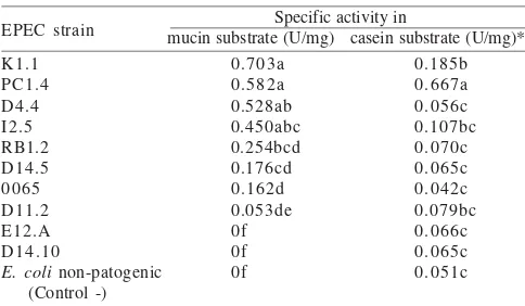
Specific activity of ten EPEC strains and non-pathogenic
E. coli as a negative control were assayed by using mucin and casein substrates. Different substrates produced specific activity (α = 0.05) (Table 1) as showed in seven strains of EPEC (K1.1, PC1.4, D4.4, I2.5, RB1.2, D14.5, and 0065). They showed different results compared to the negative control in 1% mucin substrate. Only PC1.4 and K1.1 showed a different response with control by using 0.5% casein substrate. EPEC K1.1 showed the highest specific activity (0.73 U/mg) in 0.5% mucin substrate, but PC 1.4 strain (0.667 U/mg) was the highest in 1% casein.
Non-pathogenic E. coli as a negative control did not show protease activity by using 1% casein substrate. However, positive control bacteria (Y. enterocolitica) and tentative
Table 1. Specific activity of protease EPEC to 1% mucin substrat and
0.5% casein incubated in pH 8, 37 oC
*The same alphabet followed the number in the same column was
referred to the value that has no significant difference on α 0.05
Tuckey’s Test
Figure 1. Protease activity of EPEC RB1.2 compare to
non-patho-genic E. coli (negative control), Salmonella (tentative
con-trol), and Y. enterocolitica (positive control) in 1% casein
substrate. The activity were assayed at 37 oC, pH 8.
E. coli RB1.2 Salmonella Y. enterocolitica
120
Figure 2. Protease activity of EPEC RB1.2 measured in several casein
concentrations. The activity were assayed at 37 oC, pH 8.
120
Figure 3. Protease activity of EPEC RB1.2 measured in several con-centrations of mucin. The activity were assayed at 37
oC, pH 8.
Figure 4. Protease activity of EPEC RB1.2 compared to Salmonella
and Y. enterocolitica at several mucin concentrations. The
control (Salmonella) revealed higher protease activity than that of RB1.2. Yersinia enterocolitica is a motile gram negative rod that cause acute gastroenteritis in human. The highest activity of casein degradation of Y. enterocolitica showed specificity protease activity to hydrolyze casein substrate which is the highest compound in milk protein.
The data of mucin substrate degradation might showed that protease activity of EPEC in mucin substrate was higher than those of Y. enterocolitica and Salmonella (Figure 4). The capability of mucin degraded by EPEC in vivo presumably assist EPEC to perform an aggressive colonization to the intestine surface in the first step of pathogenesis. The EPEC extracellular protease enzyme was proposed to be one of EPEC virulence factors (Salyers & Whitt 1994). Further research need to be performed to answer the whole assumption, since
in vivo condition of gastrointestinal lumen is much more complex than those of in vitro.
In several cases, in vitro condition is more conducive for bacteria growth, because it was treated in optimum pH and there is no nutrition competitor among pathogen bacteria as well as the normal flora. There is no mucosa activity such as peristaltic activity or cilia impulse that allows foreign material eliminate from mucosa support pathogen growth in vitro, as well.
Grange et al. (1998) studied the adherence mechanism of
Enterotoxigenic E. coli (ETEC) to the intestine epithelium. The result showed that the receptor of host intestinal epithelium were Intestinal Mucin-Type Glycoprotein 1 (IMTGP 1) and IMTGP 2. Entroaggregative E. coli (EAEC) was known able to adhere to mucosa tissue by using in vitro organ culture method (Czeczulin et al. 1997). This adhesion caused hyper secretion mucus on mucosa layer.
Mucosa layer consists of covalent and noncovalent chains. The later consists of glycoprotein polymer structure which were degraded by protease enzyme. However, the non-covalent chain is relatively stable to protease, hypertonic effect, and solvent such as acid and ethanol (Bell et al. 1985). Besides protease, glycosidase might degrade mucin as well. However, study on Y. enterocolitica by Mantle and Rombough (1993) showed that carbohydrates component of mucin, i.e. galactose and acetyl galactosamine labeled with radioisotope 3H was not degraded into smaller molecules. This
result supported the assumption that protease is the main enzyme that degraded mucin. Mantle and Rombough (1993) reported the ability of mucin degradation by Y. enterocolitica
harbering plasmid was much higher than those without plasmid. Presumably, the enzyme involved in this process was supposed to be regulated by plasmid.
Protease activity of another EPEC RB1.2 (K1.1 and PC1.4 isolates) showed high hyolrolysis activity in casein and mucin. Hence, both isolates are interesting to be studied in molecular aspect, such as characterization of mucin encoding gene.
ACKNOWLEDGEMENTS
This research was supported by a research grant from Hibah Bersaing VI Program (1996-1997) from Directorate General of Higher Education to Sri Budiarti. We addressed grateful acknowledgement to Maggy T. Suhartono for excellent advice and to Rini Candra Kusumaningrum for technical assistance.
REFERENCES
Bell AF et al. 1985. Properties of gastric and duodenal mucus: effect of
proteolysis, disulfide reduction, bile, acid, ethanol and hypertonicity
on mucus gel structure. Gastroenterology 88:269-280.
Bradford MM. 1976. A rapid and sensitive method for the quantitation of microgram quantities of protein utilising the principle of protein
dye binding. Anal Biochem 72:248-254.
Colina AR, Aumont F, Deslaurier N, Belhumer P, Repentigny LD. 1996. Evidence for degradation of gastrointestinal mucin by
Candida albicans secretory aspartyl proteinase. Infect Immun
64:4514-4519.
Czeczulin JR et al. 1997. Aggregative adherence fimbria II, a second
fimbrial antigen mediating aggregative adherence in
enteroaggregative Escherichia coli. Infect Immun 65:4135-4145.
Frankel G et al. 1995. Molecular characterization of carboxy-terminal
eucariotic cell-binding domain of intimin from enteropathogenic
Escherichia coli. Infect Immun 63:4323-4328.
Grange PA, Erickson AK, Anderson TJ, Francis DH. 1998. Characterization of the carbohydrate moiety of intestinal mucin-type sialoglycoprotein receptors for the K88ac fimbrial adhesin of Escherichia coli. Infect Immun 66:1613-1621.
Mantle M, Rombough C. 1993. Growth in and breakdown of purified
rabbit small intestinal mucin by Yersinia enterocolitica. Infect
Immun 61:4131-4138.
Priyanto C. 1997. Isolasi dan Karakterisasi Enzim Protease
Ekstraseluler dari EPEC (Enteropathogenic Escherichia coli)
[Skripsi]. Bogor: Fakultas Matematika dan Ilmu Pengetahuan Alam, Institut Pertanian Bogor.
Salyers AA, Whitt DD. 1994. Bacterial Pathogenesis, A Molecular
Approach. Washington: ASM Pr.
Stanley RA, Ram SP, Wilkinson RK, Roberton AM. 1986. Degradation of pig gastric and colonic mucin by bacteria isolated from the pig
colon. Appl Environ Microbiol 51:1104-1109.
Walter HE. 1984.Method with haemoglobin, casein and azocol as
substrat. In: Bergmeyer J, Graβl M (eds). Methods of Enzymatic
Analysis. Vol 5 3rd ed. Weinheim: Verlag Chemie.