Merkel cell carcinoma of right buttock in an elderly patient: a case
report
Keywords: chemotherapy, elderly patient, merkel cell carcinoma, radiotherapy
pISSN: 0853-1773 • eISSN: 2252-8083 • http://dx.doi.org/10.13181/mji.v24i3.1242 • Med J Indones. 2015;24:190-4 • Received 21 Apr 2015 • Accepted 19 Aug 2015
Correspondence author: Shiromani Debbarma, [email protected]
Copyright @ 2015 Authors. This is an open access article distributed under the terms of the Creative Commons Attribution-NonCommercial-ShareAlike 4.0 International License (http://creativecommons.org/licenses/by-nc-sa/4.0/), which permits unrestricted non-commercial use, distribution, and reproduction in any medium, provided the original author and source are properly cited.
Shiromani Debbarma, Rajesh Sharma, Dhaneshor Sharma, Th Tomcha Singh Department of Radiotherapy, Regional Institute of Medical Sciences, Lamphel, Imphal, Manipur, India
C a s e Re p o r t
ABSTRAK
Karsinoma sel merkel merupakan tumor kulit yang jarang. Sejauh ini, hanya sedikit laporan kasus mengenai penyakit ini. Kami melaporkan kasus seorang laki-laki berusia 87 tahun dengan tumor berbentuk ulkus yang bersifat proliferatif, tidak nyeri, berukuran 6x6 cm pada bokong kanan, diketahui stadium IIB (TNM, T3cNoMo). Pasien menerima kemoterapi neoadjuvan; karboplatin injeksi 420 mg (hari 1) dan etoposide 140 mg (hari 1-3) setiap tiga minggu sebanyak tiga siklus dilanjutkan dengan radioterapi eksternal menggunakan Cobalt 60. Tiga bulan setelah terapi, tidak ada sisa maupun rekurensi yang dilaporkan. Tujuan laporan kasus ini adalah menekankan pada sifat kasus yang jarang, asimtomatik sehingga bisa terlambat di diagnosis, serta diagnosis definitif untuk tujuan terapi yang terbaik.
ABSTRACT
Merkel cell carcinoma is a rare primary cutaneous tumor. So far, few cases have been reported. Herewith we report a case of an 87-years-old male with painless, ulceroproliferative growth measuring 6x6 cm, over right upper outer quadrant of buttock; stage IIB (TNM, T3cNoMo). Patient received neo-adjuvant chemotherapy, injection of carboplatin 420 mg (day one) and etoposide 140 mg (day one to three) three weekly for three cycles followed external beam radiotherapy by Cobalt 60. At three months post-treatment follow-up, clinically no evidence of residual disease or recurrences noted. The purpose of reporting this case was to emphasize to its rarity, early asymptomatic clinical course leading to possibility of delayed diagnosis and paramount importance
of high index of clinical suspicion in definitive diagnosis for
Merkel cell carcinoma (MCC) is a rare, aggressive cutaneous tumor. Due to its rarity no appropriate treatment protocol is made so far. Here we report a case treated with neoadjuvant chemotherapy followed by radiation therapy in an old debilitated patient. At three months follow-up, clinically no residual and recurrence of disease is noted.
Merkel cell carcinoma was first described by Toker.1 This rare skin tumor is also known
by various names such as primary small cell carcinoma of skin, primary neuroendocrine carcinoma of skin, primary undifferentiated carcinoma of skin, anaplastic carcinoma of skin, trabecular carcinoma of skin, and cutaneous amine precursor uptake and decarboxylation
tumor APUDoma.2 The tumor arises from
neurocrest derivative merkel cell of epidermis situated in the basal layer which contains
neuro-secretory granules. The early lesion
is often asymptomatic and may present as firm glossy, non-tender, red or bluish-purple, rapidly growing nodule at the time of presentation.2,3 There are a few reported cases
of merkel cell carcinoma so far and to the best of our knowledge this case report is the first in North-East India.
Case Illustration
A 87 year-old male was admitted to hospital with complaint of a painless ulcer measuring 6 x 6 cm located over skin of right upper outer quadrant of buttock. Unlike decubitous ulcer, the ulcer was rapidly proliferative, with reddish thickened, everted margin, violaceous hue around the ulcer for the last two months. The patient has no history of smoking, alcohol consumption, and chewing tobacco. On general physical and systemic examinations revealed no abnormality.
All routine investigation such as complete haemogram, kidney and liver function test, random blood sugar level was within normal limit, his chest X-ray, ultrasonography (USG) whole abdomen and screening bilateral inguinal
lymphnodes showed no lymphadenopathy.
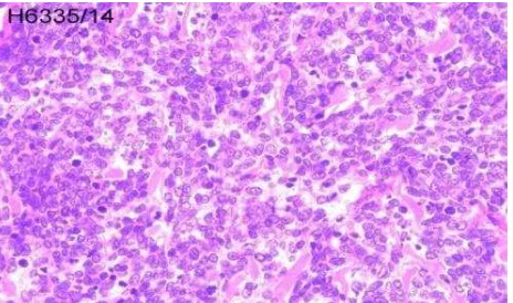
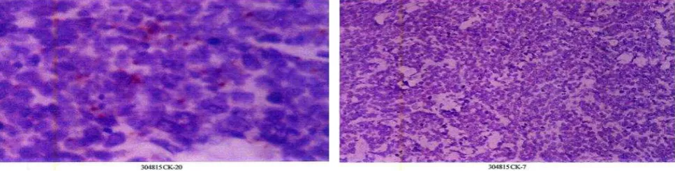
His performance status was 40% (Karnofsky scale). In view of clinically suspicious malignant ulcer, biopsy was done and reported small cell carcinoma with numerous mitotic figures and suggesting MCC. Further, immunohistochemical status of cytokeratin
(CK)7 and CK20 were investigated and found CK20 positive and CK7 negative. On the basis of clinical, imaging, histopathologic and tissue immunohistochemistry findings, the case was diagnosed as MCC; stage IIB tumor, node, metastasis (TNM) staging, T3cNoMo. Patient was explained of about different treatment modalities with their pros and cons and patient opted chemotherapy and radiation therapy.
Patient received neo-adjuvant chemotherapy (NACT) injection carboplatin 420 mg (day one) and etoposide 140 mg (day one to three), three weekly for three cycles followed conventional fractionation external beam radiotherapy giving up to a total dose of 60 Gy, 200cGy/fraction over six weeks by medial and lateral tangential fields with Cobalt 60 machine. One month following treatment, there was no residual disease clinically and patient was kept under follow-up. He was disease-free at his last check-up three months after completion of treatment.
Figure 1. Ulcero-proliferative growth
Figure 2. Biopsy report showing small round cells with numerous mitosis (MMG 40X)
Figure 3. Immunohistochemical staining showing CK20 positive and CK7 negative
DISCUSSION
Merkel cell carcinoma is an uncommon, highly malignant neuroendocrine cutaneous tumor with overall unfavorable prognosis.4 The tumor
is generally seen in sun exposed areas such as head and neck (50%), extremities (35-40%), and upper body but less than 10% cases appear in the trunk of fair elderly men.5 A rare site like pharynx,
an oral or nasal mucosa, vulva, and penile may also be involved.6
Although the exact etiology is not fully understood, there are several risk factors that contribute to its pathogenesis such as ultraviolet (UV) light, sun exposure related skin malignancy, and treatment with methoxsalen. Elderly and immunocompromised patients prone to develop MCC. Polyoma virus is incriminated as one of the causative agents.4-8
Clinically MCC may present as a non-tender, firm, painless nodule, sometimes with ulcerated skin lesion with a reddish or violaceous hue less than 2 cm at presentation.8-10 Because
of the low incidence rate and asymptomatic in its early clinical course, diagnosis is often delayed, or even missed. Around 70-80% of MCC present as localized disease, 9-26% as invasion of regional lymph nodes and 1-4% extra-nodal distant metastasis. Histologically, MCC looks small round blue cells, with scanty cytoplasm, hyperchromatic nuclei, multiple nucleoli, delicately granular chromatin, abundant mitoses, and numerous apoptotic figures.5
Diagnosis of MCC is usually confirmed by clinical, histopathologic, and immunohistochemistry
reports of the tissue biopsy. The differential diagnosis to be made between MCC from other tumors is small cell lung cancer, cutaneous lymphoma, amelanotic melanoma, and other primitive neuroendocrine tumor (PNET) category.11MCC is CK20 positive and expressed
characteristic paranuclear dot pattern. MCC is neuron specific enolase positive, S-100 (soluble-100, protein) weak positive and stain for cluster of differentiation CD117.4
Merkel cell carcinoma is a rapidly growing tumor. Nearly one-third of patients die within three years from the time of diagnosis.12 Unfavorable
prognostic factors include male gender, age more than 65 years with co-morbid diseases, distant metastatic disease, tumor of head and neck region, tumor size more than 2 cm in diameter, tumor with more than 10 mitoses per high-power field, evidence of vascular invasion, absence of an inflammatory reaction, and high expression of Kiel (Ki)-67 (proliferation index marker).4,13 Lymph node involvement decreases
survival from 88% to 50% and it shows within two years in 50% to 70% of all patients. MCC is a rapidly progressive fatal disease. Considering the high local recurrence 27-60%, regional involvement 45-91% or distant metastasis 18%-52% patients should be closely followed up after definitive treatment.5, 14
The rareness of the disease and the lack of prospective clinical studies evaluating the efficacy of therapeutic options, caused limited set of treatment guidelines.5
3 cm margin and 2.5 cm dept is sufficient, but some authors advocate less than 2-3 cm in case of 1.5-2 cm tumor without high local recurrence. An alternative to wide local resection is Mohs micrographic surgery.15
Surgical dissection of all lymph nodes is controversial, many authors advocate complete lymph node dissection in case of positive lymph node at presentation.5 One study recommended
prophylactic lymphadenectomy only if the lesion is more than six weeks and tumor more than 1.5 cm in size prior to seeking medical advice and sentinel lymph node biopsy in case of negative node.16 On the contrary, prophylactic
lymphadenectomy is neither recommended as a standard management scheme nor in sentinel lymph node (SLN)-negative biopsies; however, it is recommended in MCC with increased risk of potential recurrence and aggressive metastasis.17
Merkel cell carcinoma is predominantly a radio-sensitive malignancy. Surgery alone has very high local recurrence rates for this disease. Post-operative radiotherapy increases survival than surgery alone. Radiotherapy plays an important role in case of large primary tumor or un-attainable free surgical margin due to cosmetic or surgical difficulties and increases overall survival.12,13,18
In case of metastatic disease, treatment option consists of palliative radiotherapy and chemotherapy. MCC is also a chemo-sensitive tumor. Tumur regression and remission rates can be as high as 70% with chemotherapy, however without significant prolonged survival benefit.11 Multiple agents have been used
with different response rates.14 Those include
cyclophosphamide, doxorubicin, etoposide, cis-platin, vincristine, methotrexate, fluorouracil (FU), carboplatin, etc.5,12 Biologic agents like
anti-tumor necrosis factor and imatinib mesylate promises better result on local or systemic control of MCC.6 Radiotherapy can be used in
case of cutaneous deposits or bone and brain metastasis.5
In conclusion, MCC is a very rare skin cancer. The incidence of MCC may be increasing in future in modern society due to increase life span. Thus, there is paramount importance of awareness
among clinicians about the possibility of MCC in elderly patients. There is high need for routine skin biopsy for any painless, non-tender, proliferative, with reddish thickening margin, violaceous hue around the ulcer, and rapidly growing skin nodule to rule out MCC prior to any treatment is being suggested in this disease.
Acknowledgment
We would like to thank BABINA Diagnostic Center, Imphal, Manipur, India for the help managing the patient.
Conflict of interest
The authors affirm no conflict of interest in this study.
REFERENCES
1. Toker C. Trabecular carcinoma of the skin. Arch Dermatol. 1972;105(1):107-10.
2. Haag ML, Glass LF, Fenske NA. Merkel cell carcinoma: diagnosis and treatment. Dermatol Surg. 1995; 21(8):669-83.
3. Hitchcock CL, Bland KI, Laney RG 3rd, Franzini D, Harris B, Copeland EM 3rd. Neuroendocrine (merkel cell) carcinoma of the skin. Its natural history, diagnosis, and treatment. Ann Surg. 1988; 207(2):201-7.
4. Nghiem P, Jaimes N. Merkel cell carcinoma. In: Wolff K, Katz S, Goldsmith L, Gilchrest B, Leffell D, Paller A, editors. Fitzpatrick’s dermatology in general medicine.
7th ed. USA: McGraw-Hill; 2008.
5. Pectasides D, Pectasides M, Economopoulos T. Merkel cell cancer of skin. Ann Oncol. 2006;17(10):1489-95. 6. Dinh V, Feun L, Elgart G, Savaraj N. Merkel cell
carcinomas. Hematol Oncol Clin North Am. 2007; 21(3):527-44.
7. Feng H, Shuda M, Chang Y, Moore PS. Clonal integration of a polyomavirus in human merkel cell carcinoma. Science. 2008; 319(5866):1096-100.
8. Heath M, Jaimes N, Lemos B, Mostaghimi A, Wang LC, Peñas PF, et al. Clinical characteristics of merkel cell carcinoma at diagnosis in 195 patients: the AEIOU features. J Am Cad Dermatol. 2008; 58(3):375-81. 9. McAfee WJ, Morris CG, Mendenhall CM, Werning
JW, Mendenhall NP, Mendenhall WM. Merkel cell carcinoma: treatment and outcomes. Cancer. 2005;104(8):1761-4.
10. Gorjón PS, Morales AC, Pérez PB, González JLG, de Dios JC, Melgar AR. Merkel cell carcinoma: a presentation of 5 cases and a review of the literature. Acta Otorrinolaringol Esp. 2011; 62(4):306-10.
12. Eng TY, Boersma MG, Fuller CD, Cavanaugh SX, Valenzuela F, Herman TS. Treatment of merkel cell carcinoma. Am J Clin Oncol. 2004;27(5):510-5.
13. Veness MJ. Merkel cell carcinoma (primary cutaneous neuroendocrine carcinoma): an overview on management. Australas J Dermatol. 2006;47(3):160-5. 14. Mehrany K, Otley CC, Weenig RH, Phillips PK, Roenigk
RK, Nguyen TH. A meta-analysis of the prognostic
significance of sentinel lymph node status in merkel cell
carcinoma. Dermatol Surg.2002; 28(2):113-7.
15. Bichakjian CK, Lowe L, Lao CD, Sandler HM, Bradford CR, Johnson TM, et al. Merkel cell carcinoma: critical review
with guidelines for multidisciplinary management. Cancer. 2007; 110(1):1-12.
16. Boyse K, Foley EH, Bradley V, Scarborough D. Merkel cell carcinoma: a case report with treatment summary and updates. Cutis.2004; 74:350-6.
17. Ratner D, Nelson BR, Brown MD, Johnson TM. Merkel cell carcinoma. J Am Acad Dermatol. 1993; 29(2Pt1):143-56.
18. Meeuwissen JA, Bourne RG, Kearsley JH. The importance of post-operative radiation therapy in the treatment of merkel cell carcinoma. Int J Radiat Oncol Biol Phys.