UNIVERSITI TEKNIKAL MALAYSIA MELAKA
PREPARATION AND CHARACTERIZATION OF SOL-GEL DERIVED HYDROXYAPATITE COATING ON STAINLESS STEEL SUBSTRATE
This report submitted in accordance with requirement of the Universiti Teknikal Malaysia Melaka (UTeM) for the Bachelor Degree of Manufacturing Engineering
(Engineering Materials) (Hons.)
by
ZURIANEE BT LOKMAN LOGANATHAN B050910174
890203075596
i
ABSTRAK
Tujuan kajian ini adalah untuk menyediakan dan mencirikan salutan hydroxyapatite
ii
ABSTRACT
iii
DEDICATION
iv
ACKNOWLEDGEMENT
vi
2.6.5.1 Phytic Acid 24
CHAPTER 3 : METHODOLOGY 26
3.1 Introduction 26
3.2 Material Selection 28
3.3 Synthesis Of Hydroxyapatite Sol 28
3.3.1 Sol Without Catalyst 28
3.3.2 Sol With HCl Catalyst 29
3.4 Coating Process 29
3.4.1 Stainless steel Substrate With Conversion Coating 29 3.4.2 Stainless steel Substrate Without Conversion Coating 29
3.5 X-Ray Diffraction Analysis 31
3.6 Material Composition Analysis (SEM & EDX) 32
3.7 Optical Microscope 33
CHAPTER 4 : RESULT AND DISCUSSION 34
4.1 Final pH value of Hydroxyapatite 35
4.2 XRD analysis 36
4.3 Optical Microscope 38
4.4 Scanning Electron Microscope & EDX Analysis 42
CHAPTER 5 : CONCLUSION AND RECOMMENDATION 47
5.1 Conclusion 47
5.2 Recommendation 48
REFERENCES 50
APPENDICES 55
A Gantt Chart of PSM I 56
vii
LIST OF TABLES
2.1 Example of Biomaterials 5
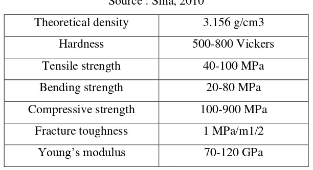
2.2 Advantages And Disadvantages Of Each Class Of Biomaterial. 6 2.3 Composition Ranges For 316L Stainless Steels. 8 2.4 Typical Mechanical Properties of Dense Hydroxyapatite Ceramics 10 2.5 Previous Studies on the Effect of Precursors towards Hydroxyapatite 18
Formed.
2.6 Comparison Of Coating Process. 23
4.1 Ph Value Of Hydroxyapatite Sol With And Without The Presence 35 Of HCl catalyst.
4.2 Composition Of Element Present In Stainless Steel Substrate Coated 43 With Phytic Acid And Hydroxyapatite Sol.
4.3 Composition Of Elements Present In Stainless Steel Substrate Coated 45 With Hydroxyapatite Sol (With Presence of HCl).
viii
LIST OF FIGURES
2.1 Structure on Hydroxyapatite 12
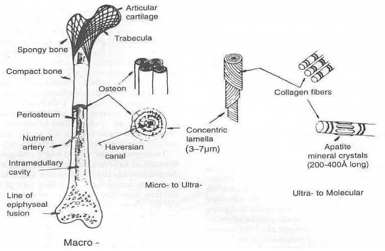
2.2 The Molecular Structure, Macrostructure and Microstructure of 14 Hydroxyapatie In Bone
2.3 Sol-gel Process 17
2.4 Steady State Dip Coating Process 22
3.1 Process Overview 27
3.2 Stirring Process 28
3.3 Dip Coating Process 30
3.4 X’pert Pro Diffractometer 31
3.5 Scanning Electron Microscope 32
3.6 Optical Microscope 33
4.1 XRD Pattern of Hydroxyapatite 36
4.2 XRD Result Of Stainless Steel Substrate Coated With Hydroxyapatite 37 4.3 XRD Result Of Stainless Steel Substrate Coated With Hydroxyapatite 37
With Presence of HCl Catalyst
4.4 Stainless Steel Substrate Coated With Hydroxyapatite In 38 Magnification 20X
4.5 Stainless Steel Substrate Coated With Phytic Acid And Hydroxyapatite 39 In Magnification 20X.
4.6 Stainless Steel Substrate Coated With Hydroxyapatite 40 (With Present of HCl) In Magnification 20X
4.7 Stainless Steel Substrate Coated With Phytic Acid And Hydroxyapatite 41 (With Present of HCl) In Magnification 20X
4.8 SEM and EDXof Stainless Steel Substrate Coated With Phytic Acid 43 And Hydroxyapatite Sol In Magnification 500X
4.9 SEM and EDX of Stainless Steel Substrate Coated With Hydroxyapatite 44 Sol (With Presence Of HCl) In Magnification 500X.
ix
LIST ABBREVIATIONS, SYMBOLS, NOMENCLATURE
Al = Aluminium
ASTM = American Society for Testing Materials
Ca = Calcium
Cu = Copper
Fe = Iron
HA = Hydroxyapatite
HCl = Hydrochloric Acid
JCPDS = Joint Committee on Powder Diffraction Standard
L = Length
ml = Milliliter
Mn = Manganese
Ni = Nickel
P = Phosphate
T = Thick
W = Width
Zn = Zinc
1
CHAPTER 1
INTRODUCTION
1.1Background of Study
Biomaterials have been use for quite a long time ago by humans. The Romans, Chinese, and Aztec used gold in dentistry more than 2000 years ago. Besides, glass eyes and wooden teeth have been used trough much of the recorded history. During that period of time, there were no medical device manufacturers, no formalized regulatory approval processes, no understanding of biocompatibility and certainly no academic courses on biomaterials (Ratner, 2004). Yet, crude biomaterials have been used, generally with poor to mixed results, throughout history. Earlier of the 21st century, biomaterials are widely used throughout medicine, dentistry and biotechnology.
2
an immune system response will be generated. When an object is incorporated into the body without any immune responses it is said to be biocompatible. In order for a device to be biocompatible, it must follow a very strict requirement from the body.
Stainless steel has enjoyed clinical success because of their superior strength, biocompatibility, durability, and resistance to corrosion in physiological environment. The high mechanical strength and toughness of these bio-metals are the most important advantages over bioactive ceramics, which are inherently weak and brittle. Upon implantation, a close contact of the metal prostheses with surrounding host tissue is required for a subsequent ingrowths of bone tissue into the pre-design cavities on the implant surface. The change of relative position between implant and the surrounding tissue is highly undesirable and therefore immobilization of a patient may be needed before the implant fixation is strong enough to bear load.
However, there are concerns about their corrosion resistance upon the body physiologic fluids and also their bioactivity, since the probability of pitting corrosion is high in stainless steel. Corrosion is constituted of the material loss that causes an implant to become weak. The more significant matter is the release of the corrosion products in the body tissues which causes some adverse effects, which increases the rate of formation of fibrous tissue around the implant. On the other hand, stainless steel are not capable of forming a suitable bond between the implant and tissues. For this reason, developing the techniques for increasing their corrosion resistance and bioactivity become significant.
3 1.2 Problem Statement
Due to high mechanical strength and toughness, stainless steel is being use as biomaterial. Besides, other factors that makes stainless steel as a success biomaterial are its durability, biocompatibility and resistance to corrosion. However, the corrosion resistance of stainless steel towards body fluid and their bioactivity become a concern. Thus, hydroxyapatite coating is being used over stainless steel substrate as hydroxyapatite is a material that has characteristic close to body tissue. Furthermore, phytic acid will be used as a conversion coating on stainless steel substrate to enhance its corrosion resistivity. In this research, stainless steel will be coated with sol-gel derived hydroxyapatite by using dip coating technique. The use of catalyst and conversion coating will be studied in order to prepare sample that can restrain corrosion and degradation.
1.3Objectives
The objectives that need to be achieved during this study include:
a. To prepare hydroxyapatite coating solution via sol-gel technique.
b. To implement the use of catalyst and conversion coating in the coating preparation.
c. To develop a coating layer on substrate by using dip coating technique.
1.4Scope
4
CHAPTER 2
LITERATURE REVIEW
2.1 Introduction
This chapter reviews on the related study based on the previous research conducted by other researchers on the hydroxyapatite derided by sol-gel method as a coating process on stainless steel substrate. Through this chapter, the study on hydroxyapatire sol-gel is being studied and discussed. Besides, the literature review will also focus on the principle of stainless steel as biomaterials and coating technique used which is dip coating.
2.2 Biomaterial
Biomaterials improve the quality of life for an ever increasing number of people each year. The range of applications is vast and includes such things as joint and limb replacements, artificial arteries and skin, contact lenses, and dentures. The biomaterials community is producing new and improved implant materials and techniques to meet this demand, but also to aid the treatment of younger patients where the necessary properties are even more demanding. To meet these urge needs it is necessary to have reliable methods of characterisation of the material and material-host tissue interactions.
5
bonding to surrounding tissue. For example, new bone growth being stimulates. Degradable or resorbable materials are incorporated into the surrounding tissue, or may even dissolve completely over a period of time. Metals are typically inert, ceramics may be inert, active or resorbable and polymers may be inert or resorbable. Some examples of biomaterials are provided in Table 2.1.
Table 2.1 : Example of Biomaterials (Rodriguez et al, 2004)
Metals Ceramics Polymers
Ultra high molecular weight polyethylene
Polyurethane
6
Table 2.2 : Advantages And Disadvantages Of Each Class Of Biomaterial. Class of
material
Advantage Disadvantage Example
7 2.2.1 Biocompatibility
According to Mihov et al (2010), biocompatibility is the ability of a material to perform with an appropriate host response in a specific application. The material should posses the quality of not having toxic or injurious effects on biological systems. There are many factors which influence implant biocompatibility such as implant size, shape, material composition, and surface wettability, roughness and charge. For a material to be considered biocompatible, any adverse reactions which may ensue at the blood/material or tissue/material interface must be minimal. This requires a biomaterial to interact as a natural material would in the presence of blood and tissue. Biomaterials should not alter plasma proteins (including enzymes) so as to trigger undesirable reactions, cause adverse immune responses, cause thrombus formations, cause cancer, produce toxic and allergic responses and destroy or sensitize the cellular elements of blood.
2.3 Corrosion extensively used in medical devices, might corrode severely in this bioenvironmental in accordance with both thermodynamic and kinetic considerations. This degradation process is undesirable because it limits the functionality and lifetime of medical devices. Besides, it releases corrosion products that may cause an adverse biological reaction in the host. Ceramics may also undergo selective leaching, although they more often fail in situ due to mechanical processes such as wear.
8
Corrosion is something that has to be avoided. Approaches available for controlling corrosion include the application of protective coatings to metal surfaces to act as a barrier or perhaps provide sacrificial protection. The coating functions to inhibit corrosion by altering the alloy chemistry to make it more resistant to corrosion (Shaw, 2006).
2.4 Stainless Steel
Stainless steel materials are resistant to a wide range of corrosive agents due to their
high Cr content (more than 12 wt%), which allows the formation of a strongly
adherent, self-healing and corrosion resistant coating oxide of Cr2O3. Several types
of stainless steel are available and the most widely used for implant manufacture is austenitic stainless steel. In order to be austenitic at room temperature, stainless steel needs to contain a certain amount of austenite stabilizing elements such as Ni or Mn. The stainless steel most widely used in clinical applications is AISI 316L that contains 0.03 wt% C, 16–18 wt% Cr, 10–14 wt% Ni, 2–3 wt% Mo and minor amounts of nitrogen, manganese, phosphorus, silicon and sulphur as can be seen in Table 2.3 below.
Table 2.3 : Composition Ranges For 316L Stainless Steels.
Grade C Mn Si P S Cr Mo Ni N
316L Min - - - 16.0 2.00 10.0 -
Max 0.03 2.0 0.75 0.045 0.03 18.0 3.00 14.0 0.10
9
N stabilizes the austenitic phase and induces an increase in both the corrosion resistance and the mechanical properties (yield stress). This is a clear example of new materials with an improved performance that have been developed chronologically during the second generation, but that from a conceptual point of view belong to the first generation.
Stainless steels are characterized by corrosion resistance higher than other steels due to the formation of a passive oxide film. That film reduces the corrosion rate by blocking the transport of metallic ions and electrons. The stainless steels are classified into three categories according to their microstructures: ferritic, martensitic and austenitic. Among them, the austenitic stainless steels (face centered cubic structure, nonmagnetic) which contains Cr (16-18 wt. (%)) and Ni (12-15 wt. (%)) in its composition are responsible for increasing corrosion strength and ensure the stability of the austenitic phase, respectively (Karimi et al, 2012).
10
extensively in medicine and dentistry for implant fabrication owing to its biocompatibility with human bone and teeth. However due to its poor mechanical properties, hydroxyapatite ceramics cannot be used for heavy load bearing applications, but common uses include bone graft substitution and coatings on metallic implants. Table 2.4 shows the mechanical properties of hydroxyapatite.
11
There are several methods to produce hydroxyapatite powder. The most popular and widely researched route is solution precipitation. Hydroxyapatite nanoparticles can be prepared using microwave irradiation. Sol-gel and hydrothermal routes are the two other important routes for hydroxyapatite synthesis. Even hydroxyapatite can be produced by mechanosynthesis route, in which case no heat treatment is required to produce crystalline nano hydroxyapatite. Some other routes for synthesis of hydroxyapatite are solid state reaction, plasma technique, hydrothermal hot pressing, ultrasonic spray pyrolysis, and emulsion system (Nath et al, 2006).
Porous hydroxyapatite has better biocompatibility as tissues can grow much faster into the available pores. The pore size can be controlled and also complex shaped materials can be fabricated. Several efforts have been made to improve the mechanical properties of hydroxyapatite (Nawawi et al, 2011). Thermal treatment is necessary to improve the mechanical properties. Even sometimes some amount of additives can be added to improve the sinterability and mechanical properties without affecting the bioactivity. Using C(OH)2 additives the sintering temperature can be
increased without any dissociation (Sina, 2010).
Hydroxyapatite is an important inorganic biomaterial which has attracted the attention of researchers related to biomaterials field in recent years. Hydroxyapatite is chemically similar to the mineral component of bones and hard tissues in mammals. It is one of few materials that are classed as bioactive, meaning that it will support bone ingrowth and osseointegration when used in orthopaedic, dental and maxillofacial applications (Rodriguez,2004). As a result, this inorganic phosphate has been studied extensively for medical applications in the form of powders, composites or even coatings. According to Agrawal et al (2011), it is also observed that dense sintered HA has many bone replacement applications and is used for repairing bone defects in dental and orthopedic sites and immediate tooth replacement. For substituting or repairing the bone, the designed material must has the ability to create a bond with the host living bone.
12
forms such as powders, porous blocks or beads to fill bone defects or voids. These may arise when large sections of bone have had to be removed for example in the case for bone cancer or when bone augmentations are required. The bone filler will provide a scaffold and encourage the rapid filling of the void by naturally forming bone and provides an alternative to bone grafts. It will also become part of the bone structure and will reduce healing times compared to the situation where no bone filler was used.
Coatings of hydroxyapatite are often applied to metallic implants (most commonly titanium or titanium alloys and stainless steels) to alter the surface properties. In this manner the body sees hydroxyapatiteas the type of material which is happy to accept. Without the coating the body would see a foreign body and work in such a way as to isolate it from surrounding tissues (Costan et al, 2011). Synthetic HA is classified as polycrystalline ceramics since its material structure is derived from individual crystals that have been fused together by a high temperature sintering. Figure 2.1 shows the structure of hydroxyapatite.
13 2.5.1 Advantages Of Hydroxyapatite
Many biocompatibility studies prove that hydroxyapatite has a very similar chemical composition as the inorganic part of human hard tissue, such as bone and teeth. According to Liu et al (2011), the most important advantage of hydroxyapatite being a bioactive material is that bone will form a direct chemical bonding to hydroxyapatite implant without forming a collagen interface layer which is usually found in many other bioinert materials after implantation. Thereby the relative micro-movement between the implant and bone is dramatically reduced by this direct bonding, and no fibrous tissue capsule can be found between the implant and bone. This is important for the patient’s recovery in the early period after implantation. Due to this chemical bonding interface, the bonding strength of hydroxyapatite and bone is much higher than other materials, such as Al2O3, ZrO2 and Titanium alloy
(Bosco et al, 2012).
2.5.2 Disadvantages Of Hydroxyapatite
The main disadvantage of hydroxyapatite is the poor mechanical property. Like most ceramics, the low toughness and impact resistance limit the clinical application of these materials (Rodriguez et. al, 2004). Hydroxyapatite cannott be used as a bulk material sustaining tension or impact. The tensile strength and compressive strength of the synthetic dense hydroxyapatite are 10-28x103 psi and 30-130x103 psi. The tensile strength and compressive strength of cortical bone are 10x103psi and 20 x103 psi. There are two problems with hydroxyapatite mechanical properties which affect the clinical use of hydroxyapatite which are:
a. Hydroxyapatite is stiffer than bone. There is a modulus difference between cortical bone and dense hydroxyapatite. The Young’s Modulus of cortical bone is 2x10⁶ psi, while 5-15x106 psi for dense hydroxyapatite.
14
and macrostructure as can be seen in Figure 2.2. These structures give bone a unique ceramic/collagen fiber composite structure, which successfully prevent bone from breaking under mechanical load. Among them the microstructure plays an important role in this protection. The osteons are composed of concentric lamellae. Each lamella is composed of collagen fibers helixes. The apatite crystals can be found both inter-and intra-fibrillarly within the collagen layer. In this way, the bone elastic modulus drops but the elasticity increases. Certainly this microstructure is an ideal model for all hydroxyapatite composite, while the synthesis methods already developed are not capable of simulating this structure (Haibo, 2004).
Figure 2.2 : The Molecular Structure, Macrostructure and Microstructure of Hydroxyapatie In Bone (Haibo, 2004)