-Vol 3, No 4, October-Decenber 1994
The
HOX-ll
Gene is
Expressed
in Leukemia Cells
Sofia Mubarika Haryana
INTRODUCTION
HOX-I1 (homeobox 11) was isolated from T cell in acute Lymphoblastic leukemia (T-ALL) with specific interchromosomal translocation
t
(10;14).,
Ap. proximately 77oof
T-ALL
posses thet
(10;14) (q24;ql1). Thel4qll
represent the alpha and beta subunit of TCR (T cell receptor).This
translocation catlses overexpression of HOX- I I in T cells and makes the cells leukemic.It
is not known whetherHOX-ll
participate in other type of leukemia.In T-ALL the expression of HOX-11 is showed by Northern blotting. However,
HOX-ll
mRNA wasnot
detectedin
any other leukemia cells without (10;la) translocation, by the same method. RT-PCR (Reverse transcriptase-Polymerase chain reaction) is a method which can amplify cDNA from a small amountof
RNA,2 which can not be detectedby
Northern hybridization. The method is quite sensitive to detect genetic abberation, and has been extensively used in the diagnosisof
genetic disorders,3 the detection of nucleic acid seguencesof
pathogenic organism in cl inical s_a mples,' the genetic identification of forensic samples) and the analysisof
mutationin
activated oar"og"n"r.6In order to elucidate the involvement of HOX-l
I
in acute leukemia from myeloid lineage, HOX-11 ex-pression was examined in AML without translocation t(10;14)(q24;qll)
using the RT-pCT method. Since c-fos as a cellular proto oncogene is expressed in a very low state in normal haematopoeitic cells, and thought to participate in leukomogenesis, this gene expression was compared to HOX-11 in this work.The Ho*I
I
Gene in LeukeniaCells
195From Myeloid
Lineage
Abstrak
Telah dilaporkan bahwa over ekspresi gen HOX- I I pada sel T akibat translokasi krotnosottt ttrcn),ebabkan leukettia. pengecekan ekspresi iuga dilakukan di sini pada rotal RNA berosal dari sel leuketnia ripe nûeloid, ntenggunakan tttetode RT- pCR. RNA HOX-It dideteksi patla 2 dari 9 penderita AML. Hasil ini tidak dapat menyingkirkan kenungkinan bahwa HOX-I I juga ntenyslabkan leuketnia tipe nieloid.
Abstract
It has been reported that overexpresslon of the HOX-II gene in T cells by choronosonrul translocation causes leukenùa. The expression was e.tatnined in lotal RNA frotn leukenia cells of ntyeloid lineage by the RT-PCR nethod. HOX-I1 RNA was detected in two out of nine AML patients. These results suggest that HOX-I
I
also causes leuketùa in ntyeloid lineage.Ke1' words : Leukenia - HOX-11 - RT-pCR
196 Haryana
MATERIALS AND METIIODS
Heparinised venôuq blood (10-15
ml)
was obtained from leukemic patients.Isolation of humân lymphocytes
Human lymphocytes were separated from heparinised
venous
bloodby
density gradient centrifugation. Briefly, the blood was diluted in saline, overlaid onFicoll
Isopaque (GIBCO, USA) and centrifuged at400 g for 40 minutes at 4oC. The mononuclear cells including lymphocytes which situated at the interface weie harvested and washed
in
phosphate buffered saline three times. After washing, total RNA was im-mediately isolated from the cells.Digoxigenin (DIG) labelled cDNA probe for human
HOX-lland
c-fosDIG \abe\\ed cDNA probe c-fos was prepared as previo
plasmid containing human HOX-I1 cDNA, a 160 bp
HOX-11 fragment was PCR-amplified with specific primers in the presence of DIG-dNTP labeling mix-ture.
A
489 bp fragment isolated from Apal site in exon-4 of the c-sfos gene was used as c-fos probe.RNA Isolation and Northern blotting
Total RNA was isolated from leukemic cells using guanidine thiocyanate.E Totul RNA (10 pg) was
size-fractionated by electrophoresis on l7o agarose gel
con-taining 0.57o formaldehyde and ethidium bromide, transfered
to
Hybond N-Nylon membranes(Amer-sham) bycapillary action in 10X standard saline citrate (IOXSSC). After baking l2OoC
for
30 minutes, themembrane was prehybridized at 42oC overnight in 5XSSC, 0.5% SDS, 2% blocktng reagent, 50 mM Na Phosphat, O.L% lauryl sacorsine and yeast RNA (10
pg/ml) and 5O7o formamide, Then the membrane was
hybridized
in
the same solution containing DIG-labelled cDNA probe (HOX-I1 or c-fos). After a 12hour hybridization, the membrane was washed twice in 2XSSC andO.I% SDS at room temperature for five
minutes then twice in 0.1 X SSC containing 0.1% SDS at 68oC for 30 minutes. Probes on the membrane were detected by chemiluminescent detection kit using anti-Digoxi genin antibody.T
Southern blotting and hybridization
The RT-PCR product of leukemia samples was
size-fractionated by electrophoresis on l% agarose gel.
Med J Univ Indon
Afterwards the gel was denatured and neutralised
as mentioned elsewhere,9 and dry blotted overnight. The filter was prehybridised at 42oC for
t
hour andhybridised
with
HOX-ll
probeat
42oC overnight. Then thefilter
was washed with 2XSSC containing0,17o SDS
for
10 minutes at room temperature, and0.lXSSC containing 0.1% SDS for 30 minutes at 68oC.
The probes were detected by the chemiluminescent
method.
Chemiluminescent detection
Blotted
membrane (HybondN,
Amershan) waswashed with 0.3 % tween 20 buffer for five minutes and
then reacted with anti-digoxigenin alkaline phospha-tase (anti-DIG-AP).
Filter was washed twice in O3% tween 20 buffer for 30 minutes and then incubated
for
5 minutes atroom temperature in lumigen PPD substrate solution (4-methoxy-4-(3-phosphate-phenyl))
spiro
(1,2-dioxetane-3,2-adamantane (1:100)). The membranewas exposed to a Kodak X-Omat AR
film
with anintensifying screen at room temperature and the film
was developed.'
Reserwe transcription and polymerase chain Reac-tion amplificaReac-tion of IIOX-11
rRNA
Oligo nucleotide primers were kindly provided by Dr. Hatano. One microgram of total RNA sediment were
diluted in 5
pl
of RNAse-free water containing 0.01OD units of random hexanucleotide primer (Takara,
Kyoto, Japan). Four microliters
of
Moloney murine leukemia virus reverse transcriptase (MMLV-RT) buffer (GIBCO, BRL, Gaithersburg, Maryland, USA),0. 1 pl of human placentae ribonuclcase inhibitor (110
u/pl, Takara), 0.1 prl
of
5 mM DTT, 5 trrlof
5 mMdNTPs and
I
pl of MMLV-RT (200u/pl, Takara) were added. Reverse transcription was performed at 42oCfor 15 minutes, 99o to inactivate RNAse inhibitor for 5 minutes, and 5oC for 5 minute to cool down. The
reaction mixture was stored at -2OoC.
Five microliters of reserve transcription products were amplified
by
TaqDNA
polymerase (Perkin Elmer Cetus, Norwalk, Connecticut, USA) in a 20 pl reaction containingl0
mM Tris-HCL (pH 8.3), 1.5mM MgCl2 and 40 pmol of each primer. The primers were chosen from exon 2 and exon 3 to amplify the
cDNA which was reverse transcripted from HOX-l HOX-lmRNA. Genomic DNA can not be a tenrplate for
Vol 3, No 4, October-Decenber 1994
RESULT
Expression of HOX-I1 mRNA in leukemic samples
In order to examine
HOX-Il
expression in leukemiacells from
AML
andALL,
HOX-Il
mRNA in totalRNA from leukemia cells was analyzed by the Nort-hern blotting method and hybridization to HOX-ll probe. For positive control total RNA from liver cell
line transfected with the HOX-11 gene was used. As
shown in Fig. la 3.8 kilo basepair (kb) HOX-l I mRNA
The Ho.t-lI Gene in Leukenia
Cells
l9'7was detected in the sample from leukemia cells did not express detectable amount
of
HOX-I1
mRNA. We also examined whether leukemia cells express other oncogenes such as c-fos. The c-fos mRNA transcriptof 2.2 kb
was expressedin
someof
the leukemic samples (Fig. lb).As an amount control of mRNA in those samples
we used the human G3PDH gene which expressed a 1.6 kb transcription as probe. Fig lc demonstrates that all of the leukemic cells examined expressed almost the same amount of G3PDH mRNA.
æs G3PDH
HOX-11;
3,6 kb
C
Filg. I : Analysis of HOX-l
l
etpression in leukeuia celLs, using the Northern blotting,ilethod.a) HOX- I l, b) c-ftts and c) G3PDH utRNA e.tpressiotLs v,ete analyzed in total RNA frotn leukemia cells.
lnne
I :ALL-LI
2: AML-M31.6 kb
t8s
A
28S
2.2kb
l8s
3: AML-Ml
5:ALL
7: AML-MS
4: ALL + Rb 6: AML
8 : HOX-11 transfected liver cell line.
ALL-LI
:
Acute lynphoblastic leukenia Lt q.pe AML MI :
Acure nyeloblastic leukenia M I 4,peAML M3
:
Acute nyektblasric leukenia Mj 4-peAML M5
:
Acute n.yeloblastic leukeuia M5 rype198 Haryana
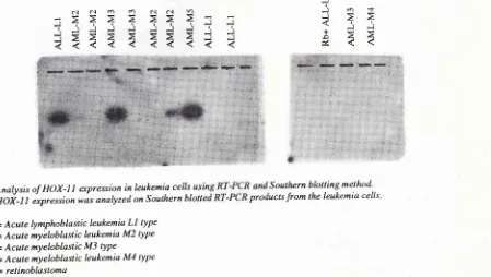
In order to confirm whether HOX- I I is expressed
in
the samples, the productof
the RT-PCRof
the samples was subjectedto
Southernblotting
andhybridized to
HOX-Il
probe. The results shown in Fig. 2 demonstrate that one(ALL-Ll)
out of4
ALL,and 2 (AML-M3 and AML-M5) out of 9 AML patients expressed HOX-l
I
mRNA.DISCUSSION
All of the leukemic samples expressed c-fos mRNA by Northern hybridization. c-fos is a cellular proto on-cogene which is induced 5 minutes after treatment with many stimulants, and the level
of
c-fos mRNA in-creases 20-30fold within
15to
30 minutes.lo'll In normal condition c-fos only expressedin
very lowstate. 10'12 c-fos is thought
to
growthand differentiatioh.l2 c-fos
is
level in normal erythroid cellsand
ia'10 Inhaematopoeitic cells of myeloid lineage, cells express high level of c-fos.'' Fig. lb alsodemonstrates that two
AML samples express c-fos mRNA. The role of c-fos in normal myelopoeisis and in leukemogenesis has not been established.la c-fos expression
is
increased in myetoid m I to terminal differentiation if there isgene-tic lesion affecting c-fos expression, c-fos might play an important role in the development of preleukemic myelodysplastic syndrome. l4 c-fos is not constitutive-ly expressed in lymphocytes. However, large amounts of c-fos RNA were detected in all of the samples from
Med J Uttiv Indott
ALL.15 These results suggest that overexpression of c-fos causes leukemogenesis of lymphocytes.
HOX-ll
(homeoboxll)
mRNA is not detected in all cells by Northern blotting method.It
is thought that abnormal expression of homeobox gene is one of the abnormalities associated with the development of murine and human leukemia. l3 Expressionof
the HOX-2 gene in transfected clone had been shown to inhibit the pathway of the myeloid differerrtiation. This abnormal expression of HOX-2 may contribute to the development of leukemia by interfering thedifferen-' 13
tratron program.
Since
HOX-ll
is one of homeobox genés,lit
is thought thatit
might play a rolein
cell growth and differentiation.tu HOX-lI
is expressedin
liver cells and alsoin
children T-ALL.I It i.
not known thatHOX-ll
play an in.rportant rolein
leukemogenesis. Our results (Figure 2) show that HOX- II
is expressed in ALL (lanel)
and AML samples (lane 4 and 8). As mentioned by Lord et al. This gene is expressed in lymphoid lineages as well as in myeloid lineages and in cell with terminal dilfcrentiatio;.ls'16CONCLUSION
HOX- I
I
is expressed is one out of 4 ALL and 2 out 9AML patients, as detected by RT-PCR method. These results suggest that
HOX-ll
causes leukenlo-genesis of myeloid lineage cells.J
i es
a 44
J:
É c.l al t.l t.) ôl a.l Yo
-=
iijjjjjill
i=
?
Fig.
"
2:
Andlysisof HOX-llescpressioninleukemiacells
Southernblottingmethod'HOi'l1
expression wo" anoly'ed on Southernbl
ucts from lhe leukemia cells'ALL-LI
= Acute lymphoblastic leukemia Ll typeAML M2 = Acute myëIoblastic leukemia M2 type
AML M3 = Acute mYeloblastic Mj tYPe
AML M4 = Acute myeloblastic leukemia M4 type
[image:4.595.103.552.481.735.2]Vol 3, No 4, Octobcr-Dccetnber 1994
Acknowledgment
We wish to thank Prof. T. Tokuhisa for reviewing and
correcting
the
manucsript andDr.
Hatano whoprovided the probe. This research is sponsored by the
JSPS-grant.
REFERENCES
l. Hatano M, Roberts CWM, Mindew M, Crist WM,
Korsmeyer SJ. Deregulation of a horneobox gene HOX-11,
by the t(10; l4) in T cell leukenria. Science l99l;253 :79-82. 2. Saiki RK, Gelfand DH, Stoffel S, Scharf SI, Higuchi R, Harn GT, et al. Prirner directed enzyrnatic arnplification of DNA with thennostable DNA polynrerase. Science 1988; 239 :
487- 8.
3. Vigilant L, Pennington H, Haçending H, Kocher TD,
Wil-son AC. Mitochondrial DNA sequences in single hair from
a Soutlrern African population. PNAS USA 1989; 86
:9350-4.
4. Kwok S, Higuchi R. Avoiding false positive with PCR.
l.lature 1989; 339 : 237 -8.
5. HiguchiR. Sirnple and rapid preparation of sanrples forPCR. In: Ehrlich HA (ed). PCR technology. New York NY:
Stoc-khlorn, 1989.
6. Neubauer A, Neubauer B, Lin E. Polynrerase chain based
assay to detect allelic loss in hurnan DNA : loss of
inter-ferase gene in chronic rnyelogenesis leukenria. Nuc Acid Res
199O,321 : 1689-95.
The Hot-I I Gene in Leukemia
Cells
1997. Boehringer Mannheim. Manual Detection of DIG labelled nucleic Acids. 1993.
8. Maniatis. RNA Isolation. In: Laboratory Manual on
Molecular Cloning. New York: Cold Spring habor, 1989,
9. Chirgwin IM, Przybyla AE, MacDonald RI, Rutter WI.
Isolation ofbiologically active ribonucleic acid from sources
enriched in Ribonuclease. Biochemistry 1979; 18 :5294-9.
10. L,ezenes R, Meneceur P, Ray D, Morean-Grachelin F. Proto oncogene expression in normal, preleukemic and leukemic murine erythroid cells and its relationship to differentiation
and proliferation. Canc.er Res 1988; 48 :3972-6.
ll.
Miller AD, Curran T, Venna IM. c-fos protein can induce cellular transfonnation : a novel mechanism ofactivation of a cellular oncogene. Cell 1984; 36 : 5l-4.12. Ianuwein T, Muller R. Structure Function analysis of fos protein : A single amino acid change activates the immor-talizing potential of v-fos. Cell 1987; 48 : 647 48.
13. Blatt C, Intam I, Sachs I. Inhibition of spesific pathways of nryeloid cell differentiation by an activated HOX 2-4 homeobox gene. Cell Growth Differ 1992; Oct.3: 671-76. 14. Lord KA, AMollahi A, Lieberrnann B, Hoffam I, Liebennan
DA. Proto oncogenes of the fos/jun fantily of transcription factom are positive regulators of myeloid differentiation.
Mol Cell Biol 1993; 13 : 841-51.
15. Haryana SM. Over expression of c-myc c-myb c-fos in
severalcases of ALL AML frorn Iogjakarta province, ICMR
1992, Unpublislred data.
16. Watson ID, Gilnran M, Witkowski I,Zoller M. (eds) Genes
that control the developrnent of Drosolthila.In: Recotrrbinant