Proceeding - ICB Pharma II
Current Breakthrough in Pharmacy Materials and Analyses ISSN : 9-772476-969006
13 | P a g e
OP
A005
PHARMACEUTICAL TECHNOLOGY
Preparation and Characterization of
Submicron Particles of PLGA Incorporating
Rifampin Using Emulsion Solvent Diffusion
Method
Mardiyanto
1*
1Department of Pharmacy, Faculty of Mathematics and Natural Science
Inderalaya-Indonesia
*E-mail: [email protected]
Abstract— The research had been performed to incorporate rifampin into PLGA submicron-sized particles. This research has a prospect to be applied to overcome the ineffectiveness use of rifampin for tuberculosis patients as rifampin was not stable in human lung macrophages, while Mycobacterium tuberculosis was able to survive in human lung macrophages. Rifampin was incorporated into submicron particles of PLGAs using the emulsion solvent diffusion method. The use of rifampin 50 mg in every batch resulted in the submicron-sized particles of 220 nm, PDI 0:12, zeta potential 21 mV and EE 37%. In the batch using rifampin 300 mg, resulted the submicron-sized particles of 410 nm, PDI 0:22, zeta potential 14 mV and EE 40%. The surface of the particles was visualized by SEM and hydrodynamic size compared to TEM. It was known that particle is spherical with a smaller diameter than the hydrodynamic size. TEM measurement revealed the size of particles with PVA was 208 nm.
Keywords—characterization; PLGA; rifampin; hydrodynamic-size; TEM; %EE
I. INTRODUCTION
In the hold of his life, one of the strategies of pathogenic bacteria is attacking the cells involved in the immune system of the human body such as macrophages. Mycobacterium tuberculosis has the ability to withstand oxidative conditions in the cell macrophages. So that he can survive the human immune system attack (1,2). This hideaway also makes the molecule drugs are not effective contact with these pathogens.
Molecular form must directly exposed to the barriers to entry in macrophages. So that
tuberculosis caused by Mycobacterium
tuberculosis is still a scourge till today (2). Uptake of particles was more effective based on the shape of particles.
Several strategies have been attempted, but the disease still be a dangerous killing machine. Until now a lot of research on the development of
treatments to immobilize the bacteria
Mycobacterium tuberculosis. During bacterial pathogens such as this is able to hide and survive in macrophages, such as what drugs are able to disable it (3).
The ability to hide and survive is also likely to make a dose of antibiotics rifampin so great because not all of the drug molecules could be in contact with the bacteria became resistant to these pathogens, and the rest of the molecule will poison the cells of the body healthy. In 2013, HIPS Saarland University in Germany has developed a method of cultivation of human lung macrophages and was developed at the Institute for the preparation of nanoparticles (NPs) are biodegradable and biocompatible (4-6). It has been reported also that sub-micron sized particles (7,8) can diuptake optimally by macrophages (8). All reported data becomes larat behind and rationale for this research to be conducted.
II. MATERIALS AND METHODS
A. Materials
Rifampin was obained from Sigma Aldrich. Poly-(lactide co glicolide) (PLGA) as resormer 70:30 was purchased from Evonik, poly (vinyl alcohol) (PVA) from Kuraray. All solvents was used as high grade of Merc.
B. Methods Preparation of NPs Kementrian Riset dan Teknologi, Isentif Riset SINAS,
14 | P a g e Proceeding - ICB Pharma II Current Breakthrough in Pharmacy Materials and Analyses
ISSN : 9-772476-969006 Emulsion solvent diffusion method was used to
prepare the PLGA NPs (9-12). In brief, the emulsion was prepared by using PLGA 40 mg/mL and rifampin 50 mg and 300 mg/batch in 2.5 mL ethyl acetate as organic phase. The aqueous phase consisted of 2.5 mL of 25 mg/mL PVA solution. The aqueous phase was dropped into ethyl acetate as organic phase. The mixture was placed in an ice bath under stirring for 1 hour at 750 RPM. To produce a nanoemulsion, both ultrathurax at 13,200 RPM for 5 minutes in an ice bath were applied. The resulting emulsion was stirred over night at 500 RPM to evaporate the organic solvent.
Purification of nanoparticles and %EE
5 mL suspension of NPs was placed in Viva-spin 100 and 300 kDa (Sartorius, Göttingen, Germany). The suspensions were purified by using a centrifuge at 14,000 x g at T = 8°C for 30 minutes. The supernatant were collected to determine the encapsulation efficiency (%EE) of extract by using Spectrometer-analysis at nm. %EE was calculated by substracting of the total amount of rifampin in formulation by the amount of rifampin in supernatant (12).
Zeta-sizer and Microscopic analysis
NPs of 1 mL was added into cuvette for measurement of hydrodynamic size and zeta-potential by using PSA. AFM measurements were done with a Bioscope with a Nanoscope IV controller (Veeco Instruments, Bruker, Germany). Samples were prepared by dropping
diluted sample on freshly cleaved mica. The NPs were investigated after drying under ambient condition with tapping mode using a tip with cantilever of k = 40 N/m and a resonance frequency of ~250 kHz. For SEM measurements, the samples were dried under ambient conditions and coated with gold using a Quorum Q 150 ES sputter coater at 40 mA for 50 s in order to render the sample conductive, improving the image quality (thickness of the gold coating was less than 20 nm). For visualization a ZEISS EVO HD 15 was used in high vacuum mode (12).
III. RESULT AND DISCUSSION
NPs preparation for medicinal purposes is recommended that involves a polymer that can be degraded and can also be accepted by the human body without any side effects, and is known as a popular term biodegradable and biocompatible polymer. The FDA has recommended poly (lactide-co-glicolide) (PLGA) as the polymer to be used in producing NPs. NPs manufacture of a drug depends on the type of materials to be used. Hydrophobic drug ingredients that will be easier to be prepared and proven high loading using
single emulsion technique. As an oposite, for drug as hydrophilic materials, this technique is not suitable, and of course nanopresipitation and double emulsion is an appropriate (3-6).
Creation of PLGA NPs with a compound which can fluoresce for imaging of a desired activity, has made as NPs. They have the leverage in answering the progress of time. How small particles that can meet its receptor in the cell can be visualized (5,6,9). So far rifampin has been used to prevent tuberculosis and rifampin be used in the form of capsules and tablets. Rifampin is a known large doses, and many reports of side effects from its use to cases of resistance (1-3).
Rifampin is used as entrapped material in the particle, still needs to develope so that future research will produce an effective drug dosage forms that are well known tackling tuberculosis skilled in medicine and avoiding attacks the body's defense system.
Rifampin readily oxidized by air and preparation with PLGA carried in vials coated with aluminum foil. After the suspension, the pengkontaminan during overnight evaporation process which will also contaminate the results of polydispersity index determination.
PVA suspension separation from the rest is done by centrifugation. Viva-spin 300 kDa are used for this process are known as the purification process. Agric after separate from PVA, suspensions characterized by PSA to determine particle size, polydispersity index and zeta-potential of particles carrier.
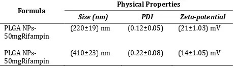
The results obtained from the PSA tabulated in Table 1 which can be seen below. Overall, the resulting particles are submicron-sized in the range of 200 to 300 nm with an average calculated by the tool. The average diameter of the particles depends on the number of particles detected by the tool. So that the particle density greatly affect the measurement.
Table 1. Particles Size Analysis of PLGA-Rifampin NPs
Formula Physical Properties
Size (nm) PDI Zeta-potential
NPs-Proceeding - ICB Pharma II
Current Breakthrough in Pharmacy Materials and Analyses ISSN : 9-772476-969006
15 | P a g e
contains the supernatant. %EE was calculated based on the amount of rifampin in this part. Spectrophometry UV-Vis methods was used to determine the amount of rifampin at max wave-lenght 283 nm. The result of %EE was showed in Fig.1.
Fig. 1. Comparison of %EE between PLGA NPs-Rifampin50mg to PLGA NPs-Rifampin 300mg
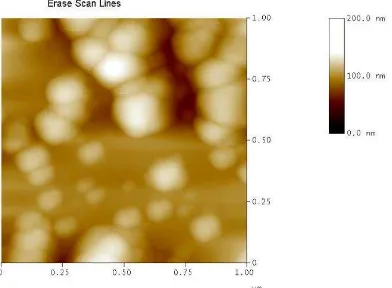
Microscopic measurements were performed by using atomic forced microscopy (AFM), scanning electron microscopy (SEM), and transmission electron microscopy (SEM). These microscope showed all particles were spheric with size in submicron range. The image of appropriate formula (PLGA NPs-Rifampin50mg) using AFM, SEM, and TEM were added into Fig.2 to Fig.4.
Figure 2. AFM image of formula PLGA NPs-Rifampin50mg
The distance of between particles were similar when we compared AFM image to the SEM image. This condition was occured because of the drying process.
Figure 3. SEM image of formula PLGA NPs-Rifampin50mg
Using the transmited electron, the whole shape of each particles were obtained and the size is rather small than the hydrodynamic size.
Figure 4. TEM image of formula PLGA NPs-Rifampin50mg
IV. CONCLUSIONS
The use of rifampin as much as 50 mg in every batch, resulted the submicron-sized particles of 220 nm, PDI 0:12, zeta potential 21 mV and EE 37%. In the batch using rifampin 300 mg, resulted the submicron-sized particles of 410 nm, PDI 0:22, zeta potential 14 mV and EE 40%.
The surface of the particles is visualized by SEM and hydrodynamic size compared to TEM. It was known that particles produced is spherical with a smaller diameter than the hydrodynamic size.
Acknowledgment
This research has funded by Indonesian Minister of Research and Technology with grand Insentive Research SINAS for academic year 2015.
References
[1] Karyadi, E., W. Schultink, "Poor micronutrient status of active pulmonary tuberculosis patients in Indonesia". Nutrition, 2000. 130(12). pp 2953-2958.
[2] Moghazeh, S. L., X. I. Pan,. "Comparative antimycobacterial activities of rifampin, rifapentine, and KRM-1648 against a collection of rifampin-resistant Mycobacterium tuberculosis isolates with known rpoB mutations" Antimicrobial Agents, 1996, 40(11). pp 2655-2657.
[3] Walters, S. B. and B. A. Hanna, "Testing of susceptibility of Mycobacterium tuberculosis to isoniazid and rifampin by mycobacterium growth indicator tube method" Clin Microb. 1996, 34(6). pp 1565-1567. [4] Barichello, J.M., Morishita, M., Takayama, K., and Nagai,
T., "Encapsulation of hydrophilic and lipophilic drugs in PLGA nanoparticles by the nanoprecipitation method" Drug Dev Ind Pharm, 1999. 25(4). pp 471-476.
[5] Jain, R.A., "The manufacturing techniques of various drug loaded biodegradable poly(lactide-co-glycolide) (PLGA) devices. Biomater", 2000. 21(23). pp 2475-2490.
[6] Ravi Kumar, M.N., Bakowsky, U., and Lehr, C.M., "Preparation and characterization of cationic PLGA nanospheres as DNA carriers" Biomater, 2004. 25(10). pp 1771-1777.
[7] Astete, C.E. and Sabliov, C.M., "Synthesis and characterization of PLGA nanoparticles" J Biomater Sci Polym Ed, 2006. 17(3). pp 247-289.
16 | P a g e Proceeding - ICB Pharma II Current Breakthrough in Pharmacy Materials and Analyses
ISSN : 9-772476-969006
nanoparticles in drug delivery: the state of the art" Crit Rev Drug Carrier Syst, 2004. 21(5). pp 387-393. [9] Cohen-Sela, E., Chorny, M., Koroukhov, N., Danenberg,
H.D., and Golomb, G., A new double emulsion solvent diffusion technique for encapsulating hydrophilic molecules in PLGA nanoparticles. J Controlled Release, 2009. 133(2). pp 90-95.
[10] Kawashima, Y., Yamamoto, H., Takeuchi, H., Hino, T., and Niwa, T., Properties of a peptide containing DL-lactide/glycolide copolymer nanospheres prepared by novel emulsion solvent diffusion methods. Eur J Pharm Biopharm, 1998. 45(1). pp 41-48.
[11] Niwa, T., Takeuchi, H., Hino, T., Kunou, N., and
Kawashima, Y., "In vitro drug release behavior of D,L-lactide/glycolide copolymer (PLGA) nanospheres with nafarelin acetate prepared by a novel spontaneous emulsification solvent diffusion method" J Pharm Sci, 1994. 83(5). pp 727-32.