Stem cell therapy in diabetic foot patients: where are we now?
Stanley Kirana, Diethelm Tschöpe, Bernd Stratmann
Diabetes Center, Heart and Diabetes Center NRW,University Clinic of Ruhr University Bochum; Bad Oeynhausen, Germany
Abstrak
Kaki Diabetik (KD) merupakan penyakit penyerta Diabetes Mellitus (DM). DM merupakan salah satu penyebab utama kasus amputasi non-traumatik di Jerman, dengan penyakit arterial perifer berat (PAP) dengan iskemia kritis tungkai yang menjadi masalah utama. Meskipun teknik modern tersedia, intervensi perkutan dan pembedahan pemulih vaskularisasi masih terbatas. Masalah ini menyebabkan peningkatan jumlah amputasi pada pasien dengan diabetes mellitus. Proses fi siologik angiogenesis, vaskulogenesis dan arteriogenesis mengarahkan ke pertumbuhan pembuluh darah kolateral pada keadaan penyakit penyumbatan pembuluh arterial penyebab iskemia tungkai. Pada praktik klinik respons angiogenik endogenik seringkali terganggu. Angiogenesis terapetik merupakan penerapan bioteknologi untuk merangsang pembentukkan pembuluh darah baru via (melalui) aplikasi lokal faktor penumbuh pro-angiogenik dalam bentuk protein rekombinan, atau terapi gen; atau dengan implantasi sel progenitor atau sel punca yang akan mensintesa sitokin angiogenik multipel. Artikel review ini merangkum fungsi endotelial dan disfungsi pada DM, mekanisme homing, metode transplantasi dan status uji klinik di bidang sel punca untuk pengobatan iskemia tungkai. (Med J Indones 2011; 20:154-60)
Abstract
Diabetic foot (DF) occurs as a concomitant illness of diabetes mellitus (DM). DM is one of the main causes of non-traumatic amputation in Germany with severe peripheral arterial disease (PAD) with critical limb ischemia (CLI) being of major concern. Although modern techniques are available surgical vascularisation and percutaneous intervention are limited. This problem leads increasing numbers of limb amputations in patients with diabetes mellitus. Thephysiological process of angiogenesis, vasculogenes is and arteriogenesis contribute to the growth of collateral vessels in response to obstructive arterial disease causing limb ischemi. In clinical practice the endogenous angiogenic response is often impaired. Therapeutic angiogenesis is an application of biotechnology to stimulate new vessel formation via local administration of pro-angiogenic growth factors in the form of recombinant protein, or gene therapy,or by implantation of progenitor cells or stem cells that will synthe size multiple angiogenic cytokines. This review summarises the endothelial function and dysfunctionin DM, the mechanism of homing, the transplantation method and the status of clinical trials in stem cell fi eld to treat limb ischemia.
(Med J Indones 2011; 20:154-60)
Key words: diabetes mellitus, endothelial progenitor cells, peripheral arterial disease, stem cells, therapeutic angiogenesis
Correspondence email to: [email protected]
Diabetes mellitus (DM) is known as an emerging chron-ic disease world wide. DM is accepted as cardiovascular risk factor and therefore plays an important role in the pathogenesis of cardiovascular disease. Peripheral ar-tery disease (PAD) is one of the major health problems resulting from macrovascular complication in diabetic patients. As a manifestation of atherosclerosis, PAD is characterized by atherosclerotic occlusive disease of the lower extremities and is a marker for atherothrombotic disease in other vascular beds. PAD is present in approx-imately one-half of all patients with foot ulcers.1-4 Along
with polyneuropathy, PAD causes foot ulceration which often leads to lower limb amputations.
DM causes almost 50% of all non traumatic lower-extremity amputations world wide. It is estimated that the life time risk for amputation in diabetic patients is 10–15%, which is 10-30 times higher in comparison to the general population.5-8 In Germany the number
of limb amputation is increasing. According to the new-est data from German Association of Angiology, which were released during the Scientifi c Meeting in Mannheim in September 2008, about 60.000 amputations are per-formed every year. Compared to european countries such
as Denmark and The Netherlands, Germany is leader in the number of limb amputation. The main factors induc-ing these amputations are diabetic foot (DF) or arterio-sclerosis. Almost every 3rd patient suffers from diabetes
mellitus. An amputation for elderly patients resembles a fatal step in their quality of life.
In the other hand, the patients are often late to seek special-ized professional care or they are not treated in a vascular center. These factors along with pathology-anatomical factors limit the possibilities for revascularisation.
The strengths and limitations of surgical revascularisa-tion in PAD are well known. Surgical revascularisarevascularisa-tion and percutaneous interventions are limited to patients with critical limb ischemia (CLI) and those with dis-abling claudication (Fontaine Stadium IIb–IV) due to discrete proximal disease.1,9
Endothelial functions and dysfunctions
For a long time two specialized endothelial functions were accepted: gas exchange in pulmonary circulation and fenestration in hepatic and splenic circulation. Un-der normal homeostatic conditions, the endothelium resists vasospasm, prevents leukocyte and platelet ad-hesion to the vessel wall, favours fi brinolysis, combats coagulation of blood, and inhibits the proliferation of vasculars mooth muscle cells.
During the last two decades, accumulating evidence has described the vascular endothelium as an active en-docrine, paracrine, and autocrine organ, indispensable for the maintenance of vascular homeostasis. Altered homeostasis induced by various stimuli may cause lo-calized alterations, or ‘endothelial dysfunction’, of the antihemostatic properties, vascular tone, heightened leukocyte adhesion, and increased production of cytok-ines and growth factors. The dramatic change of en-dothelial interactions with blood leukocyte soccurring in infl ammation provides an example of endothelial activation.10,12,13
Diabetes mellitus and endothelial dysfunction
Vascular diseases, including atherosclerosis, medial calcifi cation, and microangiopathy, are prevalent in pa-tients with DM and are the principal causes of morbid-ity and mortalmorbid-ity in these individuals. Atherosclerosis occurs earlier in patients with diabetes, frequently with greater severity and more diffuse distribution.
Hyperglycemia per se causes endothelial dysfunc-tion. Healthy humans exposed to a hyperglycaemic clamp suffi cient to increase forearm glucose concen-tration experience impaired endothelium-dependent vasodilation.10,14,15 Hyperglycemia also decreases
fl ow-mediated endothelium-dependent vasodilation of the brachial artery of healthy subjects.14,16,17 Hyperglyce-mia may decrease the bioavailability of nitric oxide (NO) through multiple mechanisms (Figure1). Ad-ditionally, hyperglycemia may increase the formation of oxygen-derived free radicals that inactivate NO or cause intracellular signalling disturbances that inhibit nitric oxide synthase (NOS) activity and thereby re-duce NO production. Hyperglycemia is also associated with increased oxidative stress. Increased inactivation of NO by oxygen-derived free radicals and decreased production of NO by NOS reduce NO levels in the vas-cular milieu.14,17-20
Rationale for stem cell therapy in diabetic foot
The essential part of normal wound healing is the for-mation of new blood vessels with in the provisional wound matrix that is referred to as granulation tissue. Neovascularisation of the granulation tissue occurs by the processes of angiogenesis or vasculogenesis, or
both.21,22 Therapeutic angiogenesis has been studied many times for the treatment of patients with periph-eral arterial occlusive disease who do not qualify for surgical revascularisation or radiologic intervention. Angiogenesis can be achieved by introducing growth factors as mature proteins or as complementary DNA carrying vector systems (cDNA-plasmid) or stem cells which contain endothelial progenitor cells (EPCs) and mesenchymal cells.23-27
The following sections discuss various methods of us-ing stem cell therapies to induce angiogenesis in DF.
Defi nition of stem cells
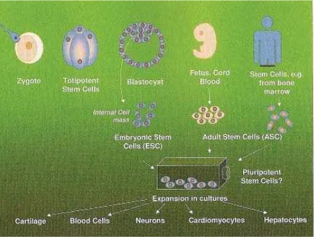
All life forms begin with a stem cell, which is defi ned as a cell that has the dual ability to self-renew and to produce progenitors and different types of specialized cells in the organism.28
Regardless of their sources, stem cells are defi ned by their indefi nite capability of self-renewal (prolifera-tion) and unlimited potential to generate specialized tissue cells (differentiation) (Figure2). Currently, scien-tists and clinical researchers are working on two major types of stem cells, embryonic and adult, which share three characteristic properties: 1. Stem cells are pre-mature, undifferentiated, or unspecialized. However, unspecialized stem cells can generate specialized cells, including heart muscle, blood vessels, blood cells, or nerve cells. 2. Stem cells can divide and renew them-selves for long periods. Unlike other cells–which nor-mally do not replicate by themselves–stem cells may replicate, or proliferate, many times. 3. Stem cells can respond to exogenous or endogenous signals by gener-ating specialized cell types. When unspecialized stem cells develop into specialized cell, the process is called differentiation.29
Adult and embryonic stem cells
Human embryonic stem cells (ESCs) used for research have been extracted from embryos created by in vitro fertilization done by the group of James Thomson in 1998 and they reported on the etablishment of human ESC lines. They concluded that the ESCs have poten-tial to form most cell types of the adult body over al-most unlimited periods.28,30-32
The adult body has a small number of adult or somatic stem cells in some tissues and organs. Such adult stem cells (ASCs) have been known to posses the ability to regenerate the corresponding tissue from which they are derived. Hematopoietic stem cells (HSCs), for ex-ample, continuously regenerate the circulating blood cells and cells of the immune system during the life span of the organism. Based on animal studies, many researchers have recently claimed that ASCs might exhibit developmental potentials comparable to those exhibited by ESCs. ASCs have the ability to regener-ate the tissue from which they are derived over the life span of the individual, while ESCs have the potential to
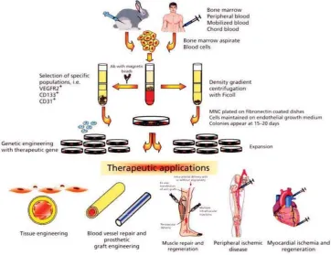
Figure 2. Isolation, cultivation and genetic engineering of endothelial progenitor cells (EPCs) for therapeutic ap-plication. EPC scan be isolated from the BM-MNC, peripheral blood or umbilical chord blood with or without further selection and purifi cation.The mononuclear cells are expanded ex vivo under endothelial-specifi c growth conditions and may be genetically modifi ed to over express one or several therapeutic genes.The differentiated cells are then used in transplantation protocols for rescue and repair of damaged tissue such as infarcted myocardium, ischemic limb or injured muscle. The cells may also be used for en-dothelialisation of damaged blood vessels and vascular prosthetic grafts and in tissue engineering. (Melo LG, Pachori AS, Kong D et al. Endothelium-targeted gene and cell-based therapy for cardiovascular dis-ease. In: De Caterina R, Libby P, eds. Endothelial Dysfunctions and Vascular Disdis-ease. Oxford: Blackwell Futura; 2007:385)
form most cell types of the adult body over very long peri-ods of in vitro cultivation.28,29,32-38 However, until now the use of human ESCs for research is prohibited in Germany. Many clinical research centers in Germany are focusing to develop the potential of ASCs.
Endothelial progenitor cells for cardiovascular re-generation
CD34 is not expressed exclusively on hematopoietic stem cells (HSC) but also on mature endothelial cells. Further studies used the more immature HSC marker CD133 also known as prominin or AC 133. Purifi ed iso-lated CD 133+ cells also differentiated to endothelial cells in vitro.41,42 However, the biological function of CD133 remains unclear. It is not well known whether CD133 only represents a surface marker or has a functional ac-tivity involved in regulation of neovascularisation.41
The characterization of EPC becomes particularly diffi -cult when cells are expanded and -cultured ex vivo, since the culture conditions (culture supplements such as fe-tal calf serum and cytokines or different plastic types) rapidly change the phenotype of the cells. Moreover, continuous cultivation was shown to increase lial differentiation, as evidenced by elevated endothe-lial marker protein expression.
Smooth muscle cells (SMC) and endothelium are two essential structures for the architecture of the blood vessels. Vascular stem cells, a group of multipotent het-erogenous stem cells, largely fall into two subgroups: endothelial and SMC progenitors. EPC develop closely from CD34+ to bone marrow stem cells, in particu-lar those committed to the monomyeloid cell lineage, whereas SMC progenitors may belong or originate to-gether in mesenchymal stem cells (MSC), which are negative for CD34. Atherosclerosis causes arterial wall damage and impairs the capacity of both vascular stem cell types to regenerate neovascular tissue, or may trig-ger abnormal proliferation or death by apoptosis under different pathological conditions.29
Mechanism of vessel formation
Angiogenesis is defi ned as the sprouting of new capil-laries from existing vascular structure, a process that is triggered by endothelial cell migration and prolifera-tion. Remodelling of the extra cellular matrix (ECM), tubule formation and expansion of the surrounding vas-cular tissue are key elements of angiogenesis.
The in situ formation of new blood vessels from ECP which differentiated into endothelial cells and fuse into luminal structures is called vasculogenesis.
Meanwhile, arteriogenesis refers to an increase in the calibre of pre-existing arteriolar collateral connections by recruitment of perivascular cells and expansion and remodelling of the extracellular matrix. Arteriogenesis increases the size and wall thickness of collateral ves-sels, and shear stress (rather than hypoxia) seems to be the main factor that stimulates arteriogenesis.9
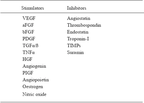
The key steps in vessel formation–namely endothelial cell activation, migration, proliferation and
reorgani-sation– are tightly regulated in a complex balance be-tween pro-and anti-angiogenic mechanisms (Table1).9
Table 1. Factors that stimulate and inhibit angiogenesis
Stimulators Inhibitors
VEGF Angiostatin
aFGF Thrombospondin
bFGF Endostatin
PDGF Troponin-I
TGFα/ß TIMPs
TNFα Suramin
HGF
Angiogenin PIGF
Angiopoietin Oestrogen
Nitric oxide
VEGF: vascular endothelial growth factor; aFGF, bFGF: acid and basic fi broblast growth factor (FGF-1 and-2, respectively); PDGF: platelet-derived growth factor; TGF: transforming growth factor; TNF: tumour necrosis factor; HGF: hepatocyte growth factor; PIGF: placental growth factor; TIMPs: tissue inhibitors of metal-loproteinases.
The most important pro-angiogenic factors are vascular endothelial growth factor (VEGF) and basic fi broblast growth factor (bFGF), also known as FGF-2. VEGF is an endothelial cell specifi c mitogen that is markedly up-regulated by hypoxia, and plays an important role in en-dothelial cell proliferation, differentiation and survival.
FGFs are non-secreted growth factors that are released only during cell death or ischemic cell injury. Clinical and biochemical factors also infl uence the formation of, and biological response to different angiogenic growth factors. Hypoxia, for example, is the most potent in-ducer of angiogenesis,44 principally via up-regulation of VEGF, whereas diabetes mellitus and increased lev-els of cholesterol and lipoprotein(a) are associated with a reduced angiogenic response.9
As bone marrow-derived stem and progenitor cells home to sites of ischemia, this may allow the local re-lease of factors acting in paracrine manner on the sur-rounding ischemic tissue. Bone marrow-derived mono-nuclear cells release angiogenic growth factors such as VEGF, bFGF and angiopoietins, thereby enhancing the local angiogenic response.9,41,44 This mechanism is named ‘homing’ and it is the most likely theory which represents the reality in ischemic process.
The idea to induce neovascularisation in the wound area or ischemic area is now a hot topic in regenerative medi-cine. There are a lot of clinical trials in this area, however only few centers world wide perform these studies.
Stem cell transplantation
Until now, there are a lot of protocols, hypotheses, de-bates and discussions about the best methods of stem cell transplantation. Most clinical studies are performing 2 methods of stem cell transplantation: intra-arterial and intra-muscular.
The concept of intra-arterial stem cell transplantation assures that the stem cells reach all stenosis sites an-tegradely. We believe that the stenosis area is the isch-emic area which stimulates the homing effect of the
cells.The intra-muscular application is based on the ischemic musculature which could be supplied with a high concentration of stem cells through several local depots. Whether the cells reached the ischemic area by blood fl ow remains unclear. In theory, it induces the homing effect and then stimulates the angiogenesis or vasculogenesis in the ischemic area.45-47
Current clinical trial
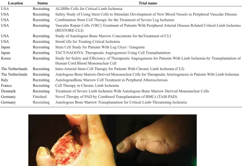
There are a lot of clinical trials running on this topic (Table 2). However, the outcomes have not been pub-lished yet. Only some studies pubpub-lished their results or case reports.25,45,46 A multi center study has not been performed to take conclusion of stem cell therapy. To what we know, the data from other centers as well as ours show positive results (Figure 3).
Table 2. List of stem cell clinical trials in critical limb ischemia as listed in Clinicaltrial.gov from September 2007
Location Status Trial name
USA Recruiting ALDHbr Cells for Critical Limb Ischemia
USA Recruiting Safety Study of Using Stem Cells to Stimulate Development of New Blood Vessels in Peripheral Vascular Disease
USA Recruiting Combination Stem Cell Therapy for the Treatment of Severe Leg Ischemia
USA Recruiting Vascular Repair Cells (VRC) Treatment of Patients With Peripheral Arterial Disease Related Critical Limb Ischemia (RESTORE-CLI)
USA Recruiting Study of Autologous Bone Marrow Concentrate for theTreatment of CLI
USA Recruiting StemCells for Treating Critical Ischemia
Japan Recruiting Stem Cell Study for Patients With Leg Ulcer / Gangrene
Japan Recruiting TACT-NAGOYA: Therapeutic Angiogenesis Using Cell Transplantation
Korea Recruiting Study for Safety and Effi ciency of Therapeutic Angiogenesis for Patients With Limb Ischemia by Transplantation of Human Cord Blood Mononuclear Cell
The Netherlands Recruiting Intra-Arterial Stem Cell Therapy for Patients With Chronic Limb Ischemia (CLI)
The Netherlands Recruiting Autologous Bone Marrow-Derived Mononuclear Cells for Therapeutic Arteriogenesis in Patients With Limb Ischemia
Italy Recruiting AutologousBone Marrow Cell Treatment in Peripheral Atherosclerosis
France Recruiting Cell Therapy in Chronic Limb Ischemia
Denmark Recruiting Treatment of Severe Limb Ischemia With Autologous Bone Marrow Derived Mononuclear Cells
Germany Finished Novel Therapy of PAD by Combined Transplantation of BMCs (TAM-PAD)
Germany Recruiting Autologous Bone Marrow Transplantation for Critical Limb-Threatening Ischemia
Figure 3. Bone marrow stem cell therapy in diabetic foot (Bad Oeynhausen Working Group, Germany). This patient had a chronic limb ischemia with forefoot necrosis. Distal stenosis A. tibialis anterior and A. dorsalis pedis without collateralisation was verifi ed by angiography. Discussion between vascular surgeon, intervention angiologist and internist revealed no surgical or inter-ventional therapeutic option. Due to the stenosis location and vascular structure, no possibilities to perform bypass or other intervention were seen. The patient agreed with the autologous bone marrow stem cell therapy.
In conclusion, bone marrow derived EPCs are the new-est cellular target that may be used to infl uence post-natal neovascularisation. The formation of new blood vessels, including collaterals, is a complex physiologi-cal process that occurs in adults in response to tissue injury or ischemia.
The stem cell transplantation to induce vessel forma-tion is promising and could be a new therapeutic opforma-tion in the future to treat limb ischemia without options of revascularization. However, there is still much to learn about the optimum treatment modality, dosing frequen-cy and route of administration, especially in patients with diabetes mellitus. We know that these patients have a reduced capability of vascular self-renewing.
Until now, there are only some reports and clinical tri-als which have already been fi nished with the whole evaluations and they show an improvement of blood perfusion in ischemic area. Moreover, multicenter stud-ies with large numbers of patients are expected to give more information about stem cell therapy.
Acknowledgments
The Bad Oeynhausen stem cell trial was supported by Aastrom Bioscience Inc., Ann Arbor–USA. We thank Dr. E. Danch of the Vascular Surgery, Bad Oeynhausen General Hospital, Germany for his critical opinion of patient recruitment. We also thank all colleagues in Di-abetes Center NRW, Bad Oeynhausen for helping us in patient recruitment and performing diagnostic.
REFERENCES
1. American Diabetes Association Editors. Peripheral Arte-rial Disease in People with Diabetes: Consensus Statement. Diabetes Care 2003; 26:12: 3333-41.
2. Prompers L, Schaper J, Apelqvist J et al. Prediction of out-come in individuals with diabetic foot ulcers: focus on the differences between individuals with and without periph-eral arterial disease. The EURODIAL Study. Diabetologia 2008; 51:747-755.
3. Prompers L, Huijberts M, Apelqvist J et al. High prevalence of ischaemia, infection and serious comorbidity in patient with diabetic foot disease in Europe. Baseline results from the Eurodiale Study. Diabetologia 2007; 50: 18-25. 4. Boulton AJ. The pathogenesis of diabetic foot problems: an
overview. Diabet Med 13 (Suppl1): S12-S16.
5. Tentolouris N, Al-Sabbagh S, Walker MG et al. Mortality in diabetic and nondiabetic patients after amputation performed from 1990 to 1995. Diabetes Care 2004; 27: 1598-604. 6. Most RS, Sinnock P. The epidemiology of lower extremity
amputations in diabetic individuals. Diabetes Care 1983; 6: 87-91.
7. Siitonen OI, Niskanen LK, Laakso M et al. Lower extrem-ity amputations in diabetic and non diabetic patients: a pop-ulation-based study in eastern Finland. Diabetes Care 1993; 16:16-20.
8. Chaturvedi N, Stevens LK, Fuller JH et al. Risk factors, eth-nic differences und mortality associated with lower extrem-ity gangrene and amputation in diabetes: the WHO multi-national study of vascular disease in diabetes. Diabetologia 2001; 44 (Suppl.2) 27:7:S65-S71.
9. Collinson DF, Donnelly R. Therapeutic angiogenesis in peripheral arterial disease: can biotechnology produce an effective collateral circulation? Eur J Vasc Endovasc Surg 2004; 28:9-23.
10. De Caterina R, Massaro M, Libby P. Endothelial function and dysfunctions. In: De Caterina R, Libby P, eds. Endothe-lial Dysfunctions and Vascular Disease. Oxford: Blackwell Futura; 2007: 1-25.
11. Fishman AP. Endothelium: a distributed organ of diverse capabilities. Ann NY Acad Sci 1982; 401: 1-8.
12. Gimbrone MA, Kume N, Cybulsky MI. Vascular endothe-lium dysfunction and the pathogenesis of atherosclerosis. In: Weber PC, Leaf A, eds. Atherosclerosis Reviews. New-York: Raven Press; 1993.
13. De Caterina R, Libby P, Peng HB et al. Nitric oxide de-creases cytokine-induced endothelial activation. J Clin In-vest 1995; 96: 60-8.
14. Creager MA, Beckman JA. Vascular function and diabetes mellitus. In: De Caterina R, Libby P, eds. Endothelial Dys-functions and Vascular Disease. Oxford: Blackwell Futura; 2007:232-44.
15. Williams SB, Goldfi ne AB, Timimi FK et al. Acute hyper-glycemia attenuates endothelium-dependent vasodilation in humans in vivo. Circulation 1998; 97: 1695-701.
16. Kawano H, Motoyama T, Hirashima O et al. Hyperglycemia rapidly suppresses fl ow-mediated endothelium-dependent vaso-dilation of brachial artery. J Am Coll Cardiol 1999; 34: 146-54. 17. Uribarri J, Stirban A, Sander D, et al. Single oral
chal-lenge by advanced glycation end products acutely impairs endothelial function in diabetic and non diabetic subjects. Diabetes Care. 2007; 30(10): 2579-82.
18. HinkU, LiH, Mollnau H et al. Mechanisms underlying en-dothelial dysfunction in diabetes mellitus. Circ Res 2001; 88: E14-E22.
19. Brownlee M. Biochemistry and molecular cell biology of diabetic complications. Nature 2000; 404: 787-90.
20. Nishikawa T, Edelstein D, Du XL et al. Normalizing mi-tocondrial superoxide production blocks three pathways of hyperglycaemic damage. Nature 2000; 404: 787-90. 21. Velazquez OC. Angiogenesis and vasculogenesis:
Induc-ing the growth of new blood vessels and wound healInduc-ing by stimulation of bone marrow–derived progenitor cells mobi-lization and homing. J Vasc Surg 2007; 45: 39A-47A. 22. Bauer SM, Bauer RJ, Velasquez OC. Angiogenesis,
vascu-logenesis, and induction of healing in chronic wounds. Vasc Endovascular Surg 2005; 39: 293-306.
23. Ausprunk DH, Folkman J. Migration and proliferation of en-dothelial cells in performed and newly formed blood vessels during tumor angiogenesis. Microvasc Res 1977;14: 53-65. 24. Kim D-I, Kim M-J, Joh J-H et al. Angiogenesis Facilitated by
Autologous Whole Bone Marrow Stem Cell Transplantation for Buerger’s Disease. Stem Cells 2006; 24(5): 1194-200. 25. Tateishi-Yuyama E, Matsubara H, Murohara T et al.
Thera-peutic angiogenesis for patients with limb ischaemia by au-tologous transplantation of bone-marrow cells: a pilot study and a randomised controlled trial. Lancet 2002; 360:427-35 26. Nikol S. Therapeutic angiogenesis for peripheral artery
27. Asahara T, Kalka C, Isner JM. Stem cell therapy and gene transfer for regeneration. Gene Ther 2000;7(6):451-7. 28. Ho AD, Wagner W. Clinical Potentials of Stem Cells: Hype
or Hope? In: Ho AD, Hoffman R, Zanjani ED, eds. Stem Cell Transplantation Biology, Processing, and Therapy. Weinheim: Wiley-Vch; 2006:3-25.
29. Geng YJ, Madonna R. Stem cells in atherosclerosis and atherosclerosis-related vascular disorder. In: De Caterina R, Libby P, eds. Endothelial Dysfunctions and Vascular Dis-ease. Oxford: Blackwell Futura; 2007:350-64.
30. Thomson JA, Itskovitz-Eldor J, Shapiro SS, Waknitz MA, Swiergiel JJ, Marshall VS et al. Embryonic stem cell lines de-rived from human blastocysts. Science 1998; 282: 1145-47. 31. Richards M, Fong CY, Chan WK, Wong PC, Bongso A.
Human feeders support prolonged undifferentiated growth of human inner cell masses and embryonic stem cells. Nat Biotechnol 2002;20:933-6.
32. Gage FH. Mammalian neural stem cells. Science 2000; 287: 837-45.
33. Weissmann IL. Translating stem and progenitor cell biol-ogy to the clinic: barriers and opportunities. Science 2000; 287:1442-6.
34. Ho AD, Punzel M. Hematopoietic stem cells: can old cells learn new tricks? J Leucocyte Biol 2003; 73:547-55. 35. Wagers AJ, Christensen JL, Weissmann IL. Cell fate
deter-mination from stem cells. Gene Ther 2002;9:606-12. 36. Terada N, Hamazaki T, Oka M et al. Bone marrow cells
adopt the phenotype of other cells by spontaneous cell fu-sion. Nature 2002; 416: 542-5.
37. Ying QL, Nichols J, Evans EP et al. Changing potency by spontaneous fusion. Nature 2002; 416: 545-8.
38. Morshead CM, Benveniste P, Iscove NN et al. Hematopoi-etic competence is a rare property of neural stem cells that may depend on genetic and epigenetic alterations. Nat Med 2002;8: 268-73.
39. Amit M, Shariki C, Margulets V et al. Feederlayer and se-rum free culture of human embryonic stem cells. Biol Re-prod 2004;70:837-45.
40. Asahara T, Murohara T, Sullivan A et al. Isolation of puta-tive progenitor endothelial cells for angiogenesis. Science 1997;275(5302):964-7.
41. Fischer-Rasokat U, Dimmeler S. Endothelial Progenitor Cells for Cardiac Regeneration. In: Ho AD, Hoffman R, Zanjani ED, eds. Stem Cell Transplantation Biology, Pro-cessing, and Therapy. Weinheim: Wiley-Vch; 2006:3-25. 42. Gehling UM, Ergün S, Schumacher U et al. In vitro
differ-entiation of endothelial cells from AC133-positive progeni-tor cells. Blood 2000;95(10):3106-12.
43. Fujiyama S, Amano K, Uehira K et al. Bone marrow cyte lineage cells adhere on injured endothelium in a mono-cyte chemoattractant protein-1-dependent manner and ac-celerate reendothelialization as endothelial progenitor cells. Circ Res. 2003; 93(10):980-9.
44. Giordano F, Johnson RS. Angiogenesis: the role of the mi-croenvironment in fl ipping the switch. Curr Opin Genet Dev 2001;11:35-40.
45. Bartsch T, Brehm M, Zeus T et al. Transplantation of au-tologous mononuclear bone marrow stem cells in patients with peripheral arterial disease (The TAM-PAD study). Clin Res Cardiol 2007; 96(12):891-9.
46. Kirana S, Stratmann B, Lammers D et al. Wound therapy with autologous bone marrow stem cells in diabetic patients with ischaemia-induced tissue ulcers affecting the lower limbs. Int J Clin Pract 2007;61(4):690-2.
47. Gastens MH, Goltry K, Prohaska W et al. Good manufac-turing practice-compliant expansion of marrow-derived stem and progenitor cells for cell therapy. Cell Transplant. 2007;16(7):685-96.
48. Oswald J, Boxberger S, Jorgensen B, Feldmann S, Eh-ninger G, Bornhauser M et al. Mesenchymal stem cells can be differentiated into endothelial cells in vitro. Stem Cells 2004;22(3):377-84.
49. Prante C, Bieback K, Funke C, Schon S, Kern S, Kuhn J et al. The formation of extracellular matrix during chondrogenic differentiation of mesenchymal stem cells correlates with in-creased levels of xylosyltransferase I. Stem Cells 2006. 50. Huang P, LiS, Han M et al. Autologous Transplantation of
Granulocyte Colony-Stimulating Factor-Mobilized Peripheral Blood Mononuclear Cells Improves Critical Limb Ischemia in Diabetes. Diabetes Care 2005;28:2155-60.
51. Rodriguez L, Azqueta C, Azzalin S, Garcia J, Querol S. Washing of cord blood grafts after thawing: high cell recovery using an automated and closed system. Vox Sang 2004; 87(3):165-172. 52. Mandalam RK, SmithAK. Ex vivo expansion of bone marrow
and cord blood cells to produce stem and progenitor cells for he-matopoietic reconstitution. Mil Med 2002;167(2 Suppl):78-81. 53. Schwartz RM, Palsson BO, Emerson SG. Rapid medium
perfusion rate signifi cantly increases the productivity and longevity of human bone marrow cultures. Proc Natl Acad Sci U S A 1991;88(15):6760-4.
54. Lemarie C, Calmels B, Malenfant C, Arneodo V, Blaise D, Viret F et al. Clinical experience with the delivery of thawed and washed autologous blood cells, with an auto-mated closed fl uid management device: Cyto Mate. Trans-fusion 2005;45(5):737-42.
55. Norgren L, Hiatt WR, Dormandy JA. Inter-Society Con-sensus for the Management of Peripheral Arterial Disease (TASCII). J Vasc Surg 2007;45(Suppl)S:S5-67.
56. Selvin E, Marinopoulos S, Berkenblit G et al. Meta-analy-sis: glycosylated hemoglobin and cardiovascular disease in diabetes mellitus. Ann Intern Med 2004;141(6):421-31. 57. Muntner P, Wildman RP, Reynolds K et al. Relationship
be-tween HbA1c level and peripheral arterial disease. Diabetes Care 2005;28(8):1981-7.