July, et al.
Clinical and radiological features of CNS lymphoma
http://mji.ui.ac.id
154 Med J Indones, Vol. 23, No. 3,
August 2014
Central nervous system lymphoma: a description and analysis
of patients’ clinical and radiological features
Abstrak
Latar belakang: Limfoma susunan saraf pusat (SSP) merupakan neoplasma otak yang jarang. Insidennya meningkat akhir-akhir ini, sehingga perlu dipertimbangkan sebagai diagnosis diferensial untuk lesi di SSP. Tujuan studi ini adalah untuk menjelaskan gambaran radiologis dan klinis pasien limfoma SSP.
Metode: Studi ini merupakan studi retrospektif yang dilakukan di RS Siloam Lippo Village, mulai dari November 2008 hingga Desember 2013. Semua pasien dengan gambaran histopatologi sesuai limfoma SSP diikutsertakan dalam studi ini. Rekam medik dan hasil MRI pasien dikumpulkan untuk dianalisis.
Hasil: 32 pasien, secara histopatologi didiagnosa dengan limfoma SSP. Rerata usia pasien adalah 54 ± 15,01 tahun, lebih banyak berjenis kelamin laki-laki. Tidak satupun pasien dengan gangguan kekebalan tubuh (CD4 > 500 sel/ µL dan leukosit 5.000-11.000 sel/µL). Rerata interval waktu antara onset gejala awal dengan diagnosis adalah 7 minggu. Gejala yang paling sering adalah sakit kepala, perubahan perilaku, dan gejala deisit neurologis yang berhubungan dengan lokasi lesi. Gambaran MRI memperlihatkan bahwa lesi disertai penyangatan kontras, iso-hipointens pada T1 weighted imaging (T1WI), iso-hipointens dengan edema perifokal pada T2 weighted imaging (T2WI), hiperintens pada diffusion weighted imaging (DWI), dengan lokasi paling sering pada substansia alba hemisfer serebri pada satu lobus atau lebih dan daerah periventrikula. Tumor dapat berupa soliter maupun multipel (24%) dengan batas tidak tegas.
Kesimpulan: Perburukan neurologis yang cepat (beberapa minggu) sebaiknya dicurigai sebagai limfoma SSP. Karakteristik gambaran MRI ditandai dengan penyangatan kontras, iso-hipointens pada T1WI dan T2WI, dan hiperintens pada DWI, melibatkan substansia alba hemisfer serebri dan daerah periventrikula.
Abstract
Background: Central nervous system (CNS) lymphoma is a rare brain neoplasm. Its incidence has increased these years, so it should be considered in the differential diagnosis for mass lesions in the CNS. The aim of the study was to describe the radiological and clinical features of patients with CNS lymphoma.
Methods: The study was a retrospective study. All
patients histopathologically conirmed to have CNS
lymphoma from November 2008 to December 2013 in Siloam Hospital Lippo Village were included in the study. Medical records and patients’ MRI results were retrieved to be analyzed.
Results: 32 patients were histopathologically diagnosed to have CNS lymphoma. The patients, mean age was 54 ± 15.01 years with slight male predominance. No patient was immuno compromised (CD4 > 500 cells/µL and leukocyte 5,000-11,000 cells/µL). The median interval between the onset of the initial symptoms and diagnosis is 7 weeks. The most common presenting symptoms were headache,
mental changes, and neurological deicits related to the
location of lesion. MR images show that most lesions were enhanced with contrast, iso-hypointense in T1 weighted imaging (T1WI), iso- to hypointense with perifocal edema in T2 weighted imaging (T2WI), hyperintense in diffusion weighted imaging (DWI), with the most common location was white matter of cerebral hemisphere on one or more lobes and periventricular area, and the tumor could be single or multiple (24%) without clear edges.
Conclusion: Short course of neurological worsening (within weeks) should lead a suspicion toward lymphoma. The characteristics of MR images are markedly enhanced by contrast, iso- to hypointense on T1WI and T2WI, and hyperintense in DWI, involving white matter of cerebral hemisphere and periventricular area.
Keywords: central nervous system lymphoma, magnetic resonance imaging
pISSN: 0853-1773 • eISSN: 2252-8083 • http://dx.doi.org/10.13181/mji.v23i3.577 • Med J Indones. 2014;23:154-7
Correspondence author: Julius July, [email protected]
C l i n i c a l R e s e a r c h
Copyright @ 2014 Authors. This is an open access article distributed under the terms of the Creative Commons Attribution-NonCommercial-ShareAlike 4.0 International License (http://creativecommons.org/licenses/by-nc-sa/4.0/), which permits unrestricted non-commercial use, distribution, and reproduction in any medium, provided the original author and source are properly cited.
Julius July,1 Andy Wijaya,2 Priscilla Muliantara,3 Mira Yuniarti4
1 Department of Neurosurgery, Faculty of Medicine, Pelita Harapan University, Neuroscience Center, Siloam Hospital Lippo Village,
Tangerang, Banten, Indonesia
2 Faculty of Medicine, Padjadjaran University, Bandung, West Java, Indonesia
July, et al. Clinical and radiological features of CNS lymphoma
http://mji.ui.ac.id 155 Med J Indones, Vol. 23, No. 3,
August 2014
Early diagnosis of central nervous system (CNS) lymphoma is important for proper treatment in both immune competent and immune compromised patients.1,2 However, according to some studies3,4 and the fact that at our department, the CNS lymphoma incidence has increased in these last couple of years. So that lymphoma should be considered in the differential diagnosis for mass lesions found in the CNS, especially who previously healthy and presented with short clinical course.
Prognosis of CNS lymphoma is determined by response to the treatment and the advance of disease. In other words, early diagnosis followed with appropriate treatment would deine the overall prognosis. Despite many challenges for early diagnosis, any clinical or radiological characteristics that can lead to early diagnosis or suspicion toward CNS lymphoma are very important.
The aim of the study was to describe the radiological indings and clinical characteristics of patients with CNS lymphoma. We hope this study would be able to help clinician when they gather the facts either to support or to rule out the CNS lymphoma.
METHODS
Medical records were analyzed retrospectively. A complete list of patients histopathologically conirmed to have CNS lymphoma in our institution from November 2008 to December 2013 was made. This study has been approved by the Siloam Hospital Lippo Village Ethics and Research Committee. Medical records were then retrieved according to the list to get the patients’ clinical data. The clinical data of the patients including the age, gender, HIV status, immune status based on the laboratory result, pathological and radiological indings, site and number of tumors, initial diagnosis, initial presenting symptoms, and duration between initial symptoms and conirmed diagnosis were obtained from the medical records. Magnetic resonance imaging (MRI) images were retrieved from the radiology department’s digital database to determine the location and number of the tumors, and the radiological characteristics of the tumors. All of the patients’ MRI results which were able to be retrieved were then reviewed again by a radiologist. For patients whose MRI results could not be retrieved, the data of the location and number of the tumors were obtained from the radiological expertise, which are available and contained in the medical records.
The retrieved data were of patients’ initial condition and indings before the patients underwent biopsy. Statistical analysis was done by using SPSS for Windows version 16.0.
RESULTS
From November 2008 to December 2013, 32 patients were diagnosed histopathologically to have CNS lymphoma. The mean of age was 54 ± 15.01 years. Of those, 19 (59%) were males and 13 (41%) were female (M/F ratio is 1.5:1). The initial symptoms presented in the patients are described in table 1. The median interval between the onset of the initial symptoms and diagnosis is 7 weeks (range = 2-55 weeks, mean = 8.37 weeks). Thirty one of the patients were HIV negative, serology testing (Combo/ELISA or rapid test) for HIV was carried out for all cases except one case, with no other acquired immune compromised status such as history of transplantation, radiotherapy, or chemotherapy recorded in the medical records. All the patients showed normal leucocyte count (5,000 -11,000 cells/ µL) and all case showed normal CD4 level (CD4 > 500 cells/µL), except one case.
Of the 32 patients, only nine patients were suspected to have CNS lymphoma prior to biopsy in our hospital based on radiological and clinical indings alone. Others initial clinical diagnosis made prior to biopsy is unspeciied tumor (7 patients) and glioma (12 patients). Two patients were suspected to have metastatic tumors, one as ependymoma, even multiple meningiomas (1 patient). Fourteen patients had solitary mass while the other had multiple patchy lesions without clear edges. The contrast enhance ment features are somehow looked like paint strokes. The patients’ initial clinical presentation can be seen in table 1. The description of the tumor locations is displayed in table 2.
July, et al.
Clinical and radiological features of CNS lymphoma
http://mji.ui.ac.id
156 Med J Indones, Vol. 23, No. 3,
August 2014
Features n
Headaches 18
Hemiparesis 17
Cognitive impairment 4
Aphasia 2
Decrease of consciousness 9
Cranial nerve deicits 8
Nausea/ vomiting 9
Neurogenic bladder 1
Cerebellar signs 2
Hemianopsia homonym 1
Seizure 2
Vertigo 1
Table 1. Patients’symptoms at presentation (n = 32)
Note: each patient may presented with more than one symptom
Anatomic site n (%)
Supratentorial 20 (80)
Cerebral hemisphere 11 (55)
Diencephalon 6 (24)
Corpus callosum 3 (12)
Infratentorial 5 (20)
Brain stem 2 (8)
Cerebellum 3 (15)
Involving leptomeninges 3 (15)
Single lesion 19 (76)
Multiple lesion 6 (24)
Table 2. Tumor location and characteristic (n = 25)
(T2WI), the lesions were iso- to hypointense with area of hyperintense surrounding the enhanced area. In diffusion weighted imaging (DWI), only one lesion were not hyperintense (Figure 1), the one which was not had no restricted area. Seven patients (28%) showed hemorrhagic lesions (intratumoral bleeding). Three showed meningeal involvement. Of these twenty ive, six patients showed more than one lesion (24%).
DISCUSSION
CNS lymphoma incidence in immune competent individuals can happen at all ages but a peak incidence is usually in the sixth decade of life with male predominance.5 Since none of our subject was immunocompromised, this corresponds well with our indings. Immunocompromised state is a well-established risk factor for CNS lymphoma development.1,6 However, in our
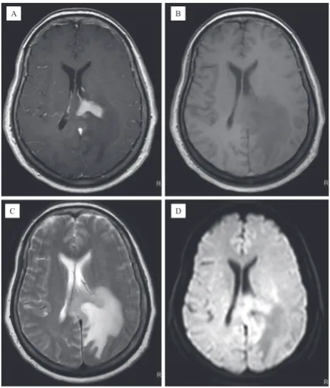
Figure 1. A 62 year-old female, which underwent biopsy and conirmed to have a CNS lymphoma, presented with headache, cognitive impairment, and right hemiparesis. The MR images enhanced with contrast (A) showed that the lesion, was located at the left trunk of corpus callosum. T1W images showed an isointense lesion (B), while T2W images showed also an isoin-tense lesion surrounded by signiicant edema (C), and high sig -nal intensity on DW images (D)
study, we are surprised that most of our cases are immunocompetent, based on the laboratory results. Moradi, et al7 also noted that in their study, of 110 subjects, none were HIV positive. This fact suggests that while immuno compromised state is a well-established risk factor for CNS lymphoma, it is not a prominent factor. It’s probably unnecessary to do routine check of CD4 count as part of our effort to determine the patient’s immune status.
In our study, the median interval time between irst symptom and diagnosis is 7 weeks. It is shorter compare to the previous report of 22 weeks.7 This short clinical course should drive our attention to the CNS lymphoma.
Since lymphoma has no distinct or characteristic clinical and radiological features, it is dificult to differentiate it with other intracranial mass lesions by those features alone.8,9 The reason might be because most intracranial pathology presents with non-speciic symptoms (increased intracranial pressure, seizure, weakness, etc.) and differ in clinical
A
D C
July, et al. Clinical and radiological features of CNS lymphoma
http://mji.ui.ac.id 157 Med J Indones, Vol. 23, No. 3,
August 2014
presentations because of the difference in location, not the type of pathology.6 Our indings suggest that CNS lymphomas are quite frequently mistaken for gliomas. The cause of this might be the clinical and radiological similarity between those two. Glioma frequently presents with headache, weakness, mental changes, and also seizure6 pretty similar with CNS lymphoma, whose most common presenting clinical conditions are symptoms of increased intracranial pressure and personality changes, followed by ataxia and hemiparesis,10 which corresponds well with our indings. Seizure is relatively rare for lymphoma compare to glioma. In addition, it is frequently almost impossible to differentiate between GBM (glioblastoma multiforme) and CNS lymphoma by conventional MR images.11 In this study, many patients were not suspected to have a CNS lymphoma before histological assessment, thus states the importance of biopsy in diagnosing CNS lymphoma. The only thing that would attract our attention is that the clinical course of lymphoma is usually much shorter compared with glioma. However, new advanced MRI techniques, such as diffusion tensor imaging, and positron-emission tomography (PET) and single photon-emission tomography (SPECT) metabolic imaging have identiied characteristic inding in CNS lymphoma that may aid in differentiation of CNS lymphomas and other CNS lesions in the near future.9,12
MR images of CNS lymphoma lesions are usually enhanced with contrast, hyperintense on diffusion-weighted imaging (DWI), in T2WI iso- to hypointense.11 The hyperintensity on DWI is related with its high cellularity, and the reduction in extracellular space caused by tumor cellularity will cause a reduction in the apparent diffusion coeficient (ADC) value.
Most tumors are located in cerebral hemisphere and periventricular location, usually involving the corpus callosum or basal ganglia.7 It’s consistent with our indings that the tumor usually involving the white matter, and following the course of the nerve ibers. It did not restrict in certain lobe of the brain, but it would follow the nerve ibers and might involve more than one lobe of the brain. It is very often on T1W1 contrast images, the tumors are looked like the paint strokes. It is more common to ind multiple tumor (more than one lesion) in lymphoma (24% in our series shows multiple lesions) compare to glioma (5% of gliomas are multicentric).13
In conclusion, consideration of the possibility of a CNS lymphoma should be done when it shows rapid progression of neurological worsening. The suspicion toward CNS lymphoma should be even higher when intracranial mass in MR images are markedly enhanced by contrast, in T1W1 show iso- to hypointense lesions while in T2W1 iso- to hypointense with marked perifocal edema, and hyperintense in DWI. It’s usually involving white matter of cerebral hemisphere and periventricular area.
Conlict of interest
All authors have nothing to disclose.
REFERENCES
1. Haldorsen IS, Espeland A, Larsen JL, Mella O. Diagnostic delay in primary central nervous system lymphoma. Acta Oncol. 2005;44(7):728-34.
2. Morris PG, Abrey LE. Therapeutic challenges in primary CNS lymphoma. Lancet Neurol. 2009;8(6):581-92. 3. Olson JE, Janney CA, Rao RD, Cerhan JR, Kurtin PJ,
Schiff D, et al. The continuing increase in the incidence of primary central nervous system non-hodgkin lymphoma. Cancer. 2002;95(7):1504-10.
4. Lister A, Abrey LE, Sandlund JT. Central nervous system lymphoma. ASH Education Book. 2002;2002(1):283-96. 5. Schabet M. Epidemiology of primary CNS lymphoma. J
neurooncol. 1999;43(3):199-201.
6. Greenberg MS. Handbook of Neurosurgery. 7th ed. New York: Thieme; 2010. p.1338.
7. Moradi A, Tajedini A, Mehrabian A, Sadeghi S, Semnani V, Khodabakhshi R, et al. Clinicopathological features of primary central nervous system lymphoma. Neurosciences (Riyadh). 2006;11(4):284-8.
8. Slone HW, Blake JJ, Shah R, Guttikonda S, Bourekas EC. CT and MRI indings of intracranial lymphoma. AJR Am J Roentgenol. 2005;184(5):1679-85.
9. Haldorsen IS, Espeland A, Larsson EM. Central nervous system lymphoma: characteristic indings on traditional and advanced imaging. AJNR Am J Neuroradiol. 2011;32(6):984-92.
10. Herrlinger U, Schabet M, Bitzer M, Petersen D, Krauseneck P. Primary central nervous system lymphoma: from clinical presentation to diagnosis. J Neurooncol. 1999;43(3):219-26.
11. Stadnik TW, Demaerel P, Luypaert RR, Chaskis C, van Rompaey KL, Michotte A, et al. Imaging tutorial: differential diagnosis of bright lesions on diffusion-weighted MR images. Radiographics. 2003;23(1):e7. 12. Toh C-H, Castillo M, Wong AM, Wei KC, Wong HF, Ng
SH, et al. Primary cerebral lymphoma and glioblastoma multiforme: differences in diffusion characteristics evaluated with diffusion tensor imaging. AJNR Am J Neuroradiol. 2008;29(3):471-5.