HAYATI Journal of Biosciences March 2010 Vol. 17 No. 1, p 1-4
EISSN: 2086-4094
Intracellular Ca
2+Regulation in Calcium Sensitive Phenotype of
Saccharomyces cerevisiae
HERMANSYAH
Chemistry Department, Faculty of Mathematic and Natural Sciences, Sriwijaya University, Jalan Raya Palembang Prabumulih Km. 35, Ogan Ilir, Inderalaya 30662, Indonesia
Phone: +62-711-580269, Fax: +62-711-580056, E-mail: [email protected] Received November 23, 2009/Accepted February 15, 2010
Intracellular cytosolic Ca2+ concentration accumulation plays an essential information in Saccharomyces
cerevisiae i.e. to explain cellular mechanism of Ca2+ sensitive phenotype. Disruption both S. cerevisiae PPase PTP2 and MSG5 genes showed an inhibited growth in the presence of Ca2+. On the other hand, by using Luminocounter with apoaequorin system, a method based upon luminescent photoprotein aequorin, intracellular Ca2+ concentration was accumulated as a consequence of calcium sensitive phenotype of S. cerevisiae. This fact indicated that PPase ptp2Δ Δ Δ Δ Δ and msg5ΔΔΔΔΔ were involved in intracellular Ca2+ transport in addition their already known pathways i.e Mitogen Activated Protein Kinase cell wall integrity pathway, high osmolarity glycerol (HOG) pathway, and pheromone response FUS3 pathway.
Key words: Saccharomyces cerevisiae, protein phosphatase, calcium sensitivity, calcium accumulation ___________________________________________________________________________
Available online at: http://journal.ipb.ac.id/index.php/hayati DOI: 10.4308/hjb.17.1.1
INTRODUCTION
Ca2+ is used by virtually every eukaryotic cell to
regulate a wide variety of cellular processes, including
gene expression. A change in the cytosolic Ca2+
concentration plays a key regulatory role in diverse cellular processes, such as T-cell activation, muscle construction and neurotransmitter release (Clapham 1995). In the yeast
Saccharomyces cerevisiae, Ca2+ has been involved in
stress-induced expression on ion transporter genes, bud formation, viability upon pheromone-induced growth (Mizunuma et al. 2001).
Recently, we observed that in the presence of high Ca2+ concentration the growth of the ptp2Δmsg5Δ double
disruptant was inhibited in G1 to S transition of cell cycle (Hermansyah et al. 2009) indicating that Ca2+ was involved
in cellular mechanism of the ptp2Δ msg5Δ double disruptant. It can be that Ca2+ plays a key role in the
transduction of external signal. Although the physiological role of Ca2+-mediated cell cycle regulation is poor
understood, this regulation has been implicated in a mechanism that may be operate under conditions where cells encounter Ca2+-caused stress. Ca2+ is required at all
stages of cell cycle except for the initiation of DNA synthesis (Shimada et al. 1991). Fast adaptation to stressing environmental changes is often requirement for cell survival, including either post transcriptional mechanism, adaptation involves remodeling gene expression and is mediated by induction or repression of a more or less specific set of genes (Estruch 2000).
PTP2 gene encodes protein tyrosine phosphatase which involved in Hog1p mitogen activated protein (MAP) kinase high-osmolarity sensing pathway and Fus3p MAP kinase pheromone, while MSG5 is dual specificity protein
tyrosine phosphatase which involved in adaptation respone to pheromone (Watanabe et al. 1995; Jacoby et al. 1997; Mattison et al. 1999; Zhan & Guan 1999; Flandez
et al. 2004).
In this study S. cerevisiae intracellular Ca2+
concentration was determined based upon its reaction with aequorin and detected using Luminocounter. Aequorin has a high specificity for Ca2+, therefore it can
be used as a biological indicator of Ca2+. Aequorin is a
photoprotein containing coelenterazine as a chromophore originally found in the jellyfish Aequorea victoria. The binding of Ca2+ to aequorin generates transient
bioluminescence, yielding as light products, CO2, and a blue fluorescent protein.
Disruption of PTP2 and MSG5 which leads to calcium sensitive phenotype caused a accumulation of intracellular Ca2+ concentration, indicating that Ca2+ transport might
implicate in the cellular mechanism of calcium sensitive phenotype of the ptp2Δ msg5Δ double disruptant S. cerevisiae.
MATERIALS AND METHODS
Yeast Strains, Plasmids, and Culture Conditions.
Yeast strains BY5209 (= FY833) MATαura3-52 his3-Ä200 leu2Ä1 lys2Ä202 trp1Ä63and BY5210 (=FY834) MAT ura3-52 his3-Ä200 leu2Ä1 lys2Ä202 trp1Ä63 (Winston
et al. 1995) were used as a wild-type and parental strains,
and the ptp2Δ msg5Δ double disruptant MATα
2 HERMANSYAH HAYATI J Biosci
the required auxotrophic supplements. SPM medium contained 0.30% potassium acetate, 0.02% raffinose and was supplemented with 10 µg/ml of adenine, arginine, histidine, isoleucine, leucine, lysine, methionine, phenylalanine, threonine, tryptophan, uracil, and valine. Unless indicated otherwise, yeast strains were grown at 30 oC. Plasmids were propagated in Escherichia coli strain
DH5α cultivated on LB medium containing 100 µg/ml ampicillin at 37 oC. Synthetic medium (SD) contained 0.67%
Bacto yeast nitrogen base without amino acids and 2% Bacto-peptone, and 2% glucose. In calcium deficient
medium (SD-Ca), CaCl2 was omitted and calcium
pantothenate was replaced by sodium pantothenate (Shimada et al. 1991). The concentration of calcium in SD–Ca medium was 0.24 µM. The ptp2Δmsg5Δ double disruptant was constructed by crossing between
ptp2Δ::CgHIS3 single disruptant and msg5Δ::CgLEU2
single disruptant (Hermansyah et al. 2009). The expression plasmid pGAPAQ1 was constructed by inserting the SacII/
EcoRI 0.6-kilobase (kb) fragment of the apoaequorin cDNA into the pYSI vector at the EcoRI site.
Introducing Plasmid pGAPAQ1 Using Yeast Transformation with Li/PEG/SS Methods. Yeast cells were inoculated on YPDA or appropriate media, the cultivation was refreshed in 5 ml of YPDA or appropriate medium. It was added 0.5 ml of culture to 5 ml of fresh medium and cultivate for 3-4 hours to reach mid mid-log phase (OD600 = 1.0). The culture was harvested in a sterile centrifuge tube at 2,000 rpm for 5 minutes. Then the medium was poured off, and washed with 5 ml of sterile water, subsequently centrifuge again. After discarded the water, cells were resuspended in 1 ml of 0.1 M LiAc and transfered to a sterile 1.5 ml tube. Pellet of cells were obtained after spinned at 12,000 rpm for 30 sec and LiAc was removed. LiAc (0.5 ml of 0.1 M) was added and store at 30 oC.
Single-strand carrier DNA was boiled for 5 min and quickly chilled in ice for 5 min. After vortex the cell suspension, 0.1 ml of sample (increase volume if cells are less dense) was pipetted into a 1.5 ml tube, and cells pellet were obtained by spinned at 10,000 xg for 30 sec, and then LiAc was remove. Then the following reagents were added carefully in the order listed; 0.24 ml of 50% (w/v) PEG 4,000, 0.036 ml of 1.0 M LiAc (overlay carefully on 50% PEG), 0.005 ml of carrier DNA (10 mg/ ml) , 0.070 ml of plasmid DNA (0.1-10 µg) and sterile water. Pellet cell was vortex vigorously until completely mixed (approximately 1 min) and was incubated for 30 min at 30 oC prior to heat shocked for
20-25 min at 42 oC. Pellet was centrifuged at 10,000 xg
for 1 min, the liquid was removed and sterile water was added (0.1 ml). Then the pellet resuspended by pipetting gently, and plate onto selective a plate. Single colonies will appear after 2-3 days incubation at 30 oC. Plasmid
DNA is a 400 µl PCR product which precipitated by a mixture of 1/10 volume 3 M NaCH3COO pH 5.2 and 2.5 volume ethanol, was centrifuged in 10,000 xg 4oC
for 1 min, subsequently dissolved in 70 µl a sterile water. Yeast transformant ptp2Δ msg5Δ containing either plasmid pGAPAQ1 or pYSI was selected in selective medium SC medium without his-, leu- and trp-, while wild
type containing either pGAPAQ1 or pYSI was selected in selective media SC medium without trp-. Each candidate disruptants were transferred into new appreciate media in order to obtain stable transformant.
Measurement of Intracellular Calcium by Luminocounter Using Apoaequorin Protein. Cells were harvested when they reached 5.106 cells/ml and cells were
counted by using hemicytometer. After centrifuge at 3,000 rpm, 30 oC, 5 min. the supernatant was discharged using
aspirator. A total of 299 µl SD medium without 1 M sorbitol was prepared and 1 µl of 1.5 mM coelenterazine was added and the solution was mixed using sonicator. This solution was added into cells prior to incubated cells solution in water bath incubator using black cover at 25 oC, 20 min.
The cells were filtered using Millipore and aspirator. The cells were washed by one and then by nine ml SD medium, respectively, and cells were resuspended with 2 x 250 µl SD medium.
Determination of Intracellular Ca2+ Concentration
Using Luminocounter. Procedure for intracellular Ca2+
concentration using Luminocounter was as follows: (i) cells sample were placed into sample cup (time 32 min), (ii) measurement was started (time 34 min), (iii) hypoosmotic stress was injected 600 µl H2O (time 35 min), (iv) 4% Triton X-100/4M CaCl2 was injected (time 39 min), and (v) measurement was finished at time 40 min.
RESULTS
Transformation of Wild Type and the ptp2ΔΔΔΔΔ msg5ΔΔΔΔΔ Double Disruptant Used pGAPAQ1 Bearing Apoaequorin Gene. The plasmid expression pGAPAQ1 was constructed by inserting the SacII/EcoRI 0.6 kilobase the apoaequorin cDNA fragment into the pYSI vector at the EcoRI site (Figure 1) (Shimada et al. 1991). Plasmid pYSI was a cloning vector based on a YEp (yeast episomal vector) and contained a S. cerevisiae glyceraldehydes-3-phosphate dehydrogenase (GAPD) as a promoter and TRP1 as a selection marker, carried 2µplasmid DNA sequence necessary for autonomous replication. The GAPD
Amp
Vol. 17, 2010 Stress-Induced Morphology of Cell Saccharomyces cerevisiae 3
promoter by which the cDNA is expressed constitutively was used because yeast cell growth is not effected by conditional expression of apoaequorin under the control of the GAL1 promoter. Before introducing the plasmid pGAPAQ1 and pYS1 into wild type and ptp2Δmsg5Δ, those plasmids (pGAPAQ1 and pYS1) was introduced into
E. coli DH5α (Figure 2).
Plasmid pYS1 size was 10.7 kb, restricted by BamH1
restriction enzymegenerated one fragment 10.7 kb. While size of plasmid pGAPAQ1 10.7 kb + 0.63 kb = 11.33 kb, and restricted by BamH1 generated two fragment, 9.9 kb and less than 1.4 kb (Figure 3).
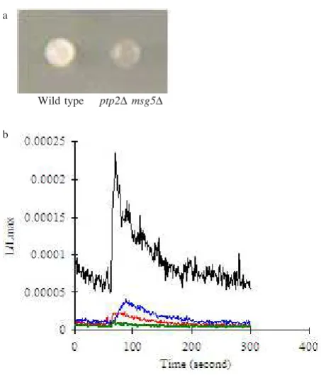
Measurement of Intracellular Ca2+ Concentration
Using Luminocounter. Intracellular Ca2+ concentration
was measured with an apoaequorin cDNA system by using Luminocounter. The result showed that in SC-trp containing Ca2+ double disruption of PTP2 and MSG5
but not in wild type due to intracellular Ca2+ accumulation.
On the other hand, in the SC-trp without Ca2+ culture either
the ptp2Δmsg5Δ double disruptant or wild type did not cause intracellular Ca2+ accumulation (Figure 4).
DISCUSSION
Limitations in the direct measurement of intracellular Ca2+ and difficulties in quantifying Ca2+ channel activity
in yeast are a problem in progress to understand Ca2+
signaling. By using Luminocounter with apoaequorin system, a method based upon luminescent photoprotein aequorin, intracellular Ca2+ concentration was changed
as a consequence of calcium sensitive phenotype of S.
cerevisiae. The result suggests that both PTP2 and MSG5
are implicated in regulation of S. cerevisiae intracellular Ca2+. Transient increases in intracellular Ca2+ regulate a
wide variety of cellular processes and Ca2+ signaling.
Overlapping Ca2+ sensitive phenotype of the ptp2Δ
msg5Δ double disruptant with its accumulation of intracellular Ca2+ concentration indicates that it has
relationship between those two phenomenon. It was reported that lacking of Pmr1p, a golgi-localized Ca2+
transporting ATPase showed growth sensitivity to Ca2+
(Yadav et al. 2007). One need to elucidate more detail whether both PPase Ptp2p and Msg5p also involved in Ca2+ transport in cellular.
Aequorin is a reaction complex of apoaequorin, coelenterazine as a substrate, and molecular oxygen. This reaction results bioluminescence triggered by ion Ca2+ by
following mechanism; the coelenterazine is oxidized to coelenteramide, and then the excited state of coelenteramide bound to apoaequorin (blue fluorescent protein) emit in the reaction. While In vitro Aequorin is regenerated by incubation with coelenterazine, molecular oxygen, 2-mercaptoethanol, pantothenate (Iida et al. 1990). Accumulation of cytosolic Ca2+ could be occurred
when two Ca2+ pumps, PMC1 and PMR1 which functions
maintaining cytosolic free Ca2+ at submicromolar levels in
budding yeast were deleted, and this cytosolic Ca2+
accumulation causes lethal (Cunningham & Fink 1994).
Figure 2. Transformation of DH5α with (a) plasmid pGAPAQ1; (b) plasmid pYS1; (c) Negative control.
a b c
1 2 3 4 5 6 7 8 9 10 11 12
9.9 kb 10.7 kb
1.43 kb
Figure 3. Agarose gel eletrophoresis analysis of pGAPAQ1 and pYS1. (a) undigested (b) Digested using BamHI restriction enzyme. Line 1, 2, 3, 7, 8, 9 is pGAPAQ1; Line 4, 5, 6, 10, 11, and 12 is pYS1. The Marker used
λ/EcoT14.
a b
Wild type ptp2Δmsg5Δ
a
Figure 4. Measurement of intracellular Ca2+ with an apoaequorin
system by using Luminocounter. Correlation between Ca2+ sensitive phenotype with intracellular Ca2+
concentration. (a) ptp2Δmsg5Δ Ca2+ showed sensitive
phenotype. (b) In SC-trp containing Ca2+ the ptp2Δ
msg5Δ double disruptant but not in wild type showed accumulation of intracellular Ca2+. While in the SC-trp
without Ca2+ culture either the ptp2Δ msg5Δ double
disruptant or wild type did not cause intracellular Ca2+
accumulation. : WT apo, : WT apo Ca, : ptp2m sg5 apo, : ptp2m sg5 apo Ca.
Further, yeast cells lacking of Pmr1p, Ca2+-ATPase pump
are unable to maintenance proper level of Ca2+ within golgi
apparatus because increase in Ca2+ uptake rate
(Kellermeyer et al. 2003).
It is previously reported that vacuole morphology of the ptp2msg5 double disruptant by staining the vacuolar membrane with FM4-64, a lypophilic styryl dye even in the absence of Ca2+ the vacuole of the Δptp2Δmsg5 double
disruptant was fragmented (Hermansyah et al. 2009). This fragmented vacuolar correlate with increase in intracellular cytosolic Ca2+ (Kellermeyer et al. 2003). Thus, PTP2 and
MSG5 may function or involved in intracellular Ca2+
transport with redundant manner because S. cerevisiae
yeast strain lacking both Ptp2p and Msg5p showed Ca2+
sensitive growth, intracellular Ca2+ concentration
accumulation and vacuole fragmentation.
ACKNOWLEDGEMENT
A grateful to Satoshi Harashima and Hidetoshi Iida for helpful and supervision in this work.
REFERENCES
Clapham DE. 1995. Calcium signaling. Cell 80:259-268. Cunningham K, Fink GR. 1994. Ca2+ transport in Saccharomyces
cerevisiae. J Exp Biol 196:157-166.
Estruch F. 2000. Stress-controlled transcription factors, stress-induced genes and stress tolerance in budding yeast. FEMS Microbiol Rev 24:469-486.
Flandez M, Cosano IC, Nombela C, Martin H, Molina M. 2004. Reciprocal regulation between Slt2 MAPK and isoforms of Msg5 dual-specificity protein phosphatase modulates the yeast cell integrity pathway. J Biol Chem 12:11027-11034.
Hermansyah, Sugiyama M, Kaneko Y, Harashima S. 2009. Yeast protein phosphatase Ptp2p and Msg5p are involved in G1-S transition, CLN2 transcription and vacuole morphogenesis.
Arch Microbiol 191:721-733.
Iida H, Sakaguchi S, Yagawa Y, Anraku Y. 1990. Cell cycle control by Ca2+ in Saccharomyces cerevisiae. J Biol Chem
265:21216-21222.
Jacoby T, Flanagan H, Faykin A, Seto AG, Mattison C, Ota I. 1997. Two protein tyrosine phosphatase inactivate the osmotic stress response pathway in yeast by targeting the mitogen-activated protein kinase in Hog1. J Biol Chem
272:17749-17755.
Kellermayer R, Aiello DP, Miseta A, Bedwell DM. 2003. Extracellular Ca2+ sensing contributes to excess Ca2+
accumulation and vacuolar fragmentation in a pmr1Δ mutant of S. cerevisiae. J Cell Sci 116:1637-1646.
Mattison CP, Spencerm SS, Kresge KA, Lee J, Ota IM. 1999. Differential regulation of the cell wall integrity mitogen-activated protein kinase pathway in the budding yeast by the protein tyrosine phosphatase Ptp2 and Ptp3. Mol Cell Biol
19:7651-7660.
Mizunuma M, Hirata D, Miyaoka R, Miyakawa T. 2001. GSK-3 kinase Mck1 and calcineurin coordinately mediate Hsll down-regulation by Ca2+ in budding yeast. EMBO J 20:1074-1085.
Shimada JN, Iida H, Tsuji FI, Anraku Y. 1991. Monitoring of intracellular calcium in Saccharomyces cerevisiae with an apoaequorin cDNA expression level. Proc Natl Acad Sci
88:6878-6882.
Watanabe Y, Irie K, Matsumoto K. 1995. Yeast RLM1 encodes a serum response factor-like protein that may function downstream of the Mpk1 (Slt2) mitogen-activated protein kinase pathway. Mol Cell Biol 15:5740-5749.
Winston F, Dollard C, Ricupero-Hovasse SL. 1995. Construction of a set of convenient Saccharomyces cerevisiae strains that are isogenic to S288C. Yeast 11:53-55.
Yadav I, Muend S, Zhang Y, Rao R. 2007. A phenomics approach in yeast links proton and calcium pump in the golgi. Mol Biol Cell 18:1480-1489.
Zhan XL, Guan KL. 1999. A specific protein-protein interaction accounts for the in vivo substrate selectivity of Ptp3 towards the Fus3 MAP kinase. Genes Dev 13:2811-2827.