Vol 6, No 3, July - September 1997 RenalHydartdDisease 187
Renal
Hydatid
Disease.
A
case
report.
R.K. Patial, D. Kapoor, H. Gupta, A. Kapoor
Abstrak
Hidatid ginjal jarang ditemukan. Dilaporkan satu kasus hidatid ginjal, yang menimbulkan keluhan nyeri daerah pinggang atas dan didiagnosis dengan CT abdomen dan pemeriksaan serologik. Dibeikan terapi albendazol 400 mg 2 kali sehai sebanyak 3 siffius, setiap siklus lamanya 28 hari. CT abdornen 6 bulan kemud.ian tidak menemukan kista lagi.
Abstract
Renal hydatid disease is uncommon even in counties where it is endemic accounting for less than 3Vo of all cases. We report a case ofrenal hydatid disease, for its raity, presenting as dull ache in left loin and diagnosed on abdominal CT and hydatid serology. It was treated with 3 cycles of Albendazole 400 mg tvvice a day, each cycle of 28 days duration. Abdominal CT done 6 months later showed disappearance of the cyst.
Keywords: Kidney, Hydatid Cyst, Albendazole
Hydatid disease is prevalent throughout the world and
is endemic in Australia, Newzealand, South America,
Africa and
Middle East. Primary hydatid diseaseof
kidney is rare even in countries where it is endemic and account for less than3Vo of all cases.l We report a case with review of literature of primary hydatid diseaseof
kidney for its rarity.Case Report
A
39 year old male, non-alcoholic, vegetarian, farmer by occupation presented in the Medical O.P.D. with a history of dull ache in the left loin for the last 30 days which was continuous, increased on walking and had no relation to meals. The pain did not interfere with hisnormal routine activity. On questioning, he was rearing
sheep and was having a pet dog in the house. Clinical examination showed mild pallor. Abdominal
examina-tion
revealed tendernessin
theleft
lumbar region without any palpable mass or lump. Rest of the physi-cal and systemic examination was non-contributory.His
haemogram showed Hb-9g%o, TLC-5600/cmmwith
Poly-72Vo,
Lympho-25%o,Mono-lZo
andEo sinophil I -27o, Biochemical investi gations
includ-ing liver and renal functions were within normal limits. Routine and microscopic examination
of
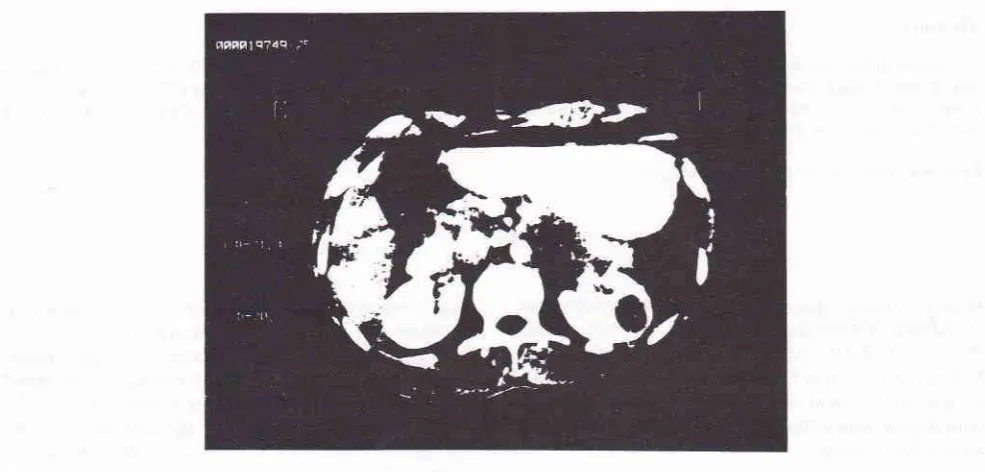
urine was also normal. X- ray chest, abdomen showed noabnor-mality. USG abdomen did not reveal any abnormal finding. CT scan Abdomen (Fig. 1) showed a cysr 2.0
X
2.1 cm
in
sizewith well
defined margins.In-travenous urography was normal. Serological tests for hydatid was positive
by
indirect haemagglutination. Amoebic serology and serology for HIV was negative.Patient was prescribed tablet Albendazole 400 mg twice a day for 28 days. The symptoms improved after 20 days of therapy. The therapy was repeated for two cycles of 28 days each. CT scan done after 6 months of therapy showed disappearance of the cyst.
DISCUSSION
Hydatid disease manifests commonly as
liver
cyst(75Vo) and lung cyst
(lïVo),
but no structure of the body,right from
the brainro
the fallopian rube is exempt.' Hydatid cystis
formed by implantationof
Echinococcus granulosus larvae. Ultrasonography andCT scan has led
to increased
recognitionof
hydatiddisease at the atypical sites. Hydatid disease of kidney are usually single and located in the cortex. Symptoms vary according to the stage of the disease. Patient may
be asymptomatic or may report flank pain, haematuria,
hypertension or a smooth palpable, often tender renal rnass.'The cyst may rupture and manifest as hydaturia.
188
Patial et al.It is the only pathognomic evidence of hydatid disease
of kidney. The skin test (Casoni's test) is accurate in 90Vo of cases. Serological tests like Indirect
haemag-glutination
areequally
sensitive. Eosinophilia is detectedin
20-50Voof
cases.'X-ray
abdomen mayshow
a soft
tissue massin
the kidney arewith
or without calcification. Ultrasonography revealstypical-ly
afluid filled
mass lesion.CT
abdomen shows acharacteristic unilocular or multilocular cyst with well
defined walls, readily enhancing after Intravenous
con-trast.3
REFERENCES
l.
Singh R, Bhadury S, Singh SK. Primary hydatid cyst in the spleen and kidney. Preoperative diagnosis. The Indian J Radiol Imaging 1993;3: I 3 l-3.2.
Nabizadeh I, Morchouse tIT, Freed SZ. Hydatid disease of kidney. Urology 1983 ;22:17 6-8.Med J Indones
Therapy with Albendazole 400 mg twice a day for 28 days and repeated for 1-8 cycles, separatedby drug free
interval
of
2-3 weeks have been shownto
be most efficacious causing reduction in the size of hydatid cystand rarely disappearance.a
The size, number, locatidn, and symptoms determine
the surgical tlrerapy which includes panial
or
total nephrectomy.' Recurrenceof
the disease is possible,since there is high risk of reexposure.
Afsar H, Yagci F, Aybasti N, Meto S. Hydatid disease of the kidney. Br J Urology 1994;73:17-22.
[image:2.595.49.542.226.462.2]Horton RI. Chemotherapy of Echinococcus infection in man with Albendazole. Aust NZ J Surg 1989;59:665.
Figure
l.
CT abdomen showing well definedfluidfilled cyst in left kidney 2.0 X 2.1 cm.