Vol. 18 No. 2, p 77-81 EISSN: 2086-4094
http://journal.ipb.ac.id/index.php/hayati DOI: 10.4308/hjb.18.2.77
Genetic Diversity of Methylotrophic Bacteria from Human Mouth
Based on Amplified Ribosomal DNA Restriction Analysis (ARDRA)
DIANA ELIZABETH WATURANGI∗∗∗∗∗, IVANA FRANCISCA, CINDY OKTAVIA SUSANTO
Faculty of Biotechnology, Atma Jaya Catholic University of Indonesia, Jalan Jenderal Sudirman 51, Jakarta Selatan 12930, Indonesia
Received August 6, 2010/Accepted June 20, 2011
Methylotrophs inhabit the human mouth. In this study, methylotrophic bacteria were isolated from the human mouth microflora of 63 subjects, especially from the tongue, gingival, and subgingival area using minimal agar supplemented with 1% methanol. The obtained isolates were subjected to biochemical assays, continued with antibiotics susceptibility testing using ampicillin (10 µµµµµg), tetracycline (20 µµµµµg), kanamycin (30 µµµµµg), trimethoprim (5 µµµµµg), and streptomycin (10 µµµµµg). Genetic diversity was analyzed using ARDRA method. Isolates varying in morphology characteristics were amplified for 16S rRNA gene and continued with DNA sequencing. As many as 21 methylotrophic bacterial isolates were purified and divided into seven groups with different phenotypic profiles. A majority of the isolates were resistant to trimethoprim but sensitive to kanamycin, streptomycin, and tetracycline. Resistance to ampicillin was variable in each isolate. ARDRA showed nine different digestion profiles. DNA sequencing analysis of the 16S rRNA gene showed that six isolates with different phenotypic and digestion profiles were closely related to Methylobacterium radiotoleran (94%), Microbacterium esteraromaticum (99%), Pseudomonas sp. (100%), and three of them were exhibited 99, 99, and 98% sequence similarity with Gordonia sp., respectively. The results of this study revealed diversity among methylotrophic bacteria particularly in human mouth.
Key words: methylotrophic bacteria, molecular characterization, restriction profile
___________________________________________________________________________
_________________
∗ ∗∗
∗∗Corresponding author. Phone: +62-21-5703306 ext 330,
Fax: +62-21-5719060, E-mail: [email protected]
INTRODUCTION
Human mouth is a habitat which is likely to be colonized by bacteria. More than 500 species of bacteria lived in human mouth (Foster et al. 2003). Those bacteria form complex interaction among them and also interact with host tissue. The oral bacteria such asstreptococci, lactobacilli, staphylococci, corynebacteria, and anaerobic bacteria, bacteroides usually called as normal mouth microbiota (Roger 2008).
Methylotrophic bacteria are a group of bacteria which is also found in human mouth (Anesti et al. 2005). Methylorophic bacteria are able to use one carbon compounds such as methane, methanol, halogenated methane, and methylated amine as sources of carbon and energy for their growth (Nishio et al. 1997). The availability of one carbon compounds in human mouth, including methylated sulphides (such as methanethiol and dimethylsulphide) supports the growth of methylotrophic bacteria. These compounds, together with hydrogen sulphide, are product of microbial degradation of free sulphur amino acids, thiol and disulphide groups which are released from proteins (Anesti et al. 2005). Volatile sulphur compound (VSC) in oral cavity caused the production of halitosis or bad breath (Ratcliff 1999).
Methylotrophic bacteria isolated from human mouth are suggested to have ability in reducing the odour of
human mouth since it can oxidize some volatile sulphur compounds such as dimetyhlsulphide and methyl mercaptan (Anesti et al. 2005; Brownlee 2005).
Methylotrophic bacteria produce some enzymes that involved in the metabolism of specific one carbon compound, such as methane monooxygenase (MMO), methanol dehydrogenase (MDH), methylamine dehydrogenase, methyltransferase, methanesulphonic acid monooxygenase (Chistoserdova et al. 2005), and dimethylsulphide oxygenase (Anesti et al. 2005). Those enzymes play important role in the oxidation of C1 compounds to formaldehyde which is further be assimilated.
The presence of methylotrophic bacteria in human mouth is diverse. It is important to investigate the diversity of metylotrophic bacteria in human mouth since it can provide more information about various metyhlotrophic bacteria lived in human mouth. Thus, it helps to study the strength and the weakness of candidate bacteria that potential to be applied in many sectors.
In this study, we analyzed the genetic diversity of methylotrophic bacteria from human mouth based on Amplified Ribosomal DNA Restriction Analysis (ARDRA). The isolates were also identified and tested for their susceptibility to some antibiotics.
MATERIALS AND METHODS
Samples were obtained from subgingivalis area (S), gingivalis area (G), and tongue (L). Samples was collected by cotton swabs and suspended into 2 ml of sterile 0.85% sodium chloride solution, for gingivalis and tongue and with sterile toothpick for subgingivalis. The samples were inoculated in minimal medium supplemented with 1% (v/v) methanol and incubated at 28 oC for approximately one week. Colonies of bacteria were then isolated. Catalase test and oxidase test were used to identify the isolates. Gram staining was also performed to differentiate the isolates.
Antibiotics Susceptibility Testing. Antibiotics susceptibility test was carried out by the agar disk diffusion method (Bauer et al. 1996). The following disks were used: ampicillin (10 µg), tetracycline (30 µg), kanamycin (30 µg), trimethoprim (5 µg), and streptomycin (10 µg). The diameters of zones of inhibition were measured to the nearest millimeter. Isolates were categorized as susceptible, intermediate and resistant based on interpretive criteria developed by the National Committee for Clinical Laboratory Standards (NCCLS, 1999).
Amplification of 16s rRNA Gene by PCR. The isolates are enriched in Luria Agar medium at 28 oC for 2 days. Genomic DNA was extracted using the CTAB method and amplified by PCR with primers 63f (52-CAGGCCTA ACACATGCAAGTC-32) and 1387r (52-GGGCGGWGTGT ACAAGGC-32, W=A or T) (Marchesi et al. 1998). The reaction mixture contained 2.5 µl enzyme buffer, 0.5 µl dNTP, 1 µl 25 pmol primer 63f, 1 µl 25 pmol primer 1387r, 0.5 µl (2.5 U) Taq polimerase (New England Biolabs, Mass., USA), and 1 µl DNA sample. PCR amplification was performed in a total volume of 25 µl using GeneAmp® PCR system 2400 (Perkin Elmer, USA). The PCR conditions were as follows: initial denaturation for 5 minutes at 94 oC and 25 cycles consisting of denaturation at 94 oC for 30 seconds, primer annealing at 55 oC for 30 seconds, and
elongation at 72 oC for 1 minute. The PCR product was checked by electrophoresis (Mini-Sub Cell GT, Bio-Rad, Richmond, CA) on a 0.8% agarose gel at 80 V for 1 hour. Genetic Diversity Analysis with ARDRA. PCR products of the isolates were digested with restriction enzyme Sau3AI (New England Biolabs, Mass., USA). The digestions were carried out in a total volume of 20 µl for 24 hours at 37 oC. The mixture contained 3 µl PCR product, 2 µl of 10× restriction buffer, 14.5 µl ddH2O and 0.5 µl unit restriction enzyme Sau3AI (New England Biolabs, Mass., USA). Digestion of 16s rRNA gene Escherichia coli DH5α was also done to compare with other isolates. The digested amplicon was visualized by electrophoresis on a 2% agarose gel at 45 V for 2 hours. Treecon computer program (Yves-Van de Peer of Department of Biochemistry, www.informatik. Unitrier.de) was employed to determine the phylogenetic tree based on the restriction fragment pattern.
DNA Sequencing Analysis. DNA fragment from six isolates were chosen to be sequenced based on the dominant group that isolated from human mouth habitat. PCR product was purified using DNA Gel Extraction Kit (V-Gene, Hangzhou, China). Purified products were sequenced using big dye terminator with ABI PRISMTM model 3130 Genetic Analyzer. The sequence data of bacterial isolates were compared to sequence from GenBank in National Center for Biotechnology Information (NCBI, www.ncbi.nlm.nih.gov) using BLASTN program to identify the isolates.
RESULTS
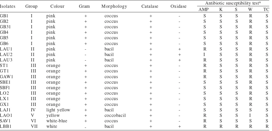
Isolation and Characterization of Methylotrophic Bacteria from Human Mouth. From 63 subjects, a total of 21 isolates of methylotrophic bacteria were successfully isolated using one carbon compound medium as selective medium (Table 1). The isolates showed various gram
Table 1. Characteristics and antibiotic susceptibility of the isolates
Antibiotic susceptibility test* AMP K S W TC
Isolates Group Colour Gram Morphology Catalase Oxidase
GB1
staining result, morphology, and pigmentation. All isolates were catalase-positive, showing that all isolates were aerobic bacteria whereas only 4 isolates were oxidase-positive. Based on its characteristic, the isolates were divided into 7 groups (Table 1).
Antibiotics Susceptibility Testing. Most of the isolates were resistant to trimethoprim, except isolate LAJ1, LAO1, and SAV1 (Table 1). Only isolate LBB1 was resistant to kanamycin, streptomycin, and tetracycline whereas other isolates were sensitive. Ampicillin susceptibility test showed various result among the isolates (Table 1).
Extraction of Genomic DNA and Amplification of 16s rRNA Gene. After the isolates were purified, the genomic DNA of the isolates was extracted using CTAB method (Marchesi et al. 1998). Genomics DNA of the isolates were used as template in 16S rRNA gene amplification. The expected amplicon size is approximately 1400 bp.
Genetic Biversity Analysis with ARDRA. Digestion with restriction enzyme Sau3AI produced different profile among the isolates (Figure 1). The isolates with different phenotypic profiles showed different restriction profile.
The isolates with similar characteristic also showed similar profile, such as isolate GB2-GB6 and isolate LAU1-LAU3. It is possible that the isolates were collected from one subject in the same isolation area. Therefore, a few isolates showed similar 16S rRNA gene restriction profile.
However, the seven orange isolates were shown to be different (Figure 1; lane 9-15). Although the isolates have similar phenotypic profiles, only 5 isolates showed similar digestion profile, whereas 2 isolates were shown to have different profile. These isolates are isolate GAW1 (lane 11) and isolate LO1 (lane 14).
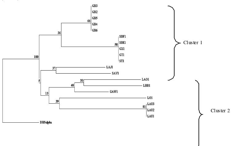
The restriction fragment pattern was used to construct phylogenetic tree (Figure 2). Phylogenetic tree showed nine groups of bacteria were separated into 2 clusters with bootstrap 100 (from 100 phylogenetic tree constructed by the program, it always separated into 2 clusters). However, the characteristic to distinguish the clusters have not yet been fully known.
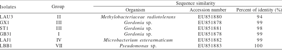
DNA Sequencing Analysis. Isolate LAU3, GX1, LAJ1, LBB1, GB31, and ST1 were chosen to be identified based on phenotypic and digestion profile. These isolates represented the dominant group of bacteria that successfully isolated in this study. The results of DNA sequencing were partial sequence of 16S rRNA gene (data was not shown). Sequence similarity of the isolates using BLASTN program was shown in Table 2. Based on sequencing analysis, some isolates were successfully identified as Methylobacterium radiotolerans, Gordonia
sp., Microbacterium esteraromaticum., and Pseudomonas
sp.
DISCUSSION
Methylotrophic bacteria are a group of bacteria that capable in using C1 compounds such as methanol as their only carbon and energy sources (Dien et al. 2003). In this M 1 2 3 4 5 6 7 8 9 10 11 12 13 14 15 16 17 18 19 20
Figure 1. Restriction pattern of PCR-amplified fragment of 16S rDNA genes digested with Sau3A1 (Lanes M: 1 kb DNA ladder, 1: GB2, 2: GB3, 3: GB4, 4: GB5, 5: GB6, 6: LAU1, 7: LAU2, 8: LAU3, 9: ST1, 10: GT1, 11: GAW1, 12: SBE1, 13: SBF1, 14: LO1, 15: GX1, 16: SAV1, 17: LBB1, 18: LAJ1, 19: LAO1, 20: E. coli DH5α).
Cluster 1
Cluster 2
study we used one carbon compound medium (minimal medium supplemented with 1% methanol) as selective medium for these bacteria and biochemical assays oxidase and catalase assay (Anesti et al. 2005).
Seven groups of the isolates with different phenotypic profiles showed the diversity of methylotrophic bacteria in human mouth (Table 1). Further, the diversity of methylotrophic bacteria in human mouth was also shown by ARDRA analysis. ARDRA analysis could also distinguish the orange coloured isolates which have similar phenotypic profile. These results suggested that ARDRA technique could be used to show the genetic diversity among the isolates with same phenotypic profiles. However, alternative techniques could be used to increase the discriminative, such as amplification of 16S-23S rDNA intergenic spacer or genome analysis with Macrorestriction Fragment Length Polymorphism (MFLP). Both of these techniques have been used by Riupassa and Suwanto (2004) to analyze the genetic diversity of pink pigmented facultative methylotroph isolated from some Indonesian edible leafy plants.
The phylogenetic tree showed that isolate GB2-GB6 was distinct from isolate LAU1-LAU3 although they produced the same pigment, indicating that the metabolism of pigment synthesis is not related to the relationship among the isolates. Isolate GAW1 and isolate LO1 were also separated into different branch in phylogenetic tree although the isolates have similar characteristics. Moreover, these isolates were distinct from the other orange isolates. These results suggested that 16S rRNA gene of these isolates were difference from 16S rRNA gene of the other orange isolates. It may possible that the species or genus of these isolate were different from the other isolates.
Further, the isolates were examined using DNA sequencing analysis. In this study partial 16S rDNA sequence was used to identify the isolates. The fact that isolate LAU3 produced pink pigment lead to the initial hypothesis that isolate LAU3 was a pink pigmented facultative methylotroph (PPFM) bacteria. Further, isolate LAU3 showed similar characteristics and morphology with PPFM bacteria. Moreover, this isolate was closely related to Methylobacterium radiotolerans (94%). Previously, a group of PPFM bacteria has been isolated from human mouth by Anesti et al. (2005). This study also supported the successful of PPFM bacteria isolation from human mouth.
Besides involved in metabolism of one carbon compounds in human mouth, PPFM bacteria also produced
micronutrient pyrrolo-quinoline quinone (PQQ). PQQ was a cofactor redox reaction and have characteristic as vitamin (Bishop et al. 1998). Thus, it is possible to apply PPFM bacteria for human being. However, for its application as odour reducing bacteria, it is still provide much more studies, such as the efficacy of the metabolism of VSC in oral cavity and its safety for human considering that PPFM bacteria has been reported as pathogen opportunistic (Hornei et al. 1999).
Isolate GX1, isolate ST1, and isolate GB31 were closely
related to Gordonia sp. (99, 98, and 99%), which
previously called Rhodococcus. Phenotypic profiles of
isolate ST1 and GX1 were similar to Gordonia that isolated byLesens et al. (2000), which is salmon-orange coloured. Moreover, Gordonia isolated by Lesens et al. (2000), also showed the same result as isolate GX1. Both isolates showed resistant to trimetoprim and sensitive to aminoglycoside group such as kanamycin and gentamycin, and to β-lactam group such as penicillin and ampicillin.
There still quite a few publications that studied the C1
metabolism of Gordonia genus. Presumptively, isolate
GX1, ST1, and GB31 use alcohol dehydrogenase enzyme while growing on minimal media supplemented with 1% (v/v) methanol. This enzyme is an enzyme possessed by
Rhodococcus genus, which plays a role in alcohol metabolism, both the short chain and long chain alcohol (Schenkels & Duine 2000). However, it should still be confirmed by detecting the presence of alcohol dehydrogenase coding gene.
Gordonia sp. has never been successfully isolated from human mouth. All this time, those bacteria are mostly isolated from clinical specimens, such as blood and hospital-origin culture, especially from patient catheters and nurses (Lesens et al. 2000), indicating that Gordonia
sp. belongs to an opportunistic bacteria group.
Isolate LAJ1 was closely related to Microbacterium
estearomaticum (99%) which is a rod shaped Gram positive bacteria, possesses yellow or orange coloured pigmentation, and catalase-positive (Funke et al. 1995).
The metabolism of C1 compounds in Microbacterium sp.
has not paid much attention in the past, but a few species, for example, M. lacticum, have been known for their ability to grow on methanol substrate (Mokashi & Paknikar 2002). However, no further explanation about the metabolism is stated.
There was also no report about the isolation of
Microbacterium sp. in the human mouth. Previously, this bacteria has been successfully isolated from soil, corn
Table 2. Sequence analysis results with BLASTN program
Sequence similarity
Organism Accession number Percent of identity (%)
Isolates Group
LAU3 GX1 ST1 GB31 LAJ1 LBB1
II III III I IV VII
Methylobacteriaceae radiotolerans Gordonia sp.
Gordonia sp. Gordonia sp. Microbacterium esterarmaticum
Pseudomonas sp.
EU851880 EU851878 EU851881 EU851878 EU851882 EU851883
liquid waste, and clinical specimens such as blood, hospital
air, and wounds (Funke et al. 1995), indicating that
Mircobacterium is an opportunistic pathogen. Susceptibility testing done by Funke et al. (1995) also showed a compatible result with this study, that this bacteria is tetracycline-sensitive.
Isolate LBB1 was identified as Pseudomonas sp. (100%
sequence similarity). A few species from Pseudomonas
sp. have been reported to be successfully isolated from the human mouth, such a P. alcalophila and P. aleovorans
(Hutter et al. 2003). Most of Pseudomonas is not included
in methylotrophic bacteria. However, a few Pseudomonas
have alcohol dehydrogenase which are able to oxidize methanol, such as P. aeruginosa, and P. putida (Vangnai
et al. 2002). Moreover, P. fluorescens has also been reported to effectively degrade VSC compounds, dimethyl disulphide (Nakagawa et al. 2007).
ACKNOWLEDGEMENT
This research was funded by Atma Jaya Research Centre 2006.
REFERENCES
Anesti V, McDonald IR, Ramaswamy M, Wade WG, Kelly DP, Wood AP. 2005. Isolation and Molecular Detection of methylotrophic bacteria occurring in the human mouth. Environ Microbiol 7:1227-1238. http://dx.doi.org/10.1111/ j.1462-2920.2005.00805.x
Bauer AW, Kirby WMM, Sherris JC, Turck M. 1966. Antibiotic susceptibility testing by a standardized single disk method. Am J Clin Pathol 45:493-496.
Bishop A, Gallop PM, Karnovsky ML. 1998. Pyrroloquinoline quinone: A novel vitamin? Nutr Rev 56:287-293. http:// dx.doi.org/10.1111/j.1753-4887.1998.tb01661.x
Brownlee C. 2005. Bacteria feed on stinky breath. Science News 168:93.
Chistoserdova L, Kalyuzhnaya G, Lidstrom ME. 2005. C1-transfer
modules: from genomics to ecology. ASM News 71:521-5 2 7 .
Dien SJV, Marx CJ, O’Brien BN, Lidstrom ME. 2003. Genetic characterization of the carotenoid biosynthesis biosynthesis pathway in Methylobacterium extorquenz AM1 and isolation of a colorless mutant. Appl Environ Microbiol 69:7563-7566. http://dx.doi.org/10.1128/AEM.69.12.7563-7566.2003
Foster JS, Palmer RJ, Kolenbrander PE. 2003. Human oral cavity as a model for the study of genome-genome interactions. Biol Bull 204:200-204. http://dx.doi.org/10.2307/1543559
Funke G, Falsen E, Barreau C. 1995. Primary identification of Microbacterium spp. encountered in clinical specimens as CDC
coryneform group A-4 and A-5 bacteria. J Clin Microbiol 33:188-192.
Hornei B, Lüneberg E, Schmidt-Rotte H, Maaβ M, Weber K, Heits F, Frosch M, Solbach W. 1999. Systemic infection of an immunocompromised patient with Methylobacterium zatmanii. J Clin Microbiol 37:248-250.
Hutter G, Schlagenhauf U, Valenza G, Horn M, Burgemeister S, Claus H, Vogel U. 2003. Molecular analysis of bacteria in periodontitis: evaluation of clone libraries, novel phylotypes, and putative pathogens. Microbiology 149:67-75. http:// dx.doi.org/10.1099/mic.0.25791-0
Lesens O, Hansmann Y, Riegel P, Heller R, Benaissa-Djellouli M, Martinot M, Petit H, Christmann D. 2000. Bacterimia and endocarditis caused by a Gordonia species in a patient with a central venous catheter. Emerging Infectious Diseases 6:382-385. http://dx.doi.org/10.3201/eid0604.000410
Marchesi JR, Sato T, Weightman AJ, Martin TA, Fry JC, Hiom SJ, Dymock D, Wade WG. 1998. Design an evaluation of useful bacterium-specific PCR primers that amplify genes coding for bacterial 16S rRNA. Appl Environ Microbiol 64:795-799. Mokashi SA, Paknikar KM. 2002. Arsenic (III) oxidizing Microbacterium lacticum and its use in the treatment of arsenic contaminated groundwater. Appl Microbiol 34:258-262. http:/ /dx.doi.org/10.1046/j.1472-765x.2002.01083.x
Nakagawa T, Fujimura S, Ito T, Miyaji T, Nakagawa T, Tomizuka N. 2007. Degradation of dimetyl disulfide by Pseudomonas fluorescens strain 76. Biosci Biotechnol Biochem 71:366-370. http://dx.doi.org/10.1271/bbb.60295
[NCCLS] National Committee for Clinical Laboratory Standards. 1999. Performance Standards for Antimicrobial Susceptibility Testing; Ninth Informational Supplement. Document M100-S9. Natl. Committee Clin. Lab. Stand., Wayne, PA. Nishio T, Yoshikura T, Itoh H. 1997. Detection of
Methylobacterium Species by 16S rRNA gene-targeted PCR. Appl Environ Microbiol 63:1594-1597.
Ratcliff PA. 1999. The relationship between oral malodor, gingivitis, and periodontitis [Review]. J Periodontol 70:485-489. http://dx.doi.org/10.1902/jop.1999.70.5.485
Riupassa PA, Suwanto A. 2004. Keragaman genetika bakteri metilotrof fakultatif berpigmen merah muda pada beberapa sayur lalaban. Hayati 11:153-158.
Rogers AH (editor). 2008. Molecular Oral Microbiology. Caister Acad Pr. ISBN 978-1-904455-24-0.
Schenkels P, Duine JA. 2000. Nicotinoprotein (NADP-containing) alcohol dehydroge-nase from Rhodococcus erythropolis DSM 1069: an efficient catalyst for coenzyme-independent oxidation of a broad spectrum of alcohols and the interconversion of alcohols an alde-hydes. Microbiology 146:775-785.
Vangnai AS, Sayavedra-Soto LA, Arp DJ. 2002. Roles for the two 1-butanol dehydrogenase of Pseudo-monas butanovora in butane and 1-butanol metabolism. J Bacteriol 184:4343-4350.