EISSN: 2086-4094
Fecal Steroid Profile of Female Javan Gibbons
(
Hylobates moloch
) Maintained in Pairing-Typed Cage
HERA MAHESHWARI1∗∗∗∗∗, LUTHFIRALDA SJAHFIRDI2, PUDJI ASTUTI3, BAMBANG PURWANTARA1, HADI SUKADI ALIKODRA4, DONDIN SAJUTHI1,5, REVIANY WIDJAJAKUSUMA1
1Faculty of Veterinary Medicine, Bogor Agricultural University, Darmaga Campus, Bogor 16680, Indonesia 2Department of Biology, Faculty of Mathematic and Natural Sciences, University of Indonesia,
UI Campus, Depok 16424, Indonesia
3Faculty of Veterinary Medicine, Gadjah Mada University, Jalan Fauna No. 2 Klebengan, Yogyakarta 55281, Indonesia 4Faculty of Forestry, Bogor Agricultural University, Darmaga Campus, Bogor 16680, Indonesia
5Primate Research Center, Institute of Research and Community Empowerment, Bogor Agricultural University, Jalan
Lodaya II No. 5, Bogor 16151, Indonesia Received November 30, 2009/Accepted Februari 18, 2010
Estrone conjugate (E1C) and pregnanediol glucuronide (PdG) were predominant steroid metabolites of estrogen and progesterone in feces of most primates and could be used to evaluate ovarian function. These metabolites were determined along with records of genital swelling throughout 3-4 months period from three female Javan Gibbons (Hylobates moloch) maintained in pairing-typed cage at Schmutzer Primate Center, Jakarta (Ullah) and at Taman Margasatwa Taman Sari, Bandung (Donna and Citah). Following methanolic extraction of lyophilized fecal powder, samples were analyzed using enzyme linked immunosorbent assay (ELISA) for E1C and PdG. In all of the three females observed, both hormone profiles did not indicate any regular cycle of ovarian function even though genital swellings were sometimes observed. In one female (Donna) the hormone patterns showed clear signs of cycle irregularities with extended luteal phase of 40 days and erratic pattern of follicular phase. Of the other two females, no ovarian cycle was found. The data indicate that the fecal steroids analysis is a practical and valuable diagnostic tool for providing reliable information on ovarian function in Javan Gibbon. Factors affected reproductive hormonal profile should be taken in consideration in trying to achieve success in captive breeding program for this species.
Key words: Javan Gibbon, fecal steroid, genital swelling
___________________________________________________________________________
DOI: 10.4308/hjb.17.1.43
_________________ ∗
∗∗
∗∗Corresponding author. Phone/Fax: +62-251-8629459, E-mail: [email protected]
INTRODUCTION
The Javan Gibbon or Owa Jawa (Hylobates moloch) is an endemic species in Java and critically threatened. This species is listed on the Red list of Threatened Animals, IUCN as ‘endangered’ and also listed on the Appendix I in CITES (Hilton-Taylor 2000). IUCN/CBSG Primate Working Group registered this species as a critically endangered species. The number of wild Javan Gibbons has reduced to about 2,000 individuals since their habitats have been degraded and fragmented as a result of extensive destruction of the native habitat, combined with illegal hunting and live capturing (Gates 1998; Rinaldi 2003). The conservation of this species is very important as its distribution is limited only to small mountain areas of west Java and a few parts of central Java (Nijman & van Ballen 1998). Javan Gibbons are arboreal animal and live in primary and secondary tropical rainforest, from sea level up to 1500m above sea level and are found only on the island of Java, and specifically only in West Java and the western parts of Central Java. They live in a group with monogamy style consisting of a prime couple of adult male and female
together with one to four juveniles and young (Chivers 1989).
Considering with the status of the Javan Gibbon, it is essential to develop and implement a conservation program, particularly for increasing their population growth as well as to manage its habitats. To date, the captive population which are mostly maintained in zoos are being managed to achieve success in promoting population growth. However, there are still numerous problems associated with breeding of this species in captivity, including high proportion of non-reproducing adult female. The causes for the difficult to breed this species are not clear, but are likely to be both behaviour and reproductive physiology basis (Leighton 1987).
Lack of information on reproductive biology of this species may contribute to the unsuccessful breeding program at present. Studies dealing with endocrinology of reproduction in gibbons (Hylobatidae) referred to
female White-handed Gibbon (Dahl & Nadler 1992; Nadler
et al. 1993; Collins et al. 1994) were also valuable for Javan Gibbon. This study was designed to evaluate the ovarian function of the female Javan Gibbon kept in pairing-typed cage by measuring their fecal steroid estrone conjugate (E1C) and pregnanediol glucuronide (PdG), and combining with the observation of genital swelling and menstruation blood. The information obtained will be valuable in improving management system of these species to achieve their captive breeding success.
MATERIALS AND METHODS
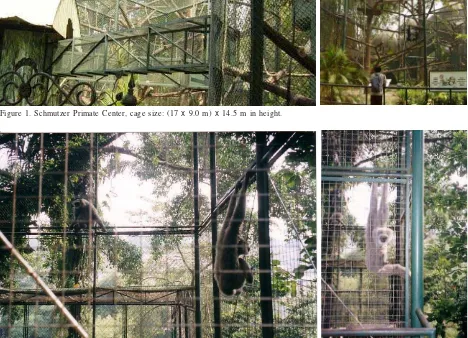
Animals and Housing. Prior to this study, survey was conducted to find the location where the female maintained. Three sexually adult female Javan Gibbons, non pregnant, 7-8 years of age and 6-8 kg of body weight with no history of breeding were used in this study. One of the female is maintained at Schmutzer Primate Center, Jakarta (Ullah, Figure 1) and the other two females (Donna & Citah, Figure 2) are kept at Taman Sari Zoo, Bandung,
all with in pairing-typed cage. The size of the cage at Schmutzer Primate Center is (17 x 9.0 m) x 14.5 m in height, whereas the cage size at Taman Sari Zoo is (4.5 x 4.0 m) x
6.0 m in height. A mixture of chopped fruits and vegetables was given twice a day, and access to water was ad libitum.
Sample Collection and Genital Observation. Fecal sample was collected between 07.00 and 08.00 am, 5-7 days per week over 3-4 months period, and following collection, samples were immediately stored at -20 oC without
preservative until assayed. Daily records of menstruation were monitored and visual inspections of the genital swelling were carried out everyday at the same time. The degree of genital swelling was scored as (0) no swelling, (1) partial swelling, no color change and no discharge, (2) relative increase in swelling, reddish but no discharge, (3) maximum swelling with discharge and red in color (Dahl & Nadler 1992; Czekala & Sicotte 2000).
Sample Preparation. Prior to analyze, the samples were extracted as described by Heistermann et al.(1993) for E1C and PdG measurements. A total amount of fecal
Figure 1. Schmutzer Primate Center, cage size: (17 x 9.0 m) x 14.5 m in height.
samples collected were first lyophilized, and the resulting dried pellets were pulverized and extracted with 3 ml of 80% methanol in water by vortex for 10 minutes followed by centrifugation at 2200 x g for 10 minutes. The supernatant was decanted into a clean glass tube and after appropriate dilution in assay buffer (5.96 g Na2HPO4, 8.50 g NaCl, and 1 g BSA Fr. V in 1 L H2O, pH 7.2) was taken directly to assay.
Assay Validation. Validations of assay were evaluated by monitoring individual extraction efficiencies, parallelism test, intra- and interassay coefficient of variation of quality control at low and high concentration, and the concentration at which 90% of the antibody binds to the conjugate (sensitivity). Individual extraction efficiencies were monitored using the recovery of [3H]-progesterone
(35,000 cpm; NEN Du Pont, Bad Homburg, Germany), which was added to the fecal powder before extraction. Parallelism test was performed to the fecal extracts in serial dilutions using E1C and PdG assays.
E1C and PdG Analysis. Microtitreplate EIAs previously characterized by Heistermann and Hodges (1995) were used to determine immunoreactive E1C and PdG in feces. The samples were diluted in assay buffer with a certain dilution depending on the reproductive status.
Data Analysis. The follicular phase was defined as the interval between the first day of menstruation until the day of the E1Cpeak, whereas the luteal phase comprised the interval from the day after the E1C peak until the day before the menstruation. A threshold value of two standard deviations (SD) above the mean of the preceding follicular phase values was taken in order to determine of the first increase in fecal PdG concentration. An increase in concentrations above this threshold value indicates a statistically significant rise with P < 0.05 (Jeffcoate 1983).
RESULTS
Validation of Assays. Recovery of [3H]-progesterone
gave the mean + SD values ranged from 78.2 + 6.6%. Serial dilutions of fecal extracts gave displacement curves parallel to those of E1C and PdG standards (Figure 3). Intra assay coefficients of variation calculated from replicate determinations of fecal quality control pools gave values of 8.6% for low concentration and 6.9% for high
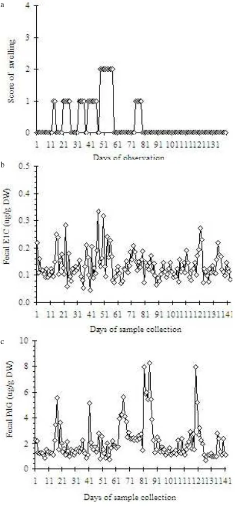
Figure 4. Profiles of genital swelling (a), E1C (b), and PdG (c)in
feces of female Javan Gibbon (Ullah) during the 4-months period of observation.
a
b
c
Figure 3. Parallelism Test of E1C (A) and PdG (B) Assays. Standard, Mimis21/8, Mimis1/9, Mimis14/9, Mimis26/9, Mimis8/10.
E1C Std concentration (pg/50 µl) a
concentration, whereas inter assay coefficients of variations were 14.2% for low concentration and 9.4% for high concentration. Sensitivity of assay was 1.56 pg for E1C and 25 pg for PdG per well.
FecalE1C and PdG Profiles. The profile of fecal E1C and PdG in individual female was depicted in Figure 4 for Ullah, showed the patterns of both hormones that did not reflect any regularity of ovarian function. The genital swelling profile also showed no correlation with the concentration of E1C throughout the period of observation. The level of E1C was fluctuated erratically closed to the basal concentration (0.07-0.13 mg/g), although several high concentration was reached at the level of 0.27-0.33 mg/g, however did not seemed to be periovulatory increase. Compare to the profile of E1C which
seemed to be more vary in fluctuations, the profile of PdG showed patterns with some fluctuation.
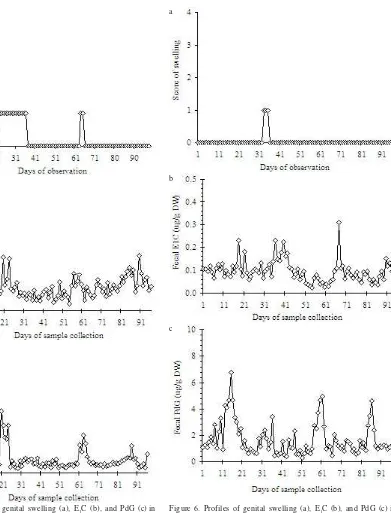
Similar to Ullah’s, the profile of E1C and PdG of Donna (Figure 5) also indicated irregularities in the cycling, with the concentration of E1C ranged from 0.03-0.15 mg/g without any significant increase. During the observation, there was two pattern of PdG indicated luteal phase with extended follicular phase. The degree of swelling and changes in genital swelling showed no fluctuation during the cycles observed which may consistent with the low level of E1C. No menstruation blood flow was seen for this female.
The profile of E1C and PdG of Citah (Figure 6) also indicated that this female was not cycling, and its E1C’s level ranged from 0.03-0.13 mg/g without any significant
Figure 6. Profiles of genital swelling (a), E1C (b), and PdG (c)in feces of female Javan Gibbon Citah during the 3-months period of observation.
a
b
c
Figure 5. Profiles of genital swelling (a), E1C (b), and PdG (c)in
feces of female Javan Gibbon (Donna) during the 3-months period of observation.
a
b
increase. Changes in genital swelling and menstruation blood flow was not seen for this female as well. Without menstruation blood flow that could be seen, the overall conclusion was difficult to draw for this female.
DISCUSSION
This study focused on E1C and PdG because these hormones are known to be primary or major metabolites in other primates (Ziegler et al. 1989; Czekala et al. 1991) and the enzyme immunoassays for this metabolite were immediately available. The present study can also be applied to examine the relationship between the pattern of female genital swelling and underlying hormonal changes during the ovarian cycle. Compare to the concentration of fecal steroid obtained from a cycling female Javan Gibbon that is kept in individual cage (Maheshwari et al. 2007), the data of this study seemed to be lower.
One of the major knowledge that has to be provided in preserving rare and endangered animal species is knowledge on the reproductive biology of the species, which is lack in the Javan Gibbon. Monitoring reproductive status is therefore one of the most important prerequisite for any work designed to enhance captive breeding (Heistermann 1996). Reproductive status could be monitored by using several possible methods such as ultrasonography, laparotomy/laparoscopy and hormone measurements, of which analysis of reproductive endocrine profile is the most effective methods in monitoring reproductive function since the characteristic changes in hormone production represent reproductive status (Hodges 1996). For endangered or wild animals that are difficult to handle and sensitive to distress condition, evaluation of reproductive hormone can also be carried out by using samples from feces or urine. The metabolites homone profile will give correlation with the blood hormone but its time lag has to taken in consideration (Hodges 1996).
Gibbons have been reported to exhibit cyclical swellings of perineal tissues, mostly with concealed form. Although the degree of the genital swelling shows considerable individual variation, Cheyne and Chivers (2006) found that the average pattern indicates a significant level of swelling for 6.3 days on average (range 4-8 days). In two studies on H. lar, genital swellings were found to reach their maxima in association with mid-cycle peaks in estrogens with ovulation, and appeared to be a useful marker for monitoring progress of the menstrual cycle (Barelli et al. 2007). In a study of H. moloch, in contrast, the sexual swellings of four females did not coincide with the fertile periods (Hodgkiss 2007), and Maheshwari et al. (2007) found less uniform of ovarian cycle with average of 21-24 days in H. moloch.
The cycle length was calculated from the highest hormone concentration of a cyclical peak to that of the next. However, the length of the ovarian cycle of the three females observed could not be deducted as the clear peak of the E1C of all females seemed to be difficult to be determined. As for the length of the cycle, Geissmann and
Anzenberger (2009) found that the average of ovarian cycle in Hylobates spp.: 20.0-25.4 days, and longer hormonal cycles in Hylobates moloch reported for an average of 25.4 ± 8.4 days, including some unusually long cycles of up to 38 days (Hodgkiss 2007). Cycle durations in gibbons could be determined using different markers such as endocrinological methods, genital swelling, menstrual bleeding, or copulation observation. Very long cycles might be resulted from determination using copulation marker as gibbons copulate in very short time and easily escape detection, and may even occur during the night (van Schaik et al. 1999; Hodgkiss 2007). Cycles may also remain undetected if menstrual bleedings are used as marker.
In captive breeding management of primates, monitoring ovarian cycles is of considerable importance. These non-cycling females might be resulted from many factors that influence their reproduction system, including external and internal factors. The external factor that had most effect to the disruption of the ovarian cycle was assumed to be the size of cage, followed by feeding condition, mating pair, social interaction, and visitors (van Schaik et al. 1999). These external factors furthermore can affect the Central Nervous System (CNS) and its axis as internal factors in which regulate reproductive steroids secretion and transportation (Leighton 1987; Ferin et al. 1993).
Even though these three females were maintained in pairing system as their natural life, there were some possibilities to bring about the unpleasant conditions of these gibbons. Recall that gibbons are arboreal animals and in its natural habitat, it rarely descends to the forest floor. It spends most of its time in the forest canopy, with locomotion in the form of brachiation, where it swings by its long arms from branch to branch (Leighton 1987), the size of the cage such in these two locations will affect very much for inducing distress condition. The existence of unwanted mating would also be a potential factor to induce stress. At some captive breeding locations other than zoo, the animals mostly encounter with keepers. In zoo, however those animals faced visitors almost everyday with different behavior. Contact with a lot of different people could also generate very stressful conditions.
adaptive significance of this finding is poorly understood, the duration of the follicular phase has been reported to be the main source of variability and diversity of ovarian cycle length (Rosen & Cedars 2004). The hypothalamic-pituitary-ovarian axis is capable of maintaining ovulatory cyclicity on its own, multiple endogenous or environment influences may impinge on the normal activity of the pulse generator usually to decrease GnRH pulse frequency and thereby induce cyclic dysfunction (Richards et al. 2002). The present study also examined the relationship between the pattern of genital swelling and underlying hormonal changes during the ovarian cycle. As the level of LH was not measured to accurately place the ovulation time, it is assumed that the approach of ovulation is signaled by an estradiol peak, followed by the fall of estradiol and the rise of progesterone. By examining the profile of E1C and PdG as independent markers of the female cycle stage and characteristic changes in genital swelling pattern, it was likely to determine the time of presumed ovulation (Ferin et al. 1993). This low concentration of E1C may be one of the causes of non cycling female, as the low level of E1C can not induce LH surge for ovulation (Johnson & Everitt 2000). Since it is known that mating system of this species is monogamous, partners would influence the time of estrus. Unwilling partners would make the female does not come into estrus, so that would affect their menstrual patterns (Leighton 1987), although those females were kept together with male. Neither report on pregnancy nor the activity of copulation for those couples.
In conclusion, the methods and data presented in this study provided the basis for a practical approach to evaluate and monitor of ovarian events in female Javan Gibbon, particularly the use of E1C and PdG profile for any effort to predict the time of ovulation. We found that the external and internal conditions of the animals should be taken in consideration in elucidating factors influenced reproductive hormone production.
ACKNOWLEDGEMENT
The study was financially supported by Hibah Bersaing XII research grant. We are particularly indebted to K. Hodges and M. Heistermann from the German Primate Center, Gottingen for providing standards, antiserum, conjugates and reagents for EIA. We are grateful to the Director of Schmutzer Primate Center, Jakarta and the Director of Taman Sari Zoo, Bandung with all staffs and keepers for generously giving permission to use the animals. We also thank the Head of the Reproduction Laboratorium, LIPI Cibinong for providing laboratory facilities during our study.
REFERENCES
Barelli C, Heistermann M, Boesch C, Reichard UH. 2007. Sexual swellings in wild white-handed gibbon females (Hylobates lar) indicate the probability of ovulation. Horm Behav 51:221-230.
Cheyne SM, Chivers DJ. 2006. Sexual swellings of female gibbons.
Folia Primatol 77:345-352.
Chivers DJ. 1989. The social behavior of the lesser apes. In: Seth PK, Seth S (eds). Perspective In Primate Biology. Vol. 3. New Delhi: Today & Tomorrow’s Printers and Publishers. p 141-155.
Collins DC, Dahl JF, Nadler RD. 1994. Metabolism of progesterone by the adult female gibbon (Hylobates (H.) lar).Am J Primatol
33:193-255.
Czekala NM, Reichard T, Lasley BL. 1991. Assessment of luteal competency by urinary hormone evaluation the captive female gorilla. Am J Primatol 24:283-288.
Czekala NM, Sicotte P. 2000. Reproductive monitoring of free-ranging female mountain gorillas by urinary hormone analysis.
Am J Primatol 51:209-215.
Dahl JF, Nadler RD. 1992. Genital swelling in females of the monogamous gibbon, Hylobates (H) lar. Am J Phys Anthropol
89:101-108.
Ferin M, Jewelewicz R, Warren M. 1993. The menstrual cycle: Physiology, reproductive disorders, and infertility. New York: Oxford Univ Pr. p 250-261.
Gates R. 1998. In situ and ex situ status of the silvery or moloch gibbon, Hylobates moloch. Int Zoo Yearb 36:81-84. Geissmann T, Anzenberger G. 2009. .Hormonal Correlates of The
Ovarian Cycle In The Yellowcheeked Crested Gibbon (Nomascus gabriellae), and a Review of Ovarian Cycles In Gibbons (Hylobatidae). Gibbon J 5:61-73.
Heistermann M, Hodges JK. 1995. Endocrine monitoring of the ovarian cycle and pregnancy in the saddle-black tamarin (Saguinus fuscicollis) by measurement of steroid conjugates in urine. Am J Primatol 35:117-127.
Heistermann M, Möhle U, Vervaecke H, van Elsacker L, Hodges JK. 1996. Application of Urinary and Fecal Steroid Measurements for Monitoring Ovarian Function and Pregnancy in the Bonobo (Pan paniscus). And Evaluation of Perineal Swelling Patterns in Relation to Endocrine Events.
Biol Reprod 55:844-853.
Heistermann M, Tari S, Hodges JK. 1993. Measurement of faecal steroid for monitoring ovarian function in New World primates, Callitrichidae. J Reprod Fertil 99:243-251. Hilton-Taylor C (compiler). 2000 IUCN Red list of threatened
species. IUCN-The World Conservation Union. United Kingdom, Cambridge.
Hodges JK. 1996. Determining and manipulating female reproductive parameters. In: Kleiman DG, Allen ME, Thompson KV, Lumpkin S (eds). Wild Mammals in Captivity.
Chicago: The University of Chicago Pr. p 418-428. Hodgkiss S. 2007. Characterising the physical and hormonal
correlates of the ovarian cycle in female Javan gibbons (Hylobates moloch) [Thesis]. Oxford Brookes University, UK.
Jeffcoate SL. 1983. Use of rapid hormone assays in the prediction of ovulation. In: Ovulation, Methods for Its Prediction and Detection. Chichester: J Wiley. p 67-82.
Johnson MH, Everitt BJ. 2000. Essential Reproduction. 5th.ed.
London: Blackwell Science.
Leighton DR. 1987. Gibbons: Territoriality and Monogamy. In: Smuts BB, Cheney DL, Seyfarth RM, Wrangham RW, Struhsaker TT (eds). Primate Societies. Chicago: University of Chicago Pr. p 135-144.
Maheshwari H, Sjahfirdi L, Astuti P, Purwantara B, Alikodra HS, Sajuthi D, Widjajakusuma R, Toelihere MR. 2007. Fecal Steroid Profile And Genital Swelling of Female Javan Gibbons (Hylobates Moloch Audebert 1797) Maintained In Individual Cage. Media Konservasi XII:16-21.
Nadler RD, Dahl JF, Collins DC. 1993. Serum and urinary concentration of sex hormones and genital swelling during the menstrual cycle of the gibbon. J Endocrinol 136:447-455.
Nijman, van Ballen. 1998. A faunal survey of the Dieng Mountains, Central Java, Indonesia: Distribution and conservation of endemic primate taxa. Oryx 32:145.
Rinaldi D. 2003. The Study of Javan Gibbon (Hylobates moloch
AUDEBERT) in Gunung Halimun National Park (Distribution, Population and Behavior). Research On Endangered Species In GHNP, Research And Conservation Of Biodiversity In Indonesia XI:30-48.
Rosen M, Cedars MI. 2004. Female Reproductive Endocrinology and Infertility. In: Greenspan FS, Gardner DG (eds). Basic and Clinical Endocrinology. 7th ed. New York: Lange Medical
Books. p 511-564.
Taylor PD, Hillier SG, Fraser HM. 2004. Effects of GnRH antagonist treatment on follicular development and angiogenesis in the primate ovary. J Endocrinol 183:1-17. van Schaik CP, van Noordwijk MA, Nunn CL. 1999. Comparative
Primate Socioecology. In: Lee PC (ed). Sex and Social Evolution in Primates, Cambridge: Univ Pr. p 204-240. Ziegler TE, Sholl SA, Scheffler G, Haggerty MA, Lasley BL. 1989.