Vol 8, No 2, April
-
June 1999 Mastectomy specimensof
breast cancer 117Histopathologic
profile
of mastectomy
specimens
of
operable breast cancer
cases
Endang
Sri Roostinin, Goi
Sakamotor, Susumu
Watanabe., Muchlis Ramli.,
Joedo prihartono$,
Santoso
Comain*, Yoshiyuki
ohno.,
Esti
Soetrisno*, Gunawan
Tjahjadi-, Idral Darwis.,
Didid
Tjindarbu
mi',
Setyawati
Bud inin gsih$,Nakako
Kubo#.Abstrak
Informasi yang adekuat mengenai
profil
histopatologik dari sediaan mastektomi memberikan ramalan yang lebih baik tentangptognosis tiap penderita kanker payudara. Sejauh ini, beLwn pemah dilaporkan data
dari
kasus-kasus di Indonesia, yang antara lain disebabkan karena pada umumnya kasus-kast'ts yang clatang sudah pada staditm lanjut yang inoperabel. Paela makalahini
akan di-Iaporkan profil histopatologik dari sediaan mastektomi kasus kanker payutlara yang operabel yang tercakup clal^am penelitian bersamaIndonesia'Jepang, tentang Etiologi dan Klinikopatologi pada Kanker Payudara. Dari tiap kasus, diperiksa masing-masing 4-6 sediaan
HE untuk dinilai jenis histopatologinya, sebukan limfosit, invasi pembuluh daralt clan limfe, invasi jaringan lemak, otot clan kulit serta petryebaran ke kelenjar getah bening. Juga dikurnpulkan data-data ketahanan hielup 5 tahun. Sebanyak 70 dari 107 kasus yang operabel tlapat dinilai secara lengkap sediaannya dan pada 20 di antaranya dianalisa untuk ketahanan hiàup 5 tahun. Mayoritas kasus adalah
jenis duktal invasif(79,9Vo),2,8Eo duktal in situ dan sisanya adalah tipe khusus. Jenis skirus
^"rupàkoryong
terbanyak di antarajenisduktal invasif (43%o) Litnapuluh kasus menunjukkan sebukan timfosit, namun separuhnya adaLah derajat ringan. Invasi vaskuler hanya ditemukan pada 4 kasus, invasi pempuluh timfe pada 16 kasus. Invasi lemak pada 36 kasus, otot pada 3 kasus dan kulit pacla I kasus.
Di antara 48 kasus dengan sediaan kelenjar getah bening, 28 kasus menunjukkan metastasis. Usia penderita dan sebukan limfosit meru-pakan gambaratxyang cukup menonjol dalam kaitannya denganketahanan hidup 5 talun. Tbrlihat kecenclerungan p"nurrnàn usia clari kelompok hidup tanPa penyakit, keLompok hidup dengan penyakit dan yang meninggal. Pacla kelompok yang hiclup tanpa penyakit se-bukan limfosit derajat sedaig sampai keras terdapat pada sebagian besar kasus, sedangkan pada kelompok hittup clengan penyakit, sebukan limfosit umumnya berderajat ringan. Pada satu kasus yang meninggal tidak ditemukan sebukan timfosit. Disarankan untuk melakukan studi serupa Lebih lanjut dengan menggunakan teknik-teknik mutakhir agar cliclapatkan informasi yang lebih akurat mengenai p rofil his to pato Io g ik y an g adekuat.
Abstract
Adequate descriptiott of histopathologic profile of mastectomy specimens offers a better prognostic prediction of inclividual breast cancer patient Strch a data hasn't been reported so far from Indonesian breast cancer cases, due to the fact that the majority of admitted cases were already in unoperable late stage.
In
this report, we present the extensive histopathologic profile of mastectomy specimen from concer cases included in Indonesia - Japan Joint Study on Etiology and Clinicopathology ofBreast Cancer Four to 6 H & E slides were res'ieweclfrom each cases; aLI data about histopathological type, lymphocytic infiLtration, bloocl and lymph vessels invasion, fat , muscle and skin invasion anel lymph node involvemeni were recorded. Five years survival data were collecte4. O'nty 70 out of 107 operable cases were availablefor
a complete histopathalogical review and 20 cases were evaluatedfor
their 5 years survival data. Most of the patients were of cluctal invasive type (79.9vo); 2.8Vo ductaL in situ and the rest were of the speciaL types. Scirrhous subtype preclominatecl the ductal invasive type (43.3vo). Fifty cases showed lymphocytic infiltration. vascular invasion were founcl only in4
cases, lymph vessel invasion in l6 cases. More than half cases showedfat invasion (36 cases), muscle invasion in3
cases ancl skin invasion in I case. Among 48 cases with dissectecl lymph node, 28 showecl metastasis. ABe of patients and lymphocytic infihration were signiJicant encounteretl in the evaluation of survival tlata. There was a tendencyfor a decreasing age among group alive without
disease, alivewith
clisease anel deceased' Almostall of
cases who were aLive without disease showecl lymphocytic infiltration which were mostly moclerate to severe; while 6 cases who were alive with disease mostly showecl mild inrthration. One case who was deceased has no lymphocytic infiltration.It
is recommendeel to 'study further with the help of ancillary techniques to get more accurate information about the histopathologic profile.Keywords : Histopathologic, breast cancet mastectomy specimen.
*.
Department of Pathology, Faculty of Meelicine, IJniversity of Inclonesia, Jakarta 10430, Indonesia
t
Department of Pathology, Cancer Institute Hospital, Tokyo 170, Japan'
Department of Surgery, Faculty of Medicine, University of Intlonesia, Jakarta 10430, Inclonesia # Department of Community Medicine, Faculty of Meclicine, IJniversity of Indonesia, JakartaRoostini et
al
INTRODUCTION
Breast cancer
isthe
secondmost frequent
malignancy
among
women in Indonesia.l Previous report on our
study
revealed
that
most
breast cancer
patients,
namely
87Vo, came
in
their late
stage
(2). So, only
few of them were
subjected
to
surgical treatment
andno information about extensive histopathologic
pro-file
was
reported
sofar in our
country.
There are
many factors known to influence
the
prog-nosis
or clinical
outcome
of
breastcancer patient.
An
extensive
histopathologic
profile
of
mastectomy
specimen
provides more
accurate
information
about
the prognosis
of the individual
breast cancer
casethan
the small biopsy
specimens.In
this
study, various histopathological
finding
onmastectomy specimen
will
bereported
andcorrelated
to
the
clinical
outcome whenever possible.
MATERIALS
AND METHODS
The
samples
of this
study were
part of
the
total
sam-ples of 300
cases
including
in the
Indonesia-Japan
Joint
Research
Project
on
Breast Cancer:
anepidemiological study.
Only 107
out
of 300
caseswere
operable.
The
mastectomy specimens, consisting 4
to
6routine
H&E slides section taken
from the tumor and
sur-rounding tissue, were reviewed histopathologically
for their histopathologically types, lymphocitic
infil-tration,
blood and lymph
vessels
invasion,
fat
andmuscle tissue invasion. Lymph nodes section
were
also
reviewed
andcounted
for
the
presenceof
metas-tatic tumor
cell. Clinical
data about their
survival
were recorded.
Histological typing
was done
according to the
Japa-neseclassification
for
breast cancer
(3); lymphocitic
infiltration was
graded according
the severity
asmild, moderate
and severe.
Tumor emboli
in
the
blood
andlymph
vesselswere
search andalso
thede-struction
of the
vessel's
wall by
the
tumor
cells.
Fat infiltration
was recorded
either within
interlobu-lar fat or
submammary
fat
tissue. Same observation
was done
for
muscle
tissue.RESULTS
Seventy
caseswere available for
acomplete
review.
The histological type is
presented
in
Thble
1.
Lym-phocyte
infiltration
were present
in 50
cases(7I.4Vo)-Med
J Indones
Most of them were of mild infiltration
(507o).
Most
of the
scirrhous
types were not or only mildly
infil-trated
by lymphocyte
compare
to papilo-tubular
andsolid-tubular which
have
more
denselymphocyte
in-filtration.
(Tabte
3).Table
1.
Histopathological type of mastectomy specimensHistological type Total cases
Ductal carcinoma in situ Papilo-tubular carcinoma Solid-tubular carcinoma Scirrhous carcinoma Mucinous carcinoma Medullary carcinoma Lobular carcinoma Adenoid cystic carcinoma
Paget's disease
2
(2.8 7o)9
(12.8 Vo)16
(22.8 %)31
(44.3 E')3
(4.3 Vo)4
(5.7 Vo)3
(4.3 %)1
(t.4
7o)t
(1.4 Vo)Total
'10
(roo.0 %)Table
2.
The histopathologic profileof
the mastectomy speci-mensHistopathologic profile
Positive
Negative
TotalLymphocytic infiltration
Vascular invasion Lymph vessel invasion
Fat invasion Muscle invasion Skin invasion
Lymph node involvement
50 (25mild,
19 mod, 6 sev)
4 16 36 J 1 28 20 66 54 JJ 67 69 20 70 70 70 70 70 70 48*
*
No lymph node was foundin
22 cases.Thble
3.
Correlation between histological type and lymphocytic infiltrationHistological type
No
Mild
Moderate Severe Total filtrationDuctal in situ Papilo-tubular Solid-tubular Scirrhous Mucinous Medullary Lobular Adenoid cystic
Paget's disease
I
2 2 13 I 0 0I
0I
2 4 12 2 2 2 0 0 0 4 8 5 0I
I 0 0 0I
2 1 0I
0 0I
2 9 16 31 J 4 3I
IVol 8, No 2,
April
-
June 1999Table
4.
Correlation between Histological type and various structure invasionMastectomy specimens
of
breastcancer
119Histological Type Vasc Lymph. Fat Muscle Skin Lymph node
Ductal in situ
Papilo-tubular Solid-tubular Scirrhous Mucinous Medullary Lobular Ad.cystic Paget's 0 U9 (ltEa) 2116 (12Vo)
Il31
(3Vo)0 0 0 0 0 0 419 (M%o) 3t16 (1880) 9131 (29Eo)
0 0 0 0 0 0
5/9 (55Vo) 10/16 (62Vo) 18t31 (58%) 1/3 (33Eo)
0 213 (667o)
0 0
0 119 (ltVo)
0
2131 (6Eo) 0 0 0 0 0 0 t/9 (11Eo)
0 0 0 0 0 0 0 0 319 (3OVo) 8/16 (507o)
t4/3t
(457o)0
2t4 (50%)
t/3 (33%) 0 0 28 36 t6 Total
Ïâble
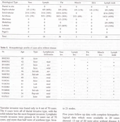
5.
Histopathologic plofile of cases alive without diseaseNr Age Type Lymphocyte
infiltration Vasc. Inv. Lymph Inv. Fat Inv. Muscle Inv. Lymph node 8902303 8900292 89 r 0090
8910224 8911250 9000785 9006042 9109241 91{J.4354 9102082 9i M800
9105836 91 0701 I
Scirr
Scir
Scirr Scirr Sol-tub Sol-tub Scirr Scirr Lob Lob Sol-tub Pap{ub Scirr mod SEV mild sev mild mod mod mod mild mod mod mild 50 42 47 62 46 50 40 44 6t 41 6r 34 5t + + +Vascular invasion
wasfound only in 4 out of 70
cases.The 4
caseswere all
of
ductal invasive
type, with the
solid-tubular
has the most frequent invasion.Lymphatic
vessels
invasion were present
in
16 cases
out
of
70cases, and
more than
half were of scirrhous
type.
Inva-sionsof fat tissue were present
in 36 out of
7O casesin
which
most of them were found in the scirrhous type.
Muscle invasion
andskin invasion were found only in
3 and
I
casesrespectively. Two
caseswho has muscle
invasion were of scirrhous
type and
I
of papilo-tubular
type; while the only
1 case with skin invasion was ofpapilo-tubular type (Table 4).
Nineteen
cases
showed involvement
of
1-3 lymph
nodesremoved
and 9 cases showed involvementof
5to
2l
nodes.Five years
follow
up data
with
complete
histopatho-logical data
which
were
available
in
20
casesshowed:
13 out
of
20 were alive without
disease, 6
cases werealive
with
diseaseand
1patient died.
Among
13 cases
that still
alive without
disease,
7 cases wereof
scirrhous
type, 3
of
solid-tubular
type,
2of lobular type and
1of papilo{ubular
type. Almost
all
cases
showed
lymphocytic
infiltration,
4
cases [image:3.595.74.570.100.604.2]Med
J
lndonesTable 6.
Roostini et aL
Histopathologic profile of cases alive with disease
Nr Age Type Lymphocyte
infiltration
Lymph node
900807 r
910933r
9 1 0s609
9107232 9lo'7430
91il03 1
3t 42 30 53 39 33
Scirr Scirr Scirr' Scirr Pap-tub Sol-tub
mild
mod mild mild
Six
casesalive
with
disease had
scirrhous type
in 4
cases,
I
has papilo-tubular
and
I
has solid-tubular
type.
Their
age wasrelatively
younger
thanthe group
who
lived
without
disease
(mean
age
39 vs.
47.3years).
Almost
all
of them
showed
lymphocyte
infil-tration, 2l'midfat invasion,
I
hadmuscle invasion
and3
hadlymph
node
involvement. The
only I
casewho
died
was
of
33 years
old with
scirrhous type,
and hadlymph
vessel and
fat
tissue invasion.
DISCUSSION
The most frequent histological type found is
ductal
type, comprising more than
half
of
the total
cases.Most of the tumors
had
lymphocyte infiltration.
The
prognosis significance
of
lymphocyte was a
subject
of
controversy. Some investigators
found it
related to
the favorable
prognosis,+
while other
group
did
nots.Looking
at
our
series,the
scirrhous
type
wasthe
lessinfiltrated by lymphocyte,
andthis type
hasthe worse
prognosis compare
to the ductal invasive
subtypeslike
papilo-tubular and solid-tubular. The
discordant
results
might
needto be clarified by further
identih-cation
of
the lymphocyte sub-population'
If the
lym-phocyte population is
B
cell, they
have
no
meaning
in
the
tumor surveillance, while if
thepopulation
is Tlymphocyte
it
has
theproperty
of tumor control.
Tbe
presence
of
B lymphocyte could be result
of
secon-dary
infection. In
breast cancer, the presenceofinfec-tion is
possible
if
ulceration occur; which
are
com-monly complicating
the advance inoperable
cases'In
operable
cases,which is
of
earlier
stageit
is
uncom-mon
to
find
this complication.
So, the
T
lymphocyte
is most
likely
to
be
the main population in this
con-dition,
and consequently
it
can be expected
asan
in-dicator
of
better
outcome.
We found
lymphatic invasion more common
thanvascular
invasion.
These
results
were similar
to
Fisher's finding.0 Structurally, vascular
vessels
aremorc resistant
to
invasion
dueto
itsthicker
layer
thanthe
lymphatic
vessels,which
arecomposed
only by
asingle
layer
of endothelial
cell.
Lymphatic emboli
have an unfàvorable prognosis
in
node
negative
pa-tients
treated
by mastectomyT
and
give
rise
to
ahigher local
recurrence rate
in
node negative patients
who
underwent breast conserving treatment.8
Blood
vesselinvasion
is also
still
asubject
of
contro-versy,
but
it was
recorded
that visceral
metastasisoc-curred
in
617o
of
caseswith
blood
vessel invasion,
compared
to only
35Vovisceral
metastasis
in
the
ab-sence
of
blood
vessel invasion
in
stage
I
cases.9Death, due to
metastasis
breast cancer was
signifi-cantly
greater
in
frequency
in
women
with blood
ves-sel
invasion
irrespective
of their total lymph
node
in-volvement in
stageII
cases.loWeigand
et alll
in
their study
revealed a
strong
cor-relation
between
blood
vessel
invasion
and
early
re-currence. The
presence
of blood vessel invasion
in
their
series
was
much higher
than
our
study
andFisher's large
series.6Several investigators claimed
the high interobserver variation
in
assessing
vesselsinvasion, either lymph
or
blood. The most
possible
source
of
this variation is the
difficulty
in
differenti-ating
between vessels and tissue
artifact
affecting
carcinomatus duct. The
useof
special
staining
isthen
highly recommended
to
overcome
this
problem,
either
histochemically
using elastic staining
or
immu-nohistochemically using endothelial
n61i$sdis5.l2-13Fat
tissue invasion
has never beenregarded
as aprog-nostic factor.
Our
series revealed
more tlran half
cases
with fat
invasion,
andthe
significance of which
couldn't be
stated
yet. Recently, several
studies
hasdone
to
correlate
the
presence
of
proteolytic
err-zymes,
like
collagenase
and cathepsins bear
by
the
tumor cells,
with their
ability to
degrade
the
base-ment
membrane and
extracellular matrix
astheir
Vol. 8, No 2,
April
- June 1999produce
a lipolytic
enzyme
besides
proteolytic
en-zymes; and
if
it
is
proced
to
be the
case,
it
might
serve
asan
additional
bad
prognostic
factor.
Cases
who
are
alive
without
disease areof
older
agecompare
to
cases
that
are
alive
with
disease. The
most
prevalence
type
in all
groups (alive
with/with-out
disease
and
died)
were ductal type, mostly
of
scirrhous
subtype.
Among the group alive
with
dis-ease,
lymphocytic infiltration
was
mostly mild, wlrile
in
the group without
disease,
the infiltration
were
mostly
moderate
to
severe.The
occurrence
of lymph
node
metastasis was
found comparably
in
the
group
with
diseaseand the
groLrpwithout
disease.A higher
yield
of
lymph
node
positivity
can be obtained
actu-ally
by performing special staining using
antibodies
against
epithelial
cells.
This
procedure
had been doneby
some investigators.lT-1e
They
had
proved
around30ûlo
positive
finding
in
negatively stated lymph
nodes
by routine
H&E
staining. This staining is
ableto find
the
micromestastases,
which is usually
over-looked by routine staining. By performing this
stain-ing,
it
will
probably
changethe
clinical
or
pathologi-cal
staging
in
certain
cases
and consequently
themanagement and prognosis
prediction.
It
seems
that
breast cancer affecting younger
agegroup
is
more
aggressive.
The
mean age
of
caseswere
decreasing
fiom
group
of
cases
alive without
disease,
alive
with
disease and
deceased case.If
we assumethat
menopausal
statusis
related
to
age, thenthis
finding
was
differed
fiom
American
breast
can-cer
cases,
in
which, post
menopausal
group whiclr
were genel'ally
of
older
age, showed
poorer survival
compare
to
the
premenopausal
group.2OHowever,
several studies
showed
that higher hormon
receptor
positivity,
another
indicator
of
better
clinical
out-come, were
found
in
post
menopausal
woment2-2tand these are
in
keeping
with
oLrrfindings. This
find-ing may
indicate the
difference of
cancer
biology
be-tween Asian and non-Asian, which might be
due to
genetic
and geographic properties.
Lymphocyte
infiltration
seemsalso
play
a
role
for
abetter
outcome among invasive ductal
carcinoma.
The
presence and
severity
of
lymphocyte infiltration
was decrcasing
from
the
groLrpalive without
disease,alive
with
disease
and the died
case.
Of
interest to
note
is
the
finding
that
noneofthe
casesin
the group
alive
with
disease showed
lymph
and
blood
u"rs"lt
invasion, unlike the
other
two
groups,
especially
thegroup alive without
disease.The explanation
of
this
phenomenon
might
bethe multisteps
processof
me-Mastectonly specimens
of
breastcancer l2l
tastasis.
After
being
ableto invade the
vessels,the
tu-mor cells
must
beable
to survive
the defense
mecha-nism
encountered
in
the circulation, and also
is
thenew environment
of
tissue
or
organ where they
areentrapped. Then, the
cell
must
be
able also
to
prolif-erate and
grow in distant
organ.22It will
bevery interesting
to design
afurtlrer
study
ona
larger
seriesof
caseswith the
help
of
anadditional
ancillary
techniques
in
order
to
get a more
accurateinformation about
the
nature
of
lymphocyte
infil-trates,
blood
andlymph
vesselsinvasion
and the pres-enceof
anoccult or
micrometastasis.
REFERENCES
l.
Cornain S, Mangunkusumo R, NasarlM,
prihartono J. Ten most f}equent cancers in Indonesia: pathology based cancer rcgistry dataof
I 988-1992. In: Cancer registry in Indonesia.National Cancer Registry Center, Jakarta Coordinating Board. 1997.
2.
'llindarbumi D, Ramli M, Watanabe S, DarwisI,
Sakamoto C, Cornain S,'Ilahjadi G, Soerrisno E, Ohnot
Roostini ES, Prihartono J, Suzuki S, Budiningsih S, WakaiK.
Clinico-pathological aspects of brcast cancer: Ajoint
study between Indonesia and Japan. Med J Indones 1995;4: 14g-53.3.
Japanese Breast Cancer Society. The gencral rules forclinicaland pathological recording of breast cancer. Jpn J Surg l9g9; 19:612-32.
4.
Stenkust B, Bengtsson E,, Dahlquist B, Eklund C, Erikson O, JakransI
NordinB.
Predicting breast cancer recurrence. Cancer I 982; 5O: 2884-93.5.
Dawson PJ, Ferguson DJ, Karrison T. Thc pathological lincl_ing of breast cancer in patient surviving 25 years aticr raclical mastectomy. Cancer 1982; -50:
2l3l-8.
6.
Fisher ER, Cregolio RM, Fisher B. The pathologyof
inva-sive breast cancer: A syllabus derived tiom finding of the Na_ tional Surgical Adjuvant Breast Cancer project, j975;36:2_85.
'7.
Nime F, Rosen PP, Thaler H, Ashikari R, Urban JA. prognos_tic significance of tumor emboli in intramammary lymphatics
in
patientswith
mammary carcinoma,Am
J
Surg pathol 1977;l:25-30.
In : Rosen pp. lnvasive duct carcinoma andmorphological prognostic markers. Breast pathology. Chapt. 1-5 LippincotRaven. Philadelphia-New
york
I 996.8.
Clemente CG. Boracchi p, Andreola S,Del
Veccchio M, Veronssi P, Rilke FO. Peritumoral lymphatic invasion in pa_ tients with node negative mammary duct carcinoma. Cancer 1992; 69 1396-403.9.
Rosen PP, Saigo PE, Braun DW Jr, Weathers E, de palo A. Predicl.orsof
recurrencein
stageI
(TlN0M0)
breasr carci-noma. Ann Surgl98l;
193: I 5-25.ln:
Rosenpp
lnvasiveduct
carcinomaand
morphological prognostic markers.122
Roostini et al10. Rosen PP, Saigo PE, Braun DW Jr, Weathers E, Kinne DW.
Prognosis
in
stage Il(TINIMO)
breast cancer. Ann Surgl98l;
,l94: 576-84.ln:
Rosen PP. lnvasive duct carcinomaand morphological prognostic markers. Breast pathology.
Chapt.t5 Lippincot-Raven. Philadelphia-New York 1996' I
l. Weigand
AR, lsenberg WM, Russo J, Brennan J, Rich MAand the Breast Cancer Prognostic Study Associates' Blood
vessels invasion arid axillary lymph node involvement as a
prognostic indicators tbr human breast cancer. Cancer, 1982;
50:962-9.
12. Oosterhuis JW Breast Cancer: Pathological Aspects. In; Cor-nain S, editor. Advanced Postgraduate Course on Oncology,
Jakarta, November I 993.
| 3. Leong
AS{
Raymond WA. Prognostic Parameters in Breast Cancer. Pathology, 1989;2l:
169-7 5.14. Murakanri Y. Decision tbr prophilactic neck dissection based
on immunohistological evaluation
of
biopsy specimen.In
:Smee R, Bridger PG eds. Procëedings of the 2nd World
con-gress on laryngeal cancer. El sevier, Amsterdam, Lausanne,
New York, Oxtbrd, Tokyo 1994;582-8.
15. Leonoe D, Nesland JM, Holm R, Sobrinko-Simoes
M.
Ex-pression of laminin, collagen
IV
Fibronectin and type IVcol
lagenase in gastric carcinoma. Cancer 1994;'13:518-2'7.
16. Aznavoorian S, Murphy AN, Settler-Srevenson WC, Liotta
Metl
J
IntlonesLA. Molecular aspects of tumor cell invasion and metastasis.
Cancer 1993; 7 l(4): 368-83.
17.
Mc
GuckinMA,
CummingsMC,
WalshMD,
Hohn BG,Bennet IC and Wright RG. Occult axillary node metastasis in breast cancer
:
their detection and prognostic significance. British J Cancer 1996; 73: 88-95.18. Sedmak DD, Meineke TA, Knechtges DS, Anderson J.
Prog-nostic significance of cytokeratin-positive bteast cancer me-tastasis. Modern Pathology 1989',2: 516-20.
19. Raymond WA, Leong ASY. Immunoperoxidase stainin-q in
the detection of lymph node metastasis in stage
I
breast can-cer. Pathology 1989;21: 1l-5.20. Sakamoto G, Sugano H, Hartman WH. Comparative
Patho-logical Study
of
Breast Carcinoma among American andJapanese Women. ln: Mccuire WL, ed. Breast Cancer.
Nash-ville USA: Plenum Pubtishing Corporation, 1981;2ll-31.
21. LesserMl, Ropsen PP, Senie RT, Duthie K, Menendez-Botet
C,
SchwartzMK.
Estrogen and progesteron receptors in breast carcinoma. Correlations with epidemiology andpa-thology. Cancer 1981; 48:299-309.
22.
Tarn D,
Metastasis; Secondary proliferationin
distant