Abstract.
To evaluate the possible role of cysteine proteases
and serine proteases, as well as their respective inhibitors and
receptors, as new prognostic factors in NSCLC, we examined,
for the first time, 10 biological parameters related to three
proteolytic systems within a homogeneous collective of 147
cases of NSCLC. Activities (cath B
AT, cath B
A7.5) and protein
levels of cath B
C, cath L
C, uPA, PAI-1, uPAR [measured by
three different assays uPAR (ADI), uPAR (HD13), uPAR
(IIIF10)] and TF were measured in homogenates of lung
tumour tissue and corresponding non-malignant lung
parenchyma. Total cath B activity (cath B
AT) and enzymatic
activity of the fraction of cath B, which is stable and active at
pH 7.5 (cath B
A7.5), were determined by a fluorogenic assay
using synthetic substrate Z-Arg-Arg-AMC. The concentrations
of cath B
C, cath L
C, uPA, PAI-1, uPAR and TF were
determined by ELISAs. uPAR was determined using three
different ELISA formats. The median levels of cath B
AT(5.1-fold), cath B
A7.5(2.5-fold), cath B
C, (8.5-fold), cath L
C(6.6-fold), uPA (6.5-(6.6-fold), PAI-1 (4.2-(6.6-fold), uPAR (ADI)
(2.2-fold), uPAR (HD13) (4.0-fold) and uPAR (IIIF10)(2.6-fold)
were higher in tumour tissue compared to the lung
parenchyma. Cath B
AT, cath B
A7.5and cath B
Cin primary
tumours correlated with lymph node metastases. Regarding
histologies, the concentration of PAI-1 seems to be associated
with the histological cell types of NSCLC. We found the
Abbreviations: NSCLC, non-small cell lung cancer; AC,adenocarcinoma; SCC, squamous cell carcinoma; cath B, cathepsin B; upa, plasminogen activator; PAI-1, plasminogenactivator-inhibitor; uPAR, plasminogenactivator-receptor; TF, tissue factor, TF; VEGF, vascular endothelial growth factor.
Correspondence to: Prof. Dr. Werner Ebert, Abteilung für Klinische Chemie und Bakteriologie, Thoraxklinik Heidelberg gGmbH, Amalienstr. 5, D-69117 Heidelberg, Germany. Tel: 49 6221 396226, Fax: 06221 396415, e-mail: [email protected]
Key Words: Cathepsin B (cath B), plasminogenactivator-inhibitor (PAI-1), plasminogenactivator-receptor (uPAR), prognosis, non-small cell lung cancer (NSCLC).
Cathepsin B, Plasminogenactivator-inhibitor (PAI-1) and
Plasminogenactivator-receptor (uPAR) are Prognostic
Factors for Patients with Non-small Cell Lung Cancer
B. WERLE
1,8, M. KOTZSCH
2, T.T. LAH
3, J. KOS
4, D. GABRIJELCIC-GEIGER
5,
E. SPIESS
6, J. SCHIRREN
7, W. EBERT
1, W. FIEHN
8, T. LUTHER
2,
V. MAGDOLEN
9, M. SCHMITT
9and N. HARBECK
91
Abteilung für Klinische Chemie und Bakteriologie,
Thoraxklinik Heidelberg gGmbH, Amalienstr. 5, D-69117 Heidelberg;
2
Institut für Pathologie, Technische Universität Dresden, Universitätsklinikum Carl Gustav Carus,
Fetscherstr. 74, D-01307 Dresden, Germany;
3
Department of Genetic Toxicology and Cancer Biology, National Institute of Biology,
Vecna pot 111, SLO-1000 Ljubljana;
4
Department of Biochemistry and Molecular Biology, Jozef Stefan-Institute, Krka d.d., R&D Division,
Department of Biochemical Research and Drug Design, Novo Mesto, SLO-8000, Ljubljana, Slovenia;
5Abteilung für Klinische Chemie und Klinische Biochemie,
Chirurgische Klinik und Poliklinik, Nußbaumstr. 20, D-80336 München;
6
AG Modellversuche zur Invasion und Metastasierung, Deutsches Krebsforschungszentrum,
INF 280, D-69120, Heidelberg;
7
Abteilung für Thoraxchirurgie, HSK Dr. Horst-Schmidt-Kliniken GmbH,
Ludwig-Erhard-Straße 100, D-65199 Wiesbaden;
8
Zentrallabor des Klinikums, Medizinische Klinik und Poliklinik, Universität Heidelberg,
Bergheimerstr. 58, D-69115 Heidelberg;
9
Klinische Forschergruppe, Frauenklinik, Klinikum rechts der Isar,
highest values of PAI-1 in large cell carcinoma > SCC, AC >
carcinoid and lowest values in metastases of primary tumours
of other organs. Only PAI-1 was significantly increased in
poorly-differentiated cells (G3) compared to well- and
moderately- differentiated cells (G1/G2). PAI-1 significantly
correlated with cath B
ATand cath B
A7.5with uPAR (ADI),
uPAR (HD13), uPAR (IIIF10) with uPA, and only weakly
with TF, but not with cath B
Cand cath L
C. Significant
correlations with overall survival in the total population of
NSCLC patients were observed in univariate analysis for cath
B
AT, cath B
C, PAI-1, uPAR (ADI), uPAR (HD13), and
uPAR (IIIF10). Cath L
Cwas not significantly associated with
poor prognosis. Regarding the histological tumour type, only in
patients with squamous cell carcinomas did cath B
A7.5and
PAI-1 remain significant prognostic factors. In multivariate
survival analysis only two proteolytic factors, PAI-1 and uPAR
(III10F), stayed significant. In conclusion, among 10
biological parameters evaluated within the same cohort of
patients, only PAI-1, uPAR (ADI), uPAR (HD13), uPAR
(IIIF10), cath B
ATand cath B
Care prognostic factors for
overall survival of NSCLC patients. Moreover, PAI-1 and
uPAR (IIIF10) add independent prognostic information with
regard to established clinical and histomorphological factors
in NSCLC.
The prognosis of patients suffering from non-small cell lung
cancer (NSCLC) is poor: the 5-year overall survival of
NSCLC patients, summarized above all TNM stages and
therapeutic strategies, is 14%. Focusing only on the
subgroup of NSCLC patients under curative surgery in
TNM-stage IA, the overall survival is 65% after 5 years. In
this respect, resection of the tumours in early stages offers
good prospects of a successful cure (1). Subsequently, for
the determination of the prognosis of NSCLC patients as
well as the choice of therapeutic strategy, the classification
of the malignant tumour is essential. Although the
TNM-staging is the most important prognostic factor, there is no
obvious association between the TNM stage and the
pathobiochemistry of malignant lung tumours. Therefore, a
search for new biochemical prognostic factors,
i.e.
biological
molecules, which play an active role during tumour
progression and metastasis, is needed. If we knew the
precise function of these biological molecules in the
progression of tumours, we would be able to develop new
therapeutic strategies. Further, low-risk patients could avoid
aggressive, adjuvant chemotherapy while high-risk patients
might benefit from the adjuvant chemotherapy. In addition,
antagonistic molecules for these biological molecules as new
chemotherapeutic reagents might be developed.
Several independent studies have demonstrated that
proteases, protease-inhibitors and their corresponding
receptors are relevant for the malignant progression of
tumours as well as formation of metastasis (2-7). Therefore,
it is quite reasonable that biological molecules, involved in
proteolysis, would be good candidates for the discrimination
of NSCLC patients with poor prognosis.
As a member of cysteine-cathepsins, cath B has been
determined on tissue sections (8-19), in tissue homogenates
(20-27) and/or in the sera/plasma (28, 29, Werle and Kos,
unpublished results) of lung cancer patients. Cysteine
protease-inhibitors,
e.g.
cystatins, have additionally been
measured in tissue homogenates (30-33, Werle, unpublished
data) and in sera/plasma (Werle and Kos, unpublished
data). From these patients, we have further localized
cystatin A, B and cystatin C on lung tumour tissue sections
(Werle and Kos, unpublished results).
So far, only a few studies have correlated cath B values
of lung tumour homogenates (23, 25- 27) and of lung
tumour tissue sections (10, 11, 14, 15) with short-term or
long-term follow-up of NSCLC patients. Univariate overall
survival analyses have shown that cath B might be a useful
prognostic marker in NSCLC. Only overexpression of cath
B on tissue sections proved to be an independent prognostic
factor in NSCLC patients (10, 11, 14).
Besides the cysteine-cathepsins and their corresponding
inhibitors, serine proteases such as uPA, serine protease
inhibitors
e.g.
PAI-1 and receptors [
e.g.
uPAR and TF]
should be considered as potential prognostic factors. The
first data on the prognostic impact of uPA, PAI-1 and
uPAR for AC and SCC patients were presented by
Pederson
et al.
(34, 35). These authors reported that high
levels of PAI-1 were associated with shorter overall survival
of patients with pulmonary AC, while high uPAR levels
were associated with shortened survival of patients with
SCC. However, a recent study by Salden
et al.
(2000) could
not confirm the prognostic impact of PAI-1 and uPAR,
either in the group of all NSCLC patients or in the
subgroups of patients with AC and SCC. In addition,
immunohistochemical studies are controversial. Pavey
et al.
(37) showed that increased expression of PAI-1, but not
uPA and uPAR, was associated with the survival probability
of SCC patients. These authors could not find a significant
correlation between the expression of PAI-1, uPA and
uPAR and survival probability in the total study group of
NSCLC patients. In contrast, Volm
et al.
(38) demonstrated
that the median survival of uPA-positive NSCLC
carcinomas was shorter than that of uPA-negative tumors.
In addition, TF, the physiological initiator of the blood
coagulation cascade, has been suggested to regulate tumour
growth and angiogenesis. By using immunohistochemistry,
Koomagi and Volm (39) demonstrated that TF correlates
with the expression of the VEGF. Moreover, these authors
showed that TF-negative tumours have a significantly better
survival than TF-positive tumours.
a few factors at a time. Yet, in order to fully evaluate the
clinical relevance of new prognostic factors, it is of the
utmost importance to study their impact on patients’
prognosis in a single, homogeneous patient collective. In
this study, we evaluated the clinical relevance of the
following 10 biological parameters belonging to three
proteolytic systems within the same tissue homogenates of
NSCLC patients: the cysteine proteases cath B (cath B
A7.5,
cath B
AT, cath B
C) and cath L
C, the serine proteases uPA,
its type-1 inhibitor PAI-1, its receptor uPAR [uPAR (ADI),
uPAR (HD13), uPAR (IIIF10)] and TF.
Patients and Methods
Patients. Tumour tissues and non-cancerous lung tissues were obtained as matched paired samples from 147 patients with lung tumours, which were resected by surgery at the Thoraxhospital Heidelberg gGmbH, Germany. For some of the analyses, sufficient tumour homogenate was available from only 95 patients. The age of the patients ranged from 15 to 81 years (median: 59 years). Whenever macroscopically possible, tumour tissue was resected in the periphery of the tumour. Necrotic parts of the tumour were always removed. The non-cancerous lung tissue was taken from areas at least 6 cm away from the tumour. Based on the predominant cell type, lung tumours were classified according to the WHO protocol (40). The tumour disease stage (pTNM) was classified according to the international staging system (41). All patients included in this study were subjected to primary surgery. None of the patients was exposed to radiation therapy or received chemotherapy prior to surgery. After curative surgery, low-stage patients were kept under surveillance, while high-stage patients additionally received adjuvant or palliative chemotherapy and/or radiation therapy according to current therapy guidelines (42, 43). The median follow-up in patients still alive at the time of analysis was 46 months; 9-89 months (3.8 years; 0.75-7.4 years). This study was carried out with ethical commitee approval.
Lung tumour and lung tissue homogenates. Tissue homogenates were prepared as previously described elsewhere (25, 44).
Cathepsin B activity (cathAT, cath BA7.5) assays. Both assays were performed as described in detail (25, 26). In brief, total cath BAT activity was measured using Z-Arg-Arg-AMC at pH 6.0 as the substrate. In order to receive the cath B fraction, which is stable and active at pH 7.5 (cath BA7.5), tissue homogenates were preincubated for 60 min at pH 7.5 and residual cath B activity was determined at the same pH. The specificity of cath BA7.5 was verified by using the synthetic inhibitor CA-074.
Cathepsin B (cath BC) and cathepsin L (cath LC)-ELISA. The human cath B protein (cath BC) in lung tissue homogenates was analysed by ELISA (Krka d.d., Novo mesto, Slovenia). The components were purified and characterized and the test was optimized as previously described (45-47).
The cathepsin L assay (cath LC) was obtained from BioAss (Dießen, Germany). The cath LC quantitative enzyme-linked immunosorbent assay (ELISA) is based on the sandwich principle. The antibodies used for cath BC-ELISA and cath LC-ELISA recognize the precursor and mature form of the enzyme as well as
enzyme-inhibitor complexes. A microplate reader (SLT Rainbow, Austria) was used to measure absorbance. Cath BCand cath LC levels were expressed as ngml-1of the tissue homogenates.
uPA, PAI-1, uPAR (ADI), uPAR (HD13), uPAR (IIIF10)-ELISA. For detection of uPAR in tissue extracts, two recently developed sandwich ELISA formats were applied. Both use polyclonal antibody (pAb) HU277 as the catcher antibody and either monoclonal antibody (mAb) HD13 or IIIF10 as detecting antibody, as described by Kotzschet al.(48). Briefly, immunoassay plates (MaxiSorbì; Nunc, Wiesbaden, Germany) were coated overnight at 4ÆC with 50 Ìl/well of pAb HU277 IgY (5 Ìgml-1). After washing, the plates were incubated with 50 Ìl/well of test samples diluted in sample buffer (50 mM Tris-HCl, 100 mM NaCl, 0.2%[v/v] Triton X-100, 1% [w/v] BSA, pH 7.6, for 2 h, 37ÆC).
Two-fold serial dilutions of recombinant CHO-suPAR (glycosylated, soluble recombinant human uPAR) in sample buffer, covering a concentration range of 0.15 to 10 ngml-1, served as uPAR standard. After washing, wells were incubated with 50 Ìl/well of peroxidase-labelled mAbs HD 13.1 or IIIF10 (5 Ìgml-1in blocking buffer) for 90 min, 37ÆC. Finally, the peroxidase reaction was initiated by addition of 50 Ìl/well of 3,3’,5,5’-tetramethylbenzidine (TMB)/H2O2as substrate solution (K&P Laboratories, Gaithersburg, MD, USA) and stopped after 20 min at room temperature, by addition of 200 Ìl/well of 0.5 M H2SO4. The absorbance was measured at 450 nm with a multichannel microtiter plate reader (ICN, Eschwege, Germany). In parallel to these new uPAR ELISA formats, uPAR in tissue extracts was determined by a commercially available kit (#893 Imubind ADI-ELISA; American Diagnostica Inc., Greenwich, CT, USA). The uPA antigen content in tissue extracts was determined by uPA-ELISA (#894 Imubind, American Diagnostica Inc.) and PAI-1 antigen by PAI-1-ELISA (#821 Imubind, American Diagnostica Inc.).
TF-ELISA. The TF antigen content of tissue extracts was determined using a sandwich-type ELISA with two mAbs as described previously, with slight modifications (49, 50). Briefly, microtiter plates (Maxisorpì, Nunc) coated with purified anti-TF mAb VIC7 (2.5 Ìgml-1) were incubated with test samples diluted in sample buffer (50 mM Tris-HCl, 100mM NaCl, 0.2% [v/v] Triton X-100, 1% [w/v] BSA, pH 7.6) for 2 h at 37ÆC. Two-fold serial dilutions of standard recombinant TF (American Diagnostica Inc.) in sample buffer were added as a reference standard. Following incubation with peroxidase-labelled anti-TF mAb IIID8 (90 min at 37ÆC) and subsequent substrate reaction with TMB / H2O2 (K & P Laboratories) for 20 min, the absorbance was measured at 450 nm with a multichannel photometer.
Protein concentration. The total protein concentration of the tissue homogenates was determined according to Bradford (51). Bovine serum albumin was used as a standard.
Table I. Cathepsin B and cathepsin L in lung tumor homogenates.
Cath BAT Cath BA7.5 Cath BC Cath LC
[ngmg-1protein] [ngmg-1protein] [ngmg-1protein] [ngmg-1 protein]
Tumour Normal Tumour Normal Tumour Normal Tumour Normal n median median Tu/Lu** median median Tu/Lu** median median Tu/Lu** median median Tu/Lu
(5%, (5%, median (5%, (5%, median (5%, (5%, median (5%, (5%, median
95%) 95%) 95%) 95%) 95%) 95%) 95%) 95%)
Primary tumours (total) 147 1197 236 5.1 306 125 2.5 1130 133 8.5 408 62 6.6 vslung tissue (total) (286, 3100) (56, 1027) (0.5, 89.4) (0.5, 8.1) (270, 2682)(36, 368) (49, 833) (9, 158)
- Squamous cell 56 1169 330 1229 409
carcinoma (234, 2724) (17, 1856) (532, 2702) (136, 772)
- Adenocarcinoma 55 1282 397 1233 435
(439, 2585) (124, 1772) (331, 2682) (100, 1017)
- Large cell carcinoma 6 2087 261 1131 565
(1568, 4217) (75, 910) (783, 2812) (368, 1748)
- Carcinoid 6 509 84 501 199
(309, 1239) (15, 174) (252, 873) (49, 526)
Small cell 4 1247 811 1226 472
carcinoma (221, 5248) (65, 3304) (92, 2119) (47, 557)
Secondary 20 1066 171 429 239
tumours (360, 3126 (46, 718) (179, 2017 (2, 707)
pT1 23 1184 308 1233 408
(249, 4772) (14, 1856) (301, 2425) (2, 1017)
pT2 76 1278 414 1213 410
(321, 3100) (55, 1721) (529, 2702) (100, 690)
pT3 17 1168 396 1379 448
(219, 2547) (9, 1893) (644, 2283) (241, 771)
pT4 11 1094 324 1090 412
(560, 2388) (39, 1624) (593, 3585) (205, 1018)
pN0 45 1598 292 1147 409
(321, 3807) (30, 1749) (298, 2283) (2, 771)
pN1 40 1124 414 1373 411
(218, 2149) (17, 1624) (616, 3549) (100, 1018)
pN2 26 970 444 1150 443
(514, 2597) (117, 1893) (556, 2682) (176, 772)
pN3 16 1820 760 1764 424
(353, 2508) (190, 1772) (864, 3382) (241, 1017)
pTNM I 38 1613 301 1147 409
(321, 6820 (30, 1856) (270, 2615) (2, 833)
pTNM II 24 1277 421 1388 398
(218, 2149) (56, 1577) (616, 2702) (76, 1031)
pTNM IIIa 32 1047 308 1200 446
(271, 2597) (27, 1893) (556, 2283) (176, 749)
pTNM IIIb 20 1214 707 1970 572
(457, 2335) (39, 1772) (593, 3585) (205, 1018)
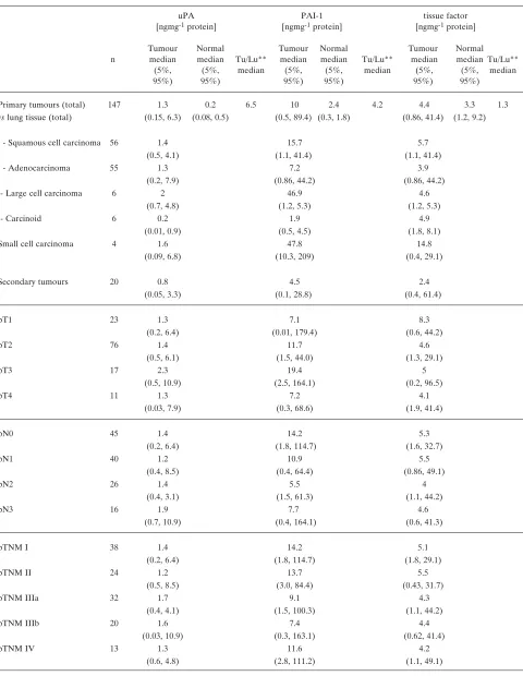
pTNM IV 13 1515 408 1301 359
Table II.Plasminogen activator (uPA), plasminogenactivator-inhibitor (PAI-1) and plasminogenactivator-receptor (uPAR). in lung tumor tissue homogenates.
uPA PAI-1 tissue factor
[ngmg-1protein] [ngmg-1protein] [ngmg-1protein]
Tumour Normal Tumour Normal Tumour Normal
n median median Tu/Lu** median median Tu/Lu** median median Tu/Lu** (5%, (5%, median (5%, (5%, median (5%, (5%, median
95%) 95%) 95%) 95%) 95%) 95%)
Primary tumours (total) 147 1.3 0.2 6.5 10 2.4 4.2 4.4 3.3 1.3
vs lung tissue (total) (0.15, 6.3) (0.08, 0.5) (0.5, 89.4) (0.3, 1.8) (0.86, 41.4) (1.2, 9.2)
- Squamous cell carcinoma 56 1.4 15.7 5.7
(0.5, 4.1) (1.1, 41.4) (1.1, 41.4)
- Adenocarcinoma 55 1.3 7.2 3.9
(0.2, 7.9) (0.86, 44.2) (0.86, 44.2)
- Large cell carcinoma 6 2 46.9 4.6
(0.7, 4.8) (1.2, 5.3) (1.2, 5.3)
- Carcinoid 6 0.2 1.9 4.9
(0.01, 0.9) (0.5, 4.5) (1.8, 8.1)
Small cell carcinoma 4 1.6 47.8 14.8
(0.09, 6.8) (10.3, 209) (0.4, 29.1)
Secondary tumours 20 0.8 4.5 2.4
(0.05, 3.3) (0.1, 28.8) (0.4, 61.4)
pT1 23 1.3 7.1 8.3
(0.2, 6.4) (0.01, 179.4) (0.6, 44.2)
pT2 76 1.4 11.7 4.6
(0.5, 6.1) (1.5, 44.0) (1.3, 29.1)
pT3 17 2.3 19.4 5
(0.5, 10.9) (2.5, 164.1) (0.2, 96.5)
pT4 11 1.3 7.2 4.1
(0.03, 7.9) (0.3, 68.6) (1.9, 41.4)
pN0 45 1.4 14.2 5.3
(0.2, 6.4) (1.8, 114.7) (1.6, 32.7)
pN1 40 1.2 10.9 5.5
(0.4, 8.5) (0.4, 64.4) (0.86, 49.1)
pN2 26 1.4 5.5 4
(0.4, 3.1) (1.5, 61.3) (1.1, 44.2)
pN3 16 1.9 7.7 4.6
(0.7, 10.9) (0.4, 164.1) (0.6, 41.3)
pTNM I 38 1.4 14.2 5.1
(0.2, 6.4) (1.8, 114.7) (1.8, 29.1)
pTNM II 24 1.2 13.7 5.5
(0.5, 8.5) (3.0, 84.4) (0.43, 31.7)
pTNM IIIa 32 1.7 9.1 4.3
(0.4, 4.1) (1.5, 100.3) (1.1, 44.2)
pTNM IIIb 20 1.6 7.4 4.4
(0.03, 10.9) (0.3, 163.1) (0.62, 41.4)
pTNM IV 13 1.3 11.6 4.2
Table III.Plasminogenactivator-receptor [uPAR (HD13, IIIF10, ADI)] in lung tumor homogenates.
uPAR (HD13) uPAR (IIIF10) uPAR (ADI)
[ngmg-1protein] [ngmg-1protein] [ngmg-1protein]
Tumour Normal Tumour Normal Tumour Normal
n median median Tu/Lu** median median Tu/Lu** median median Tu/Lu** (5%, (5%, median (5%, (5%, median (5%, (5%, median
95%) 95%) 95%) 95%) 95%) 95%)
Primary tumours (total) 95 2.8 0.7 4 3.9 1.5 2.6 2.8 1.3 2.2
vslung tissue (total) (0.4, 10.1) (0.3, 1.8) (1.3, 8.2) (0.63, 3.1) (0.46, 8.8) (0.57, 2.9)
- Squamous cell carcinoma 38 3.1 3.6 2.5
(0.49, 14.4) (1.3, 11.8) (0.53, 9.9)
- Adenocarcinoma 34 2.4 3.4 2.9
(0.46, 9.8) (1.2, 9.6) (0.43, 7.2)
- Large cell carcinoma 4 6.3 6.2 7.1
(1.8, 8.1) 3.8, 8.0 (6.4, 12.7)
- Carcinoid 2 1 2.3 0.56
(0.28, 1.8) (0.42, 4.2) (0.2, 3.3)
Small cell carcinoma 2 4.5 5.7 3.8
(3.0, 6.1) (2.4, 9.1 (0.8, 8.4)
Secondary tumours 15 1.6 2.4 2
(0.35, 13.5) (1.3, 7.3) (0.12, 8.0)
pT1 11 2.8 4.1 2.6
(0.49, 29.6) (1.6, 16.4) (0.4, 18.9)
pT2 46 3 3.5 2.6
(0.83, 9.1) (1.3, 9.6) (0.53, 7.4)
pT3 16 3.6 3.3 3.3
(0.19, 14.4) (0.55, 11.9) (1.5, 11.4)
pT4 7 2.2 2.9 2.8
(0.42, 7.2) (0.45, 7.1 (0.7, 7.9)
pN0 25 3.1 3.7 2.9
(0.42, 9.1) (0.55, 7.5) (0.4, 8.8)
pN1 27 3.2 3.8 3.2
(1.2, 10.1) (1.8, 9.6) (0.7,11.3)
pN2 17 2.8 2.6 2.2
(0.46, 14.4) (1.6, 11.8) (1.1, 7.2)
pN3 11 2.3 3.3 2.4
(0.49, 7.2) (1.6, 8.2) (0.5, 7.9)
pTNM I 18 3 3.9 2.7
(0.61, 29.6) (1.2, 16.4) (0.4, 8.8)
pTNM II 17 3.2 3.8 2.7
(1.2, 8.3) (1.8, 8.0) (0.9, 9.9)
pTNM IIIa 24 2.9 3 3
(0.46, 10.7) (1.3, 11.8) (1.1, 11.3)
pTNM IIIb 12 2.3 3.3 2.2
(0.42, 7.2) (0.45, 8.2) (0.48, 7.9)
pTNM IV 9 3 4.5 3.6
of the Spearman rank correlation coefficient was evaluated by analysis of variance (ANOVA).
Univariate analysis of survival probability was performed by Kaplan and Meier analysis (52), using the log-rank test for the determination of statistical significance between the survival curves. Multivariate analysis was performed using the Cox proportional hazard model (53) and a stepwise forward logistic regression approach. The discrimination levels to differentiate between subgroups of patients were calculated by the Critlevel programme (54). Several statistical packages (PC-Statistik by TOPSOFT, Hannover, Germany; Statistika by Statsoft, Hamburg, Germany, SPSS by SPSS Inc, Chicago, IL, USA) were applied.
Results
The results of the determination of cath B
Aactivity (cath
B
AT, cath B
A7.5) and of the protein levels of five biological
parameters cath B
C, cath L
C, uPA, PAI-1, and uPAR (ADI)
in cancer tissue and in adjacent lung parenchyma of the
same patient (n=147) are listed in Tables I and II.
Furthermore, we measured two additional uPAR variants,
uPAR (HD13), uPAR (IIIF10) and TF in 95 out of 147
tissues as shown in Table III. Altogether, 10 biological
parameters were analyzed.
For all parameters, we found increased median values in
tumour tissue (Tu) of various histologies among NSCLC
compared with lung parenchyma (Lu). The biological
parameters are listed from highest to lowest ratio: cath
B
CTu/cath B
CLu: 8.5:1; cath L
CTu/cath L
CLu: 6.6:1;
uPA
Tu/uPA
Lu: 6.5:1; cath B
ATTu/cath B
ATLu: 5.1:1;
PAI-1
Tu/PAI-1
Lu: 4.2:1, uPAR
Tu/uPAR
Lu(HD13): 4:1;
uPAR
Tu/uPAR
Lu(IIIF10): 2.6:1; cath B
A7.5Tu/cath B
A7.5Lu:
2.5:1; uPAR
Tu(ADI) /uPAR
Lu(ADI): 2.2:1, TF
Tu/TF
Lu1.3:1. With the exception of TF, this increase in tumour
tissue compared to its lung counterpart was statistically
significant (
p
<0.01) in all cases.
Correlation and associations with clinicopathological parameters.
In addition to the paired comparison between lung cancer
tissue and corresponding lung parenchyma, all 10 parameters
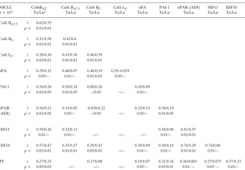
Table IV. Correlations.NSCLC CathBAT Cath BA7.5 Cath BC Cath LC uPA PAI-1 uPAR (ADI) HD13 IIIF10
n = 147 Tu/Lua Tu/Lu Tu/Lu Tu/Lu Tu/Lu Tu/Lu Tu/Lu Tu/Lu Tu/Lu
Cath BA7.5 r 0.62/0.55 p < 0.01/0.01
Cath BC r 0.31/0.58 0.42/0.6 p < 0.01/0.01 0.01/0.01
Cath LC r 0.28/0.30 0.43/0.36 0.46/0.39 p < 0.05/0.01 0.01/0.01 0.01/0.01
uPA r 0.29/0.15 0.40/0.07 0.48/0.19 0.29/-0.019 p < 0.05/-- 0.01/-- 0.01/0.05
0.05/--PAI-1 r 0.56/0.26 0.58/0.24 0.08/0.28 0.43/0.09 p < 0.01/0.05 0.01/0.05 --/0.05 --/--
0.01/--uPAR r 0.56/0.21 0.19/0.05 -0.038/0.22 0.23/0.15 0.58/0.19 (ADI) p < 0.01/0.05 0.05/-- --/0.05 --/-- 0.05/-- 0.01/0.05
HD13 r 0.58/0.26 0.32/0.12 0.56/0.06 0.81/0.55
p < 0.01/-- 0.01/-- --/-- --/-- --/-- 0.01/-- 0.01/0.01
IIIF10 r 0.57/0.47 0.35/0.37 0.29/0.47 0.39/0.09 0.49/0.18 0.74/0.29 0.74/0.06 p < 0.01/0.01 0.01/0.01 0.05/0.01 --/-- 0.01/-- 0.01/-- 0.01/0.01
0.01/--TF r 0.27/0.23 0.17/0.08 0.19/0.07 0.21/0.34 0.36/0.083 0.27/0.075 0.37/0.15 p < 0.05/0.05 --/-- --/-- --/-- 0.05/-- 0.05/0.01 0.01/ -- 0.05/ --
were analyzed for correlation to the histological cell type,
tumour stage (TNM-stage) and tumor cell differentiation (G).
The results are summarized in Tables I-III.
There was no significant difference between the median
levels of both cath B, enzymatic activities (cath B
AT, cath
B
A7.5) and protein level (cath B
C) and histological cell
types. We also found no correlation of either type of cath B
activity with the primary tumour size (pT-stage), whereas
cath B
AT, cath B
A7.5and cath B
Cof primary tumours
correlated with the presence of lymph node metastases (pN,
p
=0.06,
p
=0.07,
p
<0.05, respectively).
Only PAI-1 levels differed with the histological type of
NSCLC. We found the highest values of PAI-1 in large cell
carcinoma > SCC, AC, > carcinoid and the lowest values
in metastases of primary tumours of other origin.
Furthermore, in large cell carcinoma, uPAR (ADI),
uPAR (HD13) and uPAR (IIIF10) showed higher values
than in other histological types. There was no significant
relationship among different histological types with regard
to cath L
C, uPA, or TF. Remarkably, carcinoids, which are
low-grade neoplasms, showed low PAI-1, uPA, uPAR (HD
13, IIIF10) and low cath L
Clevels.
With the exception of PAI-1, there was no significant
association between all 10 biological parameters and either
tumour stage (pTNM) or grading (G). Only PAI-1 was
significantly increased in poorly-differentiated cells (G3)
compared to well- or moderately-differentiated cells
(G1/G2) (
p
<0.05). Furthermore, PAI-1, cath L
Cand uPAR
(ADI) were significantly higher in smokers compared to
non-smokers (each
p
<0.01).
Correlations among all 10 biological parameters related to
proteolysis.
Correlations among all 10 biological parameters
in NSCLC patients are listed in Table IV. Cath B
AT, cath
B
A7.5and cath B
Cwere correlated with each other, as
already described earlier (Werle
et al.
, 1999). Significant
correlations between cath B
AT, cathB
A7.5, cath B
Cand cath
L
Cwere found. PAI-1 significantly correlated with cath B
ATand cath B
A7.5, with uPAR (ADI), uPAR (HD13), uPAR
(IIIF10), with uPA, and weakly with TF, but not with cath
B
Cand cath L
C.
The three different ELISA formats for measuring uPAR,
which used antibodies against different epitopes of the same
uPAR molecule, rendered excellent correlations between
the test results for uPAR. All three forms of uPAR,
detected by specific ELISA formats, correlated well with
cath B
A7.5. The correlations were less pronounced for cath
B
ATand there was no correlation to cath B
C. Finally, uPA
correlated with cath B
AT, cathB
A7.5, cath B
Cand cath L
C.
Impact of biological parameters (activities and protein levels)
on patient survival.
For prognostic assessment of NSCLC
patients, the stage of the disease and histological cell type
are the most important prognostic factors used in clinical
routine. In order to evaluate the utility of biological
parameters as new prognostic factors, we compared them by
performing univariate and multivariate survival analysis.
Univariate survival analysis.
Table V presents the prognostic
significance of 10 biological parameters,
i.e.
cysteine-proteases and serine-cysteine-proteases, their inhibitors and
receptors in comparison with established prognostic factors
such as pTNM-stage, age and sex of the patients.
Measured in tumour homogenates of NSCLC patients,
cath B
ATand cath B
Cshowed significant prognostic value
for a 7-year overall survival (median follow-up of 3.8 years;
Table V and Figure 1). Of note, patients with high levels of
cath B
Chad a significantly better prognosis than patients
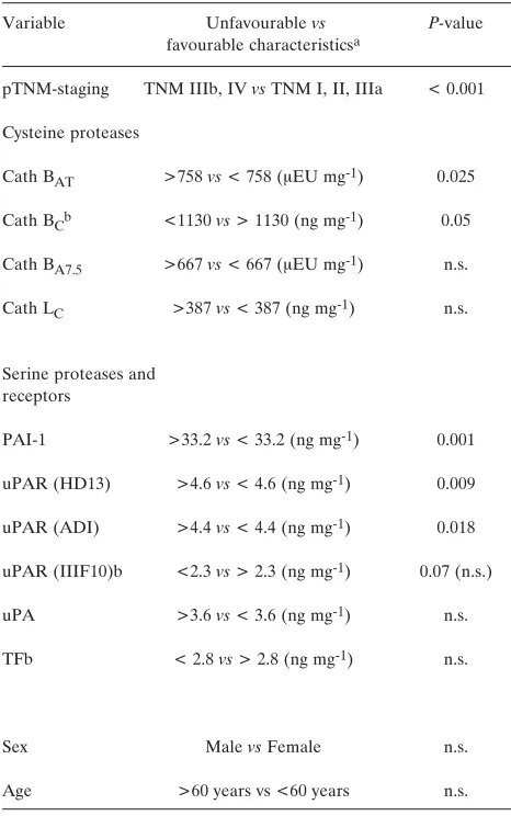
Table V.Univariate survival analyses of NSCLC.Variable Unfavourablevs P-value
favourable characteristicsa
pTNM-staging TNM IIIb, IV vs TNM I, II, IIIa < 0.001
Cysteine proteases
Cath BAT >758 vs < 758 (ÌEU mg-1) 0.025
Cath BCb <1130 vs > 1130 (ng mg-1) 0.05
Cath BA7.5 >667 vs < 667 (ÌEU mg-1) n.s.
Cath LC >387 vs < 387 (ng mg-1) n.s.
Serine proteases and receptors
PAI-1 >33.2 vs < 33.2 (ng mg-1) 0.001
uPAR (HD13) >4.6 vs < 4.6 (ng mg-1) 0.009
uPAR (ADI) >4.4 vs < 4.4 (ng mg-1) 0.018
uPAR (IIIF10)b <2.3 vs > 2.3 (ng mg-1) 0.07 (n.s.)
uPA >3.6 vs < 3.6 (ng mg-1) n.s.
TFb < 2.8 vs > 2.8 (ng mg-1) n.s.
Sex Male vs Female n.s.
Age >60 years vs <60 years n.s.
aCut-off values were calculated by a computer program developed by Abel et al.(53).
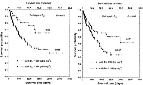
Figure 1. Probability of overall survival of patients with NSCLC in relation to cath BATand cath BC. The Critlevel program (54) calculated discriminative values of 758 (ÌEUmg-1) for cath B
ATand 1130 (ngmg-1) cath BC. Altogether, 118 patients out of 147 were included in the survival analyses. In NSCLC, 47 out of 85 patients with cath BAThigher than 758 (ÌEUmg-1) had a significantly shorter overall survival (-●- deceased patients (uncensored data), -/- still living patients (censored data)) than 9 out of 33 patients with cath BATbelow the cut off value (-■- deceased patients (uncensored data), -/- still living patients). In NSCLC, 33 out of 57 patients with cath BClower than 1130 (ngmg-1) had a shorter overall survival (-■- deceased patients, -/- still living patients) than 23 out of 61 patients with cath BCabove the cut-off value (-●- deceased patients, -/- still living patients).
Table VI.Univariate survival analyses of squamous cell and adenocarcinoma.
Variable Unfavourablevs
favourable characteristicsa
Squamous cell P-value Adenocarcinoma P-value
carcinoma
Cysteine proteases
Cath BAT >1647vs< 1647 (ÌEU mg-1) n.s. >1647vs< 1647 (ÌEU mg-1) n.s. Cath BA7.5 > 278vs< 278 (ÌEU mg-1) < 0.05 > 512vs< 512 (ÌEU mg-1) n.s. Cath BC > 512vs< 512 (ng mg-1) n.s. > 512vs< 512 (ng mg-1) n.s. Cath LC > 317vs< 317 (ng mg-1) n.s. > 403vs< 403 (ng mg-1) n.s.
Serine proteases and receptors
PAI-1 > 32.8vs< 32.8 (ng mg-1) 0.01 > 21.5vs< 21.5 (ng mg-1) n.s. uPAR (ADI) > 4.4vs< 4.4 (ng mg-1) n.s. > 4.5vs< 4.5 (ng mg-1) n.s. uPAR (HD13) > 4.6vs< 4.6 (ng mg-1) n.s. > 2.5vs< 2.5 (ng mg-1) n.s. uPAR (IIIF10) < 4.3vs> 4.3 (ng mg-1) n.s. < 3.6vs> 3.6 (ng mg-1) n.s. uPA > 3.6vs< 3.6 (ng mg-1) n.s. > 3.1vs< 3.1 (ng mg-1) n.s. TF > 4.5vs< 4.5 (ng mg-1) n.s. > 4.5vs< 4.5 (ng mg-1) n.s.
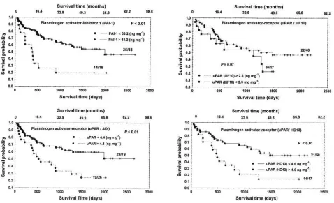
Figure 2.Prognostic significance of PAI-1 and uPAR (ADI, HD13, IIIF10) for overall survival of patients with NSCLC. For PAI-1, altogether 106 patients out of 147 were included in the survival analyses. Fourteen out of 18 patients with PAI-1 levels higher than 33.20 (ngmg-1) died in the observation period of 7 years (-●- deceased patients (uncensored data), -/- still living patients (censored data)) compared to 35 out of 88 patients with PAI-1 levels below the cut-off value (-■- deceased patients, -/- still living patients). For uPAR (ADI, HD13), altogether 105 patients out of 147, 67 out of 95 and 63 out of 95 , respectively, were included in the survival analyses. In NSCLC, 19 out of 26 patients with uPAR (ADI) higher than 4.4 (ngmg-1) and 14 out of 17 with uPAR (HD13) higher than 4.6 (ngmg-1) had a significantly shorter overall survival (-●- deceased patients (uncensored data), -/- still living patients (censored data)) than 29 out of 79 patients with uPAR (ADI) and 21 out of 50 with uPAR (HD13) below the cut-off value (-■- deceased patients, -/- still living patients). In contrast, 10 out of 17 patients with uPAR (IIIF10) lower than 2.3 (ngmg-1) had a shorter overall survival probability than 22 out of 46 patients with uPAR (IIIF10).
Table VII.Cox regression analyses of NSCLC.
Variable Unfavourable vs Relative P-value
favourable characteristicsa risk
pTNM-staging TNM IIIb, IV vsTNM I, II, IIIa -- < 0.0001
pT pT1 vspT2 vspT3 vspT4 -- n.s.
pN pN0 vspN1 vspN2 vspN3 -- n.s.
Histology Squamous cell -, Adeno -, Large cell carcinoma -- n.s.
Cysteine proteases
--Cath BAT >1460 vs< 1460 (ÌEU/mg) -- n.s.
Cath BA7.5 >555 vs< 555 (ÌEU/mg) -- n.s.
Cath BCc <1302 vs> 1302 (ng/mg) -- n.s.
Cath LC > 407 vs> 407 (ng/mg) -- n.s.
Serine proteases and receptors
PAI-1b >33.2 vs< 33.2 (ng/mg) 2.6 (6.6 - 17.3) < 0.001
uPAR (ADI) >3.6 vs< 3.6 (ng/mg) -- n.s.
uPAR (HD13) >4.1 vs< 4.1 (ng/mg) -- n.s.
uPAR (IIIF10)b,c < 2.3 vs> 2.3 (ng/mg) 0.24 (0.08 - 0.67) < 0.01
uPA >2.1 vs< 2.1 (ng/mg) -- n.s.
TFc < 11 vs> 11 (ng/mg) -- n.s.
Chemotherapy (+) vs(-) -- n.s.
with low cath B
Clevels (Table V and Figure 1). In contrast,
cath B
A7.5had no significant prognostic impact in the total
study population of NSCLC but it became significant in the
subgroup of SCC patients (Table VI;
p
<0.05). In AC no
significant correlation with survival could be found.
Regarding the plasminogen-activator system, we found,
in the total study group of NSCLC, significant correlations
with overall survival probability for PAI-1, uPAR (ADI),
uPAR (HD13) and uPAR (IIIF10) (Table V, Figures 2 - 4;
p
<0.01,
p
<0.05,
p
<0.01,
p
=0.07, respectively). In squamous
cell carcinomas, only PAI-1 significantly correlated with
overall survival probability (Table VI). In patients with AC
no significant correlation with overall survival could be
found. There was no correlation between survival
probability and uPA or TF, either in the total study
population of NSCLC or in the subgroups of SCC and AC.
Multivariate analysis.
In multivariate analysis, only those
proteolytic factors with a significant impact on overall
survival probability in univariate analysis, additional
systemic treatment (chemotherapy) as well as established
prognostic factors, were included.
PAI-1 and uPAR (IIIF10) remained the best independent
prognostic factors for patients with NSCLC (Table VII).
Discussion
This study clearly showed that the median levels of cath
B
AT, cath B
A7.5, cath B
C, cath L
C, uPA, PAI-1, and uPAR
[uPAR (ADI), uPAR (HD13), uPAR (IIIF10)] were
significantly higher in tumour tissue of NSCLC patients
compared to the corresponding lung parenchyma. All
reports in the literature agree on that point that the
biological parameters studied here are significantly
increased in tumour homogenates or are highly expressed
in tumour cells on tumour tissue sections compared to their
respective lung counterpart (2, 6, 10, 14, 20, 21, 23, 34, 35).
Moreover, after long-term follow-up of seven years (median
follow-up of 3.8 years), the activity of cath B (cath B
AT) and
protein level (cath B
C) of the cysteine protease cath B as
well as the concentrations of PAI-1 and uPAR [uPAR
(ADI), uPAR (HD13), uPAR (IIIF10)], two members of
the plasminogen activator system, provide significant
univariate prognostic information for patients with NSCLC.
Out of these three proteins, only PAI-1 and uPAR (IIIF10)
proved to be independent prognostic factors.
Cysteine proteases.
Our results for cath B are in agreement
with earlier studies by Ebert
et al.
(23) and Werle
et al.
(26).
Cath B
ATand cath B
7.5were of prognostic significance in the
observation period of 6 months (cath B
AT,
p<
0.05; 23), 15
months (both,
p<
0.05; unpublished) and 24 months (cath
B
A7.5,
p=
0.06, 26). In the subgroups of histologies, the
prognostic relevance of cath B
A7.5was found for patients
suffering from SCC after 24 months (
p<
0.05, 25, 26) and
after 7 years (
p<
0.05). In contrast, in AC no prognostic
impact of cath B
AT, cath B
A7.5and cath B
Cwas found in a
short (6, 15 and 24 months) and long (5 years) observation
period. However, a discrepancy exists regarding the
prognostic information of cath B in tumour tissue
homogenates. After a five-year observation period, we found
that cath B activities, cath B
AT, cath B
A7.5and the
concentration of cath B
Cmeasured in tissue homogenates
had no prognostic relevance in the total population of
NSCLC (14). In addition, several immunohistochemical
studies showed that the overexpression of cath B in tumour
cells only provides prognostic information, as could be
demonstrated by univariate and multivariate survival analysis
(9-11) Our findings (14) agree with IHC analyses of cath B
in tumour cells. In contrast to these studies, Mori
et al.
(13)
could not demonstrate a significant correlation between cath
B overexpression and overall survival probability of 31 lung
cancer patients, all in TNM stage I.
The discrepancy observed in the results of survival
analyses are probably due to: i) different extraction
procedures, ii) semi-quantitative analytical methods for
tumour tissue sections, iii) prolonged observation periods,
iv) a set of various cut-off values, v) divergent antibodies
resulting in altered ELISA-formats or changed staining
pattern on tissue sections for the detection of biological
parameters, vi) different treatment of patients after primary
surgery and vii) the cellular heterogeneity of the tumour.
This is particularly true for lung tumours, which are
heavily infiltrated by inflammatory cells (55, 56). Kayser
et
al.
(57) clearly showed that infiltrations of lung tumours are
in close correlation to tumour volume, containing besides
tumour cells also surrounding stromal cells
i.e.
fibroblasts,
pneumocytes type I and II, lymphocytes and histiocytes.
Each cell type, particularly the histiocytes, contribute to the
protease level in a solid tumour. On one hand, cathepsins
or other proteases derived from histiocytes may assist
tumour cells in degrading interstitial matrix components and
the basement membrane (58) or they may, on the other
hand, also destroy the tumour. This may explain the fact
that, in spite of greatly increased levels, cath B was at
borderline significance for prognosis of patient survival,
contrary to the reports on other types of carcinomas such as
head and neck (59, 60), in oral squamous cell carcinoma
(61), laryngeal carcinoma (62, 63), breast (64, 65),
pancreatic adenocarcinoma (66), gastric carcinoma (67),
colorectal carcinoma (68), chondrosarcoma (69) and
melanomas (70, 71).
survival, while in patients with AC the expression of cath B
points to shorter overall survival. Our findings were further
supported by a recent paper of Kayser
et al.
(19), which
described that NSCLC patients whose tumour cells stained cath
B-negative and macrophages-positive had a significantly better
prognosis than the opposite,
i.e.
tumour cell with positive and
macrophage with negative cath B expression. Furthermore, it
has been suggested that only a minor fraction of tumour
cell-associated cath B, perhaps a malignant, extracellular and/or
intracellular cath B isoform, seems to be involved in the
degradation of the extracellular matrix (44, 72-76).
Cysteine proteases of the cathepsin-type were controlled
by their natural inhibitors: the cystatins. Tissue
homogenates,
i.e.
tumour and host cells, consist of a mixture
of cathepsins and cystatins. Because the inhibition constant
(K
i) between cath B and cystatins A, B and C (stefins A and
B) is rather low, cath B will tightly bind cystatins and form
cath B-cystatin complexes. In such cath B-cystatin
complexes cath B is deactivated. Using the same study
population, we found that in tumour tissue the
concentration of cystatins A and B was significantly
increased as compared to the normal counterparts (31,
Werle
et al.
, unpublished). This might, at least partly,
explain, why we found a controversial role of cath B
Cand
cath B
ATand survival probability. In a study published by
Knoch
et al.
(30), we showed that the imbalance between
cath B and cystatins provides prognostic information.
Although we could demonstrate that the concentrations of
cystatins were increased in tissue homogenates (30, 31,
Werle
et al.
, unpublished), cystatins may be less effective in
binding cath B, which facilitates the invasion process of
tumour cells (29, 77). Therefore, the cellular origin of
proteases, malignant isoform(s), their subcellular
distribution as well as their corresponding endogenous
inhibitors, may be decisive factors in the relationship
between the protease levels and the survival probability of
lung cancer patients.
Serine proteases.
Pederson
et al.
(34, 35) measured uPA,
PAI-1 and uPAR in 106 patients with AC and in 84 patients
with SCC of the lung. These authors found that high levels
of PAI-1 in AC and high levels of uPAR in SCC were
significantly associated with overall survival. No conclusion
concerning PAI-1 and uPAR in the total study population
of NSCLC has been made.
In the present study, we describe that PAI-1 and uPAR,
measured as uPAR (ADI), uPAR (HD13) and uPAR
(IIIF10) format, have prognostic impact in patients with
NSCLC. Furthermore, PAI-1 proved to be a prognostic
factor in SCC. Salden
et al.
(36) could not find a significant
relationship with survival probability of PAI-1 and uPAR,
determined in tumour tissue extracts, either in the group of
all patients or in the subgroups of patients with SCC or AC.
Immunohistochemical studies of Pavey
et al.
(37) showed
that PAI-1 and not uPA was associated with the survival
probability of SCC patients, while Volm
et al.
(38) could
demonstrate that only uPA and not PAI-1-positive
carcinomas correlated with survival probability.
However, for uPAR antigen determination, we used three
different ELISA formats. As shown in previous studies (48),
all three ELISA formats detect recombinant human
CHO-suPAR and human uPAR antigen in lysates of non-malignant
keratinocytes, mammary epithelial cells, omental cells and
peripheal blood mononuclear cells with similar sensitivity (48).
Interestingly, when analysing a series of breast cancer cell
lines, the ELISA using mAb IIIF10 detected
tumour-associated uPAR antigen with higher sensitivity. Furthermore,
uPAR (HD13) was directed to a conformational epitope of
glycosylated uPAR and uPAR (IIIF10) react with domain 1
of both glycosylated and non-glycosylated uPAR. Therefore,
we determined three distinct isoform patterns of uPAR in
lung tumour homogenates and control lung parenchyma. In
control lung parenchyma we found two-fold higher levels of
uPAR (IIIF10) compared to uPAR (HD13). This holds true
for lung tumour tissue, where we found 1.4-fold higher protein
levels, of uPAR (IIIF10) compared to uPAR (HD13). This
was in contrast to the study of Kotzsch
et al.
(48), who found
a similar uPAR concentration in cell lysates of non-malignant
epithelial cells as determined by all three ELISA formats.
These authors also found higher levels of uPAR (ADI), uPAR
(HD13) and uPAR (IIIF10) in breast cancer tissue. However,
only for uPAR (IIIF10) could a correlation with prognosis be
determined. Elevated uPAR (IIIF10) levels were associated
with poor prognosis, while for the other two types of uPAR
protein levels, no significant correlation with survival could be
found. In the study presented here, we found the opposite for
uPAR (IIIF10) and a significant association with overall
survival using uPAR (ADI) and uPAR (HD13). Therefore,
we suggest that the significant differences in the
pathomechanism of tumour invasion and metastasis between
breast and lung cancer is most probably due to various cell
types of the host, which seem to be involved (see also
discussion in the paragraph "cysteine proteases").
uPA, PAI-1 and uPAR have been reported as prognostic
factors in a variety of solid tumours (64). In breast cancer,
tumour tissue levels of uPA and PAI-1 have reached the
highest level of evidence for their prognostic impact in a
prospective therapy trial as well as a large meta-analysis and
are thus ready to be used for clinical decision making (78).
Also, in our collective of NSCLC-patients, PAI-1 and uPAR
measured by mAb IIIF10 have a very strong independent
prognostic impact.
In conclusion, out of the 10 biological parameters
evaluated here, only cath B
AT, cath B
C, PAI-1 and uPAR
[uPAR (HD13), uPAR (IIIF10)] in tumour homogenates of
NSCLC patients had significant impact on patient prognosis
after long-term overall survival. PAI-1 and uPAR (IIIF10)
proved to be independent prognostic factors, enabling
better prognostic discrimination together with established
clinical and histopathological factors. This is consistent with
evidence from other solid tumours such as breast cancer
(48, 78). Further clinical studies are now needed in order to
fully validate the role of these proteolytic factors for clinical
decision making in NSCLC.
Acknowledgements
The authors are grateful to Wolfgang Klein, Beate Schauffler and Theresia Trull for their excellent technical assistance, and to Marta Krasovec for the measurement of the cath BC concentration. Furthermore, we are grateful to Dr. Hans Knoch, Dr. Clemens Kraft, Britta Jülke, Alexander Staib and Johannes Schumacher for providing tissue homogenates and clinical data of lung cancer patients. We thank Prof. Dr. Dr. Dres. h. c. Klaus Kayser for pathological assessment of tumour material as well as Dr. Heinrich Bülzebruck, Thoraxklinik Heidelberg gGmbH and Dr. Werner Rittgen, Deutsches Krebsforschungszentrum, for their support with the statistical evaluation.
References
1 Hoffmann H, Bülzebruck H and Dienemann: Chirurgische Therapie des Nicht-kleinzelligen Bronchialkarzionms (NSCLC). Onkologe8: 425-433, 2002.
2 Schmitt M, Jänicke F and Graeff H: Tumor-associate proteases. Fibrinolysis 6: 3-26, 1992.
3 Elliot E and Sloane BF: The cysteine protease cathepsin B in cancer. Perspect Drug Discov Design 6: 12-32, 1996.
4 Lah T and Kos J: Cysteine proteinases in cancer progression and their clinical relevance for prognosis. Biol Chem 379: 125-130, 1998.
5 Yan SQ, Sameni M and Sloane BF: Cathepsin B and human tumor progression. Biol Chem 379: 113-123, 1998.
6 Kos J, Werle B, Lah TT and Brunner N: Cysteine proteinases and their inhibitors in extracellular fluids: markers for diagnosis and prognosis in cancer. Int J Biol Markers 15: 84-89, 2000. 7 Yan S and Sloane BF: Molecular regulation of human cathepsin
B: implication inpathologies. Biol Chem 384: 845-854, 2003. 8 Higashiyama M, Doi O, Kodama K, Yokouchi H and Tateishi
R: Cathepsin B expression in tumour cells and laminin distribution in pulmonary adenocarcinoma. J Clin Pathol 46: 18-22, 1993.
9 Ozeki Y, Takishima K, Takagi K, Aida S, Tamai S, Mamiya G and Oagata T: Immunohistochemical analysis of cathepsin B expression in human lung adenocarcinoma: the role in cancer progression. Jpn J Cancer Res 84: 972-975, 1993.
10 Sukoh N, Abe S, Ogura S, Isobe H, Takekawa H, Inoue K and Kawakami Y: Immunohistochemical study of cathepsin B. Prognostic significance in human lung cancer. Cancer 74: 46-51, 1994.
11 Sukoh N, Abe S, Nakajima I, Ogura S, Isobe H, Inoue K and Kawakami Y: Immunohistochemical distributions of cathepsin B and basement membrane antigens in human lung adenocarcinoma: association with invasion and metastasis. Virchows Arch 424: 33-38, 1994.
12 Inoue T, Ishida T, Sugio K and Sugimachi K: Cathepsin B expression and laminin degradation as factors influencing prognosis of surgically treated patients with lung adenocarcinoma. Cancer Res 54: 6133-6136, 1994.
13 Mori M, Kohli A, Baker SP, Savas L and Fraire AE: Laminin and cathepsin B as prognostic factors in stage I non-small cell lung cancer: are they useful? Mod Pathol 10: 572-577, 1997. 14 Werle B, Lötterle H, Schanzenbächer U, Lah TT, Kalman E,
Kayser K, Bülzebruck H, Schirren J, Krasovec M, Kos J and Spiess E: Immunochemical analysis of cathepsin B in lung tumours: an independent prognostic factor for squamous cell carcinoma patients. Br J Cancer 81: 510-519, 1999.
15 Fujise N, Nanashim A, Taniguchi Y, Matsuo S, Hatano K, Matsumoto Y, Tagawa Y and Ayabe H: Prognostic impact of cathepsin B and matrix metalloproteinase-9 in pulmonary adenocarcinomas by immunohistochemical study. Lung Cancer 27: 19-26, 2000.
16 Delebecq TJ, Porte H, Zerimech F, Copin MC, Gouyer V, Dacquembronne E, Balduyck M, Wurtz A and Huet G: Overexpression level of stromelysin 3 is related to the lymph node involvement in non-small cell lung cancer. Clin Cancer Res 6: 1086-1092, 2000.
17 Baumhäkel JD, Kayser K, Kalman E, Kos J, Lah TT, Spiess E, Ebert W, Fiehn W and Werle B: Histological and thermodynamic features of cathepsin B-positive tumors of non-small cell lung cancer obtained by syntactic structure analysis. Electronic J Pathol Histol 7: 011-06, 2001.
18 Werle B: Immunohistochemical analysis of cathepsin B and cathepsin S in tumors, parenchyma and regional lymphnodes of the lung. Radiol Oncol 36: 183-184, 2002.
19 Kayser K, Richter N, Hufnagl P, Kayser G, Kos J and Werle B: Expression of cathepsin B and cathepsin L and proliferation activity in operated lung cancer, and their clinical significance. Anticancer Res 23: 2767-2772, 2003.
20 Trefz G, Lüthgens K, Erdel M, Spiess E and Ebert W: Plasminogen activator and cathepsin B in normal and malignant human lung tissue. J Cancer Res Clin Oncol 115: S50, 1989. 21 Krepela E, Kasafirek E, Novak K and Viklicky J: Increased
cathepsin B activity in human lung tumors. Neoplasma 37: 61-70, 1990.
22 Sedo A, Krepela E and Kasafirek E: Dipeptidylpeptidase IV, prolylendopeptidase and cathepsin B activities in primary human lung tumors and lung parenchyma. J Cancer Res Clin Oncol 117: 249-253, 1991.
23 Ebert W, Knoch H, Werle B, Trefz G, Muley Th and Spiess E: Prognostic value of increased lung tumor tissue cathepsin B. Anticancer Res 14: 895-900, 1994.
24 Ledakis P, Tester, WT, Rosenberg N, Romero-Fischmann D, Daskal I, and Lah TT: Cathepsins D, B, and L in malignant human lung tissue. Clin Cancer Res 2: 561-568, 1996.
26 Werle B, Jülke B, Lah T, Spiess E and Ebert W: Cathepsin B fraction active at physiological pH of 7.5 is of prognostic significance in squamous cell carcinoma of human lung. Br J Cancer 75: 1137-1143, 1997.
27 Werle B, Kraft C, Lah TT, Kos J, Schanzenbächer U, Kayser K, Ebert W and Spiess E: Cathepsin B in infiltrated lymphnodes is of prognostic significance for non-small cell lung cancer patients. Cancer 89: 2282-2291, 2000.
28 Hirano T, Manabe T and Takeuchi S: Serum cathepsin B levels and urinary excretion of cathepsin B in the cancer patients with remote metastasis. Cancer Lett 70: 41-44, 1993.
29 Zore I, Krasovec M, Cimerman N, Kuhelj R, Werle B, Nielsen HJ, Brunner N and Kos J: Cathepsin B/cystatin C complex levels in sera from patients with lung and colorectal cancer. Biol Chem 382: 805-810, 2001.
30 Knoch H, Werle B, Ebert W and Spiess E: Imbalance between cathepsin B and cysteine proteinase inhibitors is of prognostic significance in human lung cancer. Int J Oncol 5: 77-85, 1994.
31 Ebert E, Werle B, Jülke B, Kopitar-Jerala N, Lah TT, Abrahamson M, Spiess E and Ebert W: Expression of cysteine protease inhibitors stefin A, stefin B, and cystatin C in human lung tumor tissue. Adv Exp Med Biol 421: 259-265, 1997.
32 Krepela E, Prochazka J, Karova B, Cermak J and Roubkova H: Cathepsin B, thiols and cysteine protease inhibitors in squamous-cell lung cancer. Neoplasma 44: 219-239, 1997. 33 Krepela E, Prochazka J, Karova B, Cermak J and Roubkova H:
Cysteine proteases and cysteine protease inhibitors in non-small cell lung cancer. Neoplasma 45: 318-331, 1998.
34 Pederson H, Brunner N, Francis D, Osterlind K, Ronne E, Hansen HH, Dano K and Grondahl-Hansen J: Prognostic impact of urokinase, urokinase receptor, and type 1 plasminogen activator inhibitor in squamous and large cell lung cancer tissue. Cancer Res 54: 4671-4675, 1994.
35 Pederson H, Grondahl-Hansen J, Francis D, Osterlind K, Hansen HH, Dano K and Brunner N: Urokinase and plasminogen activator inhibitor type 1 in pulmonary adenocarcinoma. Cancer Res 54: 120-123, 1994.
36 Salden M, Splinter TAW, Peters HA, Look MP, Timmermans M, van Meerbeeck JPAM and Foekens JA: The urokinase-type plasminogen activator system in resected non-small cell lung cancer. Ann Oncol 11: 327-332, 2000.
37 Pavey SJ, Marsh NA, Ray MJ, Butler D, Dare AJ and Hawson GA: Changes in plasminogen activator inhibitor-1 levels in non-small cell lung cancer. Boll Soc Ital Biol Sper 72: 331-340, 1996.
38 Volm M, Mattern J and Koomagi R: Relationship of urokinase and urokinase receptor in non-small cell lung cancer to proliferation, angiogenesis, metastasis and patient survival. Oncol Rep 6: 611-615, 1999.
39 Koomagi R and Volm M: Tissue-factor expression in human non-small cell lung carcinoma measured by immunohistochemistry: correlation between tissue factor and angiogenesis. Int J Cancer 79: 19-22, 1998.
40 World Health Organization: Histological Classification of Lung Tumors. WHO: Geneva, 1981.
41 Hermanek P and Sobin L: TNM Classification of Malignant Tumors. International Union against Cancer (UICC), fourth edn. Springer: Berlin, 1987.
42 Manegold C and Drings P: Chemotherapie des nichtkleinzelligen Lungenkarzinoms. In: Thoraxtumoren: Diagnostik Staging -gegenwärtiges Therapiekonzept (Drings P, Vogt- Moykopf I, eds), Springer Verlag: Heidelberg, Germany , 1998, pp. 310-327. 43 Schraube P, Kimming B, Latz D, Flentje M and Wannenmacher
M: Radiotherapie des Bronchialkarzionms. In: Thoraxtumoren: Diagnostik - Staging - gegenwärtiges Therapiekonzept, (Drings P, Vogt-Moykopf I, eds). Springer Verlag Heidelberg, Germany, 1998, pp. 277-295.
44 Werle B, Ebert W, Klein W and Spiess E: Cathepsin B in tumors, normal tissue and isolated cells from the human lung. Anticancer Res 14: 1169-1176, 1994.
45 Gabrijelcic D, Annan-Prah A, Rodic B, Rozman B, Cotic V and Turk V: Determinations of cathepsins B and H in sera and synovial fluids of patients with different joint diseases. J Clin Chem Clin Biochem 28: 149-153, 1990.
46 Kos J,Sˇmid L, Krasovec M, Svetic B, Lenarcic B, Vrhovec I, Sˇkrk J and Turk V: Lysosomal proteases cathepsins D, B, H, L and their inhibitors stefins A and B in head and neck cancer. Biol Chem Hoppe-Seyler 376: 401-405, 1995.
47 Kos J, Sˇtabuc B, Schweiger A, Krasovec M, Cimerman N, Kopitar-Jerala N and Vrhovec I: Cathepsins B, H, L and their inhibitors stefin A and cystatin C in sera of melanoma patients. Clin Cancer Res 3: 1815-1822, 1997.
48 Kotzsch M, Luther T, Harbeck N, Ockert D, Lutz V, Noack F, Grossmann D, Albrecht S, Kramer MD, Lossnitzer A, Grosser M, Schmitt M, and Magdolen V: New ELISA for quantification of human urokinase receptor (CD87) in cancer. Int J Oncol 17: 827-834, 2000.
49 Albrecht S, Luther T, Grossmann H, Flossel C, Kotzsch M and Müller M: An ELISA for tissue factor using monoclonal antibodies. Blood Coagul Fibrinolysis 3: 263-270, 1992. 50 Albrecht S, Kotzsch M, Siegert G, Luther T, Grossmann H,
Grosser M and Müller M: Detection of circulating tissue factor and factor VII in a normal population. Thromb Haemost 75: 772-777, 1996.
51 Bradford MM: A rapid and sensitive method for the quantitation of microgramm quantities of protein utilizing the principle of protein-dye binding. Anal Biochem 72: 248-254, 1976.
52 Kaplan EL and Meier P: Nonparametric estimation from incomplete observations. J Am Stat Assoc 53: 457-481, 1958. 53 Cox DR: Regression models and life tables (with discussion). J
R Stat Soc 187: 187-220, 1972.
54 Abel U, Berger J and Wiebelt H: Critlevel. An exploratory procedure for the evaluation of quantitative prognostic factors. Methods Inf Med 23: 154-156, 1984.
55 Ioachim HL, Dorsett BH and Paluch E: The immune response at the tumour site in lung carcinoma. Cancer 38: 2296-2309, 1976.
56 Kayser K, Toomes H, Vollhaber HH and Burkhardt HU: Tumor volume and macroscopic growth pattern of bronchogenic carcinoma. Virchow Arch [A] 405: 387-397, 1985. 57 Kayser K, Bülzebruck H, Ebert W, Merkle NM and Vogt-Moykopf I: Local tumor inflammation, lymph node metastasis, and survival of operated bronchus carcinoma patients. JNCI 77: 77-81, 1986.
59 Budihna M, Strojan P,Sˇmid L,Sˇkrk J, Vrhovec I, Zupevc A, Rudolf Z, Zargi M, Krasovec M, Svetic B, Kopitar-Jerala N and Kos J: Prognostic value of cathepsins B, H, L, D and their endogenous inhibitors stefins A and B in head and neck carcinoma. Biol Chem Hoppe-Seyler 377: 385-390, 1996. 60 Strojan P, Budhina M, Smid L, Vrhovec L, Kos J and Skrk J:
Prognostic significance of cysteine proteinases cathepsins B and L and their endogenous inhibitors stefins A and B in patients with squamous cell carcinoma of the head and neck. Clin Cancer Res 6: 1052-1062, 2000.
61 Kawasaki G, Kato Y and Mizuno A: Cathepsin expression in oral squamous cell carcinoma: relationship with clinicopathologic factors. Oral Surg Oral Med Oral Pathol Oral Radiol Endod 4: 446-454, 2002.
62 Smid L, Strojan P, Budihna M, Skrk J, Vrhovec I, Zargi M and Kos J: Prognostic value of cathepsins B, D and stefins A and B in laryngeal carcinoma. Eur Arch Otorhinolaryngol 254: 150-153, 1997.
63 Russo A, Bazan V, Gebbia N, Pizzolanti G, Tumminello FM, Dardanoni G, Ingria F, Restivo S, Tomasino RM and Leto G: Flow cytometric DNA analysis and lysosomal cathepsin B and L in locally advanced laryngeal cancer. Relationship with clinicopathologic parameters and prognostic significance. Cancer 76: 1757-1764, 1995. 64 Schmitt M, Harbeck N, Thomssen C, Wilhem O, Magdolen V, Reuning U, Ulm K, Hofler H, Jänicke F and Graeff H: Clinical impact of the plasminogen activation system in tumor invasion and metastasis: prognostic relevance and target for therapy. Thromb Haemost 78: 285-296, 1997.
65 Levicar N, Kos J, Blejec A, Golouh R, Vrhovec I, Frkovic-Grazio S and Lah TT: Comparison of potential biological markers cathepsin B, cathepsin L, stefin A and stefin B with urokinase and plasminogen activator inhibitor-1 and clinicopathological data of breast carcinoma patients. Cancer Detec Prev 26: 42-49, 2002
66 Niedergethmann M, Hildenbrand R, Wolf G, Verbecke CS, Richter A and Post S: Angiogenesis and cathepsin expression are prognostic factors in pancreatic adenocarcinoma after curative resection. Int J Pancreatol 28: 31-39, 2000
67 Russo A, Bazan V, Migliavacca M, Zanna I, Tubiolo C, Tumminello FM, Dardanoni G, Cajozzo M, Bazan P, Modica G, Latteri M, Tomasino RM, Colucci G, Gebbia N and Leto G: Prognostic significance of DNA ploidy, S-phase fraction and tissue levels of aspartic, cysteine, and serine proteases in operable gastric carcinoma. Clin Cancer Res 6: 178-184, 2000. 68 Herszenyi L, Plebani M, Carraro P, de Paoli M, Roveroni G,
Cardin R, Foschia F, Tulassay Z, Naccarato R and Farinati F: Proteases in gastrointestinal neoplastic diseases. Clin Chim Acta 291: 171-187, 2000.
69 Hackel CG, Krueger S, Grote HJ, Oshiro Y, Hodges S, Johnston DA, Johnson ME, Roessner A, Ayala AG and Czerniak B: Overexpression of cathepsin B and urokinase plasminogenactivator is associated with increased risk of recurrence and metastasis in patients with chondrosarcoma. Cancer 89: 995-1003, 2000. 70 Otto FJ, Goldmann T, Biess B, Lippold A, Suter L and
Westhoff U: Prognostic classification of malignant melanomas by combining clinical, histological, and immunohistochemical parameters. Oncology 56: 208-214, 1999.
71 Yoshii A, Kageshita T, Tsushima H and Ono T: Clinical relevance of cathepsin B-like enzyme activity and cysteine proteinase inhibitor in melanocytic tumours. Arch Dermatol Res 287: 209-213, 1995.
72 Sloane BF, Moin K, Sameni M, Tait LR, Rozhin J and Ziegler G: Membrane association of cathepsin B can be induced by transfection of human breast epithelial cells with c- Ha-ras oncogene. J Cell Sci 107: 373-384, 1994.
73 Werle B, Ebert W, Klein W and Spiess E: Charge polymorphism in human lung cell pro-cathepsin B. Anticancer Res 16: 49-54, 1996.
74 Strohmaier AR, Porwol T, Acker H and Spiess E: Tomography of cells by confocal laser scanning microscopy and computer-assisted three-dimensional image reconstruction: localization of cathepsin B in tumor cells penetrating collagen gels in vitro. J Histochem Cytochem 45: 975-983, 1997
75 Hughes SJ, Glover TW, Zhu XX, Kuick R, Thoraval D, Orringer MB, Beer DG and Hanash S: A novel amplicon at 8p22-23 results in overexpression of cathepsin B in esophageal adenocarcinoma. Proc Natl Acad Sci USA 95: 12410-12415, 1998. 76 Szpaderska AM and Frankfater A: An intracellular form of cathepsin B contributes to invasiveness in cancer. Cancer Res 61: 3493-3500, 2001.
77 Lah TT, Clifford JL, Helmer K, Day N, Moin K, Honn KV, Crissman JD and Sloane BF: Inhibitory properties of low molecular weight cysteine proteinase inhibitors from human sarcoma. Biochim Biophys Acta 993: 63-73, 1989.
78 Harbeck N, Schmitt M, Kates RE, Kiechle M, Zemzoum I, Jänicke F and Thomssen C: Clinical utility of uPA/PAI-1 determination in primary breast cancer tissue for individualized therapy concepts. Clin Breast Cancer 3: 196-200, 2002.

![Table III. Plasminogenactivator-receptor [uPAR (HD13, IIIF10, ADI)] in lung tumor homogenates](https://thumb-ap.123doks.com/thumbv2/123dok/3197048.1736694/6.595.52.538.113.738/table-iii-plasminogenactivator-receptor-upar-iiif-tumor-homogenates.webp)