STUDY OF EXPRESSION OF SUGARCANE SUCROSE
TRANSPORTER cDNA IN YEAST
Slameto1*), Bambang Sugiharto2), Nur Basuki3) and Liliek Sulistyowati3) 1*)
Faculty of Agriculture University of Jember Jl. Kalimantan 37 Jember East Java 68121 Indonesia 2)
Faculty of Math and Natural Science University of Jember Jl. Kalimantan 37 Jember East Java 68121 Indonesia 3)
Faculty of Agriculture University of Brawijaya Jl. Veteran Malang 65145 East Java Indonesia
*)
Corresponding author Phone : +62- 331- 334054 E-mail: [email protected]
Received: November 4, 2010/ Accepted: July 12, 2011
Revisi Daftar pustaka banyak yg tidak ada. Mohon dicek di References
ABSTRACTStudies in transgenic sugarcane (Saccharum officinarum L.) demonstrated that sucrose transporter (SUT) genes were essential in sucrose translocations. Sucrose transporter gene isolated from sugarcane were designated as SoSUT1 and SoSOSUT2 respectively as encode protein of 518 and 747 amino acids. The genes were constructed into plasmid pYES2 for SoSUT1, and pYX112 for SoSOSUT2. cDNA SoSOSUT2 had also constructed into plasmid pBIN-At-GFP which was possible to trace the gene inserted. The constructed plasmid was transformed into yeast (Saccharomyces cerevi-siae) and grown in minimal medium (SD–urasil) as selection medium. The transformed colony was confirmed using PCR. The functional expression was studied by growing yeast in YPD medium with 2% Sucrose, then the sucrose uptake was measured in number interval time using resorcinol method. The result showed that yeast INVSc1-pYES2-SoSUT1 and BF264-pYX112-SoSOSUT2 had higher ability in sucrose transport compared to the control-INVSc1 for SoSUT1 and control-BF264 for SoUT2. Moreover, the result showed that SoSUT1 had higher ability to transport sucrose than SoSOSUT2. Confocal microscope observation showed that trans-formation gene SoSOSUT2 was successful, which was indicated by green exposure of GFP protein.
Keywords: sugarcane, sucrose transporter, yeast INTRODUCTION
Photosynthesis represents the major source of energy required for biological processes. As a primary product of
synthesis carbohydrates are formed in photo-synthetically active cells (Paul and Foyer, 2001). Long-distance transport of the disaccharide from source leaves to sink tissues takes place in the sieve tube/companion cell complex (phloem) and may require apoplastic loading and unloading steps. Passage of Suc through membranes is facilitated by energy dependent, cargo-specific transport proteins (Shakya and Sturm, 1998).
In higher plants leaves, and to a certain extent, other parts of the plant, e.g. stem tissue, represent the primary sites for photosynthesis. In contrast, other parts of the plant e.g. roots, seeds or tubers, do not contribute significantly to the energy gained by photosynthesis but rather are dependent on carbon dioxide fixed in photosynthetically active parts of the plant.
Sucrose is the major product of photosynthesis in many higher plants and is transported from the source tissue (mature leaves) through the phloem to various sink tissues to support plant growth and develop-ment (Rae et al., 2005). Most plants studied contain multiple sucrose transporters (SUTs), also known as sucrose carriers (SUCs), which likely have different functions in phloem loading and/or unloading or in the import of sucrose into sink tissues. Based on phylogenetic analysis (Barker et al., 2000) it was shown that sucrose and sucrose transporter like proteins were divided into three families, namely SUT1, SUT 2, and SUT 4. According to homolog sequences, SUT 1 and SUT 3 were found only in tobacco (Kuhn et al., 1999). In plant, sucrose uptake kinetics involve high- and low-affinity components. A family of low- and high-affinity sucrose transporters (SUT) was identified. SUT1 serves as a high affinity transporter essential for phloem loading and long-distance transport.
228
Slameto et al.: Study of Expression of Sugarcane Sucrose………...
SUT4 is a low-affinity transporter with an expression pattern overlapping that of SUT1 (Riesmeier et al., 1993; Kuhn 2003). Using Saccharomyces cerevisiae strain,a cDNA from sugarcane encoding a sucrose carrier was identified as functional expression. Yeast strains that allow the phenotypic recognition of a sucrose carrier activity were constructed into yeast. Previous study in transgenic plants demonstrated that sucrose transporters (SUT) were essential for sucrose translocations. Green fluorescent protein (GFP) from the jellyfish Aequorea victoria was used to analyze two important functional aspects of an unknown gene product: (1) determination of the subcellular localization of the protein and (2) expression level of the unknown protein. The GFP emits green fluorescence when excited by blue light. In contrast to other bioluminescent systems, GFP works without exogenously added proteins, substrates or co-factors, allowing expression to be monitored in vivo (Niedenthal et al., 1996).Sucrose transporter was isolated from sugarcane (Saccharum officinarum L.). Recently, a number of genes which encode enzymes of sugar metabolism or sugar transport in sugarcane have been identified. Along with a functional analysis of these enzymes, information on the localization of their expression and activity will enable us to build a model of the pathways and control points for sugar translocation into the storage sink. According to the studies above, a study to clone the cDNAs encoding sucrose transporter proteins from sugarcane is needed.
MATERIALS AND METHODS
The following bacterial and yeast strains were used: DH5α, INVSc1 and BF264. Construction ofSoSUT1 cDNA in pYES2 plasmid (Invitrogen) started with the formation of Kozak consensus nucleotide sequence for translation initiation. cDNA-SoSUT1 was amplified by PCR using forward primer containing the nucleotide sequence of Kozak consensus, 5'-ggtaccatggctcgcggcgacggcg-3’ and reverse primer 5'-tcagtggccgcccgcgctgac-3'. DNA PCR products were inserted into pGEMT plasmid and
transformed into E. coli DH5α. After the restriction
using PstI and SphI enzyme at N-and C-terminal, cDNA SoSUT1 restriction products were inserted into pYES2 plasmid and transformed back into E. coli DH5α. After digestion with SalI and XhoI, cDNA SoSUT2 fragments were cloned into pYX112. Another plasmid pBin-AtBI-GFP, after digestion using SalI and NcoI, was constructed by inserting cDNA SoSUT2 coding region site of SalI and NcoI. Transformation constructed pYES2-SoSUT1 into the yeast cell strains INVSc1, and pYX112-SoSUT2 and pBin-pYX112-SoSUT2-GFP into yeast cell strain BF264 conducted using ion Li (lithium) in accordance with the methods mentioned in the instructions from Invitrogen Inc. Yeast trans-formants were plated on SD-glucose-Ura ¯ medium and incubated at 30oC for two days.
To confirm the transformants, the cloned yeast was inoculated and cultured in proper medium. 3 ml overnight cultures of the respective yeast strains were harvested, washed in yeast lysis buffer resuspended in 100 ul of the same buffer and after addition of an equal volume of glass beads (0.5 mm diameter) the suspension was processed in vortex five times for 15 s, centrifuged, and the supernatant was analyzed using PCR and :forward primer agcctcggcaggctcatcctc-3’ and reverse primer 5’-ggagatcttgggcagcaggaa-3’for SoSUT1, whereas for SoSOSUT2 using forward primer gatggcttgtcagcaaagggc-3’ and reverse primer 5’-gcgacctgttgaaggatgtg-3’ .
In order to study the expression, yeast was grown in YPD medium with 2% sucrose. Then, the sucrose absorption was measured in number interval time using resorcinol method. Yeast cells of BF264 harboring pBin-SoSUT2-GFP were cultured for two days in SD-glucose-Ura¯ . After two days, GFP fluorescence was observed using a Confocal fluorescence microscope (Zeiss, Germany) with a 488-nm excitation wavelength.
RESULTS AND DISCUSSION
Kpn1 enzyme produced SoSUT1 cDNA which contained start and stop codon with the size of 1.5 kb and Kozak consensus. Furthermore, in order to confirm the results of transformation of SoSUT1 gene into pGEMT, the plasmid was digested using PstI and SphI. As shown in Figure 1, there were two bands with different size where the upper band indicated DNA of plasmid and lower band indicated DNA target. This result showed that transformation into pGEMT was successful and it can be used for further experiment.
Figure 1. Separation using electrophoresis gel agarose (1%) on pYES2-SoSUT1 after digestion using PstI and SphI
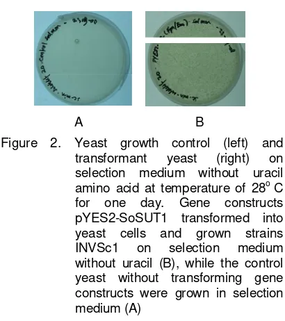
In order to study the function of SoSUT1 genes isolated from sugarcane, gene pYES2- SoSUT1 constructs were transformed and expressed into yeast cells INVSc1 strains. Transformation was performed using Li + ion as mentioned in the Invitrogen manual. Yeast transformantswere selected on minimal selection media that did not contain amino acids uracil. As
shown in Figure 2, transformant yeast gene constructs contained pYES2-SoSUT1 grew on media selection (B) and control yeast that did not contain constructs were not able to grow on selection medium (A).
Figure 2. Yeast growth control (left) and transformant yeast (right) on selection medium without uracil amino acid at temperature of 28o C for one day. Gene constructs pYES2-SoSUT1 transformed into yeast cells and grown strains INVSc1 on selection medium without uracil (B), while the control yeast without transforming gene constructs were grown in selection medium (A)
Tagged cDNA SoSUT2 using PCR and primer which contained sequence nucleotide of restriction site of SalI and XhoI was done, and PCR product was shown in Figure 3 that PCR product had size about 2.3 kb and it indicates that cDNA SoSUT2 which had been tagged could be used in transformation into yeast.
A B
Figure 3. PCR product cDNA SOSUT2 was tagged using SalI and XhoI before being used in tranformation into yeast (A); Restriction of plasmid pBin-AtBI-GFP using SalI/NcoI and pYX112 using SalI /XhoI (B)
230
Slameto et al.: Study of Expression of Sugarcane Sucrose………...
As shown in Figure 3, lane 2-3 showed the result of digestion of pBIN-AtBI-GFP using restriction enzyme SalI and NcoI, whereas lane 4-7 showed the digestion result of pYX112 using SalI and XhoI. These results of digestion were then isolated from agarose gel and used for transformation.
Figure 4. Transformant yeast on selection medium without uracil amino acid at 28oC for one day.Gene constructs pYX112-SoSUT2 were transformed into yeast cells grown on selection medium without uracil Yeast cell containing pYX112-SoSUT2 could grow in selection medium without uracil. Therefore, yeast cell which did not contain plasmid suppressed the growth of yeast cell. It can be determined that colonies growth on selection medium was transformant yeast cells. To confirm that yeast cell was transformant, single colony of yeast transformant was cultured in liquid medium. Culture was harvested and plasmid was isolated using vortex after the addition of glass bead. Supernatant was analyzed using PCR and primer for amplification of fragments of cDNA SoSUT1 and SoSUT2.
In order to observe DNA inserted, transformant yeast grown in YPD liquid medium containing 2% sucrose was cultured at 28oC. Incubation time was adjusted as treatment time, so harvesting depended on the desired incubation time.
Observation of sucrose transport indi-cated transport/sucrose absorption by both yeast transformant yeast strain INVS or BF 264. Sucrose absorption measurements used the method of Resocinol yeast transformant strains INVS-SoSUT1 with pYES2-interval 6-hours incubation.
Figure 6A showed that absorbance for supernatant of yeast transformant SoSUT1
increased in 24 hours after culture, while absorbance of supernatant of wild type of INVS remained flat during all interval time of incubation. Observation of sucrose content in medium used in culture showed that medium for the culture of yeast transformant revealed the decrease of absorbance at 6 hours after culture, whereas medium wild type culture showed that absorbance decreased after 24 hours. Thus, yeast transformant SoSUT1 had higher transport sucrose from medium than wild type yeast. It can be concluded that gene SoSUT1 has physiological activity in sucrose transport. SUT1 plays an important role for sugar transport, changes in Suc- and starch-modifying enzyme activities, and metabolite profiles are consistent with the developmental switch in unloading mechanisms. Altogether, the findings may suggest a role of SUT1 in retrieval of Suc from the apoplasm, thereby regulating the osmotic potential in the extracellular space, or a direct role in phloem unloading acting as a phloem exporter transferring Suc from the sieve elements. Although alanine and glucose influenced the transport, Tzyy and Bush (1998) showed the fact that there was no specific effect on the sucrose symporter. Their insensitivity to sucrose treatments also suggests that generic changes in membrane integrity cannot account for altered sucrose transport activity.
A B
231 Slameto et al.: Study of Expression of Sugarcane Sucrose………...
A B
Figure 6. Sucrose uptake by wild type and transformantyeast (pINVS-SoSUT1) (A); transformant yeast (pYX112-SoSUT2) (B). Duration interval of incubation of yeast for 6 hours.
A B C D
Figure 7. The cellular localization of SoSUT2-GFP in yeast cell. The fluorescence of SoSUT2-GFP was examined using Confocal microscope (A) light (B); Green big spot representing fluorescence distribution of GFP (C) light (D)
Observation on gene SoSUT2 (Figure 6. B) showed that there was no increasing absorbance of SoSUT2. It was equal absorbance value between SoSUT2 and wild type yeast BF 264. It tended to indicate that there was no physiological activity in gene SoSUT2. This result indicates that SoSUT2 was cDNA putative sucrose transporter type 2A so that there was no physiological capability in sucrose transport (Casu et al., 2003).
Further observations should be performed to explore intracellular localization of SoSUT2 in yeast cells. pBin plasmid-AtBI-GFP is used to observe the location of the SoSUT2 cDNA-transformed into yeast. Plasmid was cut with restriction enzyme NcoI and SalI. Restriction using these enzymes allowed ligation of cDNA SoSUT2 which had been tagged using nucleotide sequencing according to the site and region SalI NcoI enzymes. After transformant
yeast was cultured in liquid medium, fluorescence pBin-SoSUT2-GFP was observed using Confocal microscope. Fluorescence observations showed that the SoSUT2 was transformed into yeast cells located in the mitochondria. The presence of SoSUT2 in mitochondria indicated that transformation SoSUT2 ran well, and it suggests that it can increase sucrose absorption in medium.
CONCLUSSIONS AND SUGGESTIONS
In summary, we have identified and deter-mined the localization of sucrose transporter, where SoSUT1 is active sucrose transport process in yeast. Identification of sucrose transporter showed that SoSUT2 was putative. We hope that this research will have important implications for understanding of sugar trans-portation in sugarcane.
-0,2 0 0,2 0,4 0,6 0,8 1 1,2 1,4 1,6
0 5 10 1 5 20 25 30
A
Tim e int ervalWT SUT1-02 M WT M SUT1-02
0 0,2 0,4 0,6 0,8 1 1,2 1,4 1,6
0 20 40 60 80
A
Time Interval232
Slameto et al.: Study of Expression of Sugarcane Sucrose………...
ACKNOWLEDGMENTS
We thank Kim Kyung Min (Chief of Department of Environmental Horticulture, School of Applied Ecological Resources, Kyungpook National University Korea) for gifts of either plasmid or strains, and for technical assistance.
REFERENCES
Barker, L., C. Kuhn, A. Weise, A. Schulz, C. Gebhardt, B. Hirner, H. Hellmann, W. Schulze, J.M. Ward and W.B. Frommer. 2000. SoSUT2, a putative sucrose sensor in sieve elements. Plant Cell 12:1153-1164.
Casu, R.E, C.P.L. Grof, A.L. Rae, C.L. McIntyre, C.M. Dimmock and J.M. Manners. 2003. Identification of a novel sugar transporter homologue strongly expressed in maturing stem vascular tissues of sugarcane by expressed sequence tag and microarray analysis. Plant Molecular Biology 0:1-16. Kluwer Academic Publishers.
Kuhn, C., L. Barker, L. Burkle and W.B. Frommer. 1999. Update on sucrose transport in higher plants. Exp. Bot. 50: 935-953.
Kuhn, C. 2003. A Comparison of the sucrose transporter systems of different plants species. Plant Biol. 5 (2003): 215-232.
Niedenthal R. K., L. Riles, M. Johnston and J. H. Hegemann.1996. Yeast functional analysis reports green fluorescent protein as a marker for gene expression and subcellular localization in budding yeast. Yast Vol. 12: 773-786.
Paul M.J. and C. H. Foyer. 2001. Sink regulation of photosynthesis. J.Exp.Bot.52.(360): 1383-1400.
Rae, A.L., J.M. Perroux and C.P.L. Grof. 2005. Sucrose partitioning between vascular bundles and storage parenchyma in the sugarcane stem: a potential role for the shsut1 sucrose transporter. Planta. 220:817-825.
Riesmeier, J.W., L. Willmitzer and W.B. Frommer. 1993. Potato sucrose transporter express-ion in minor veinslndicates a role in phloem loading. The Plant Cell 5: 1591-1598. Shakya, R. and A. Sturm. 1998. Characterization
of source and sink specific sucrose/h1 symporters from carrot. Plant Physiol. 118: 1473–1480.