S
tudies of the normal structure and functions of the
body are the basis for all medical sciences. It is only
from understanding the normal that one can analyze what
is going wrong in cases of disease. These studies give one
an appreciation for the design and balance of the human
body and for living organisms in general.
◗
Studies of the Human Body
The scientific term for the study of body structure is
anatomy
(ah-NAT-o-me). The –tomy part of this word in
Latin means “cutting,” because a fundamental way to
learn about the human body is to cut it apart, or dissect
(dis-sekt) it. Physiology
(fiz-e-OL-o-je) is the term for the
study of how the body functions, and is based on a Latin
term meaning “nature.” Anatomy and physiology are
closely related—that is, form and function are
inter-twined. The stomach, for example, has a pouch-like
shape because it stores food during digestion. The cells in
the lining of the stomach are tightly packed to prevent
strong digestive juices from harming underlying tissue.
Anything that upsets the normal structure or working of
the body is considered a disease
and is studied as the
sci-ence of pathology
(pah-THOL-o-je).
Levels of Organization
All living things are organized from very simple levels to
more complex levels (Fig. 1-1).
Living matter is derived
from simple chemicals. These chemicals are formed into
the complex substances that make living cells—the basic
units of all life. Specialized groups of cells form tissues,
and tissues may function together as organs.
Organs
working together for the same general purpose make up
the body systems.
All of the systems work together to
maintain the body as a whole organism.
◗
Body Systems
Most studies of the human body are organized according
to the individual systems, as listed below, grouped
ac-cording to their general functions.
◗
Protection, support, and movement
◗
The
integumentary
(in-teg-u-MEN-tar-e)
system.
The word integument
(in-TEG-u-ment)
means skin.
The skin with its associated structures is considered a
separate body system. The structures associated with
the skin include the hair, the nails, and the sweat and
oil glands.
◗
The
skeletal system.
The basic framework of the
body is a system of 206 bones and the joints between
them, collectively known as the skeleton.
◗
The muscular system.
The muscles in this system
are attached to the bones and produce movement
of the skeleton. These skeletal muscles also give
the body structure, protect organs, and maintain
posture. The two other types of muscles are
smooth muscle, present in the walls of body
or-Figure 1-1 Levels of organization. The organ shown is the stomach, which is part of the digestive system.Chemicals
Cell
Tissue
Organ (stomach)
Organ system (digestive)
Body as a whole
gans, such as the stomach and intestine, and
car-diac muscle, which makes up the wall of the heart.
◗Coordination and control
◗
The nervous system.
The brain, the spinal cord, and the
nerves make up this complex system by which the body
is controlled and coordinated. The organs of special
sense (the eyes, ears, taste buds, and organs of smell),
together with the receptors for pain, touch, and other
generalized senses, receive stimuli from the outside
world. These stimuli are converted into impulses that
are transmitted to the brain. The brain directs the body’s
responses to these outside stimuli and also to stimuli
coming from within the body. Such higher functions as
memory and reasoning also occur in the brain.
◗The endocrine
(EN-do-krin) system.
The scattered
or-gans known as endocrine glands are grouped together
because they share a similar function. All produce
spe-cial substances called hormones,
which regulate such
body activities as growth, food utilization within the
cells, and reproduction. Examples of endocrine glands
are the thyroid, pituitary, and adrenal glands.
◗
Circulation
◗
The
cardiovascular system.
The heart and blood
vessels make up the system that pumps blood to all
the body tissues, bringing with it nutrients, oxygen,
and other needed substances. This system then
car-ries waste materials away from the tissues to points
where they can be eliminated.
◗
The lymphatic system. Lymphatic vessels assist in
circulation by bringing fluids from the tissues back
to the blood. Organs of the lymphatic system, such
as the tonsils, thymus gland, and the spleen, play a
role in immunity, protecting against disease. The
lymphatic system also aids in the absorption of
di-gested fats through special vessels in the intestine.
The fluid that circulates in the lymphatic system is
called lymph. The lymphatic and cardiovascular
systems together make up the circulatory system.
◗Nutrition and fluid balance
◗
The
respiratory system.
This system includes the
lungs and the passages leading to and from the
lungs. The purpose of this system is to take in air
and conduct it to the areas designed for gas
ex-change. Oxygen passes from the air into the blood
and is carried to all tissues by the cardiovascular
sys-tem. In like manner, carbon dioxide, a gaseous waste
product, is taken by the circulation from the tissues
back to the lungs to be expelled.
◗
The digestive system.
This system comprises all the
or-gans that are involved with taking in nutrients (foods),
converting them into a form that body cells can use,
and absorbing these nutrients into the circulation.
Or-gans of the digestive system include the mouth,
esoph-agus, stomach, intestine, liver, and pancreas.
◗
The urinary system.
The chief purpose of the urinary
system is to rid the body of waste products and excess
water. The main components of this system are the
kidneys, the ureters, the bladder, and the urethra.
(Note that some waste products are also eliminated by
the digestive and respiratory systems and by the skin.)
◗Production of offspring
◗
The reproductive system.
This system includes the
external sex organs and all related internal structures
that are concerned with the production of offspring.
The number of systems may vary in different lists.
Some, for example, show the sensory system as separate
from the nervous system. Others have a separate entry for
the immune system, which protects the body from
for-eign matter and invading organisms. The immune system
is identified by its function rather than its structure and
includes elements of both the cardiovascular and
lym-phatic systems. Bear in mind that even though the
sys-tems are studied as separate units, they are interrelated
and must cooperate to maintain health.
◗
Metabolism and Its Regulation
All the life-sustaining reactions that go on within the body
systems together make up metabolism (meh-TAB-o-lizm).
Metabolism can be divided into two types of activities:
◗In
catabolism
(kah-TAB-o-lizm), complex substances
are broken down into simpler compounds (Fig. 1-2).
The breakdown of the nutrients in food yields simple
chemical building blocks and energy to power cell
ac-tivities.
◗
In anabolism
(ah-NAB-o-lizm), simple compounds are
used to manufacture materials needed for growth,
func-tion, and repair of tissues. Anabolism is the building
phase of metabolism.
The energy obtained from the breakdown of nutrients
is used to form a compound often described as the
“en-ergy currency” of the cell. It has the long name of
adeno-sine triphosphate
(ah-DEN-o-sene tri-FOS-fate), but is
Anabolism Catabolism
commonly abbreviated ATP.
Chapter 20 has more
infor-mation on metabolism and ATP.
Homeostasis
Normal body function maintains a state of internal
bal-ance, an important characteristic of all living things. Such
conditions as body temperature, the composition of body
fluids, heart rate, respiration rate, and blood pressure
must be kept within set limits to maintain health. (See
Box 1-1, Homeostatic Imbalance: When Feedback Fails.)
This steady state within the organism is called
homeosta-sis
(ho-me-o-STA-sis), which literally means “staying
(stasis) the same (homeo).”
Fluid Balance
Our bodies are composed of large
amounts of fluids. The amount and composition of these
fluids must be regulated at all times. One type of fluid
bathes the cells, carries nutrient substances to and from
the cells, and transports the nutrients into and out of the
cells. This type is called extracellular fluid
because it
in-cludes all body fluids outside the cells. Examples of
extra-cellular fluids are blood, lymph, and the fluid between the
cells in tissues. A second type of fluid, intracellular fluid,
is contained within the cells. Extracellular and
intracellu-lar fluids account for about 60% of an adult’s weight. Body
fluids are discussed in more detail in Chapter 21.
Feedback
The main method for maintaining homeostasis
is feedback, a control system based on information
return-ing to a source. We are all accustomed to gettreturn-ing feedback
about the results of our actions and using that information
to regulate our behavior. Grades on tests and assignments,
for example, may inspire us to work harder if they’re not so
great or “keep up the good work” if they are good.
E
ach body structure contributes in some way to homeosta-sis, often through feedback mechanisms. The nervous and endocrine systems are particularly important in feedback. The nervous system’s electrical signals react quickly to changes in homeostasis, while the endocrine system’s chemical signals (hormones) react more slowly but over a longer time. Often both systems work together to maintain homeostasis.As long as feedback keeps conditions within normal limits, the body remains healthy, but if feedback cannot maintain these conditions, the body enters a state of homeostatic imbal-ance. Moderate imbalance causes illness and disease, while se-vere imbalance causes death. At some level, all illnesses and diseases can be linked to homeostatic imbalance.
For example, feedback mechanisms closely monitor and main-tain normal blood pressure. When blood pressure rises, negative feedback mechanisms lower it to normal limits. If these
mecha-nisms fail, hypertension (high blood pressure) develops. Hyper-tension further damages the cardiovascular system and, if un-treated, may lead to death. With mild hypertension, lifestyle changes in diet, exercise, and stress management may lower blood pressure sufficiently, whereas severe hypertension often requires drug therapy. The various types of antihypertensive medication all help negative feedback mechanisms lower blood pressure.
Feedback mechanisms also regulate body temperature. When body temperature falls, negative feedback mechanisms raise it back to normal limits, but if these mechanisms fail and body tem-perature continues to drop, hypothermiadevelops. Its main ef-fects are uncontrolled shivering, lack of coordination, decreased heart and respiratory rates, and, if left untreated, death. Cardiac surgeons use hypothermia to their advantage during open-heart surgery by cooling the body. This stops the heart and decreases its blood flow, creating a motionless and bloodless surgical field.
Homeostatic Imbalance: When Feedback Fails
Box 1-1 Clinical Perspectives
Homeostatic Imbalance: When Feedback Fails
Room temperature rises to 68°F (20°C)
Thermostat shuts off furnace
Heat output Room cools
down
Room temperature falls to 64°F (18°C)
Thermostat activates furnace
Most feedback systems keep body conditions within a
set normal range by reversing any upward or downward
shift. This form of feedback is called negative feedback,
because actions are reversed. A familiar example of
nega-tive feedback is the thermostat in a house (Fig. 1-3).
When the house temperature falls, the thermostat triggers
the furnace to turn on and increase the temperature;
when the house temperature reaches an upper limit, the
furnace is shut off. In the body, a center in the brain
de-tects changes in temperature and starts mechanisms for
cooling or warming if the temperature is above or below
the average set point of 37º
C (98.6º
F) (Fig. 1-4).
As another example, when glucose (a sugar) increases
in the blood, the pancreas secretes insulin, which causes
body cells to use more glucose. Increased uptake of
glu-cose and the subsequent drop in blood sugar level serves
as a signal to the pancreas to reduce insulin secretion
(Fig. 1-5). As a result of insulin’s
ac-tion, the secretion of insulin is
re-versed. This type of self-regulating
feedback loop is used in the endocrine
system to maintain proper levels of
hormones, as described in Chapter 12.
A few activities involve positive
feedback, in which a given action
pro-motes more of the same. The process
of childbirth illustrates positive
feed-back. As the contractions of labor
begin, the muscles of the uterus are
stretched. The stretching sends
nerv-ous signals to the pituitary gland to release the hormone
oxytocin into the blood. This hormone stimulates further
contractions of the uterus. As contractions increase in
force, the uterine muscles are stretched even more,
caus-ing further release of oxytocin. The escalatcaus-ing
contrac-tions and hormone release continue until the baby is born.
In positive feedback, activity continues until the stimulus
is removed or some outside force interrupts the activity.
Warming mechanisms activatedCooling mechanisms activated
Set point 37°C (98.6°F)
Time
Body temper
ature
°
C
Figure 1-4 Negative feedback and body temperature. Body temperature is kept at a set point of 37º C by negative feedback acting on a center in the brain.
Negative effect on insulin secretion Food Intake
Blood glucose level increases
Pancreatic cells activated
Body cells take up glucose
Insulin released into blood Blood glucose
level decreases
–
Figure 1-5 Negative feedback in the endocrine system. Glucose utilization regulates insulin production by means of negative feedback.
A
Action
Substance produced
or Condition
changed
Negative feedback to reverse action
–
B
Action
Substance produced
or Condition
changed
Stimulus removed
or Outside
control Positive
feedback to continue action
+
Positive and negative feedback are compared in Figure
1-6.
The Effects of Aging
With age, changes occur gradually in all body systems.
Some of these changes, such as wrinkles and gray hair,
are obvious. Others, such as decreased kidney function,
loss of bone mass, and formation of deposits within
blood vessels, are not visible. However, they may make
a person more subject to injury and disease. Changes
due to aging will be described in chapters on the body
systems.
◗
Directions in the Body
Because it would be awkward and inaccurate to speak of
bandaging the “southwest part” of the chest, a number of
terms are used universally to designate position and
di-rections in the body. For consistency, all descriptions
as-sume that the body is in the anatomical position. In this
posture, the subject is standing upright with face front,
arms at the sides with palms forward, and feet parallel, as
shown by the smaller illustration in Figure 1-7.
Directional Terms
The main terms for describing directions in the body are
as follows (see Fig. 1-7):
◗
Superior
is a term meaning above, or in a higher
posi-tion. Its opposite, inferior,
means below, or lower. The
heart, for example, is superior to the intestine.
◗
Ventral
and
anterior
have the same meaning in
hu-mans: located toward the belly surface or front of the
body. Their corresponding opposites, dorsal
and
poste-rior,
refer to locations nearer the back.
◗
Cranial
means nearer to the head. Caudal
means nearer
to the sacral region of the spinal column (i.e.,
where the
tail is located in lower animals), or, in humans, in an
in-ferior direction.
◗
Medial
means nearer to an imaginary plane that passes
through the midline of the body, dividing it into left
and right portions. Lateral,
its opposite, means farther
away from the midline, toward the side.
◗
Proximal
means nearer the origin of a structure; distal,
farther from that point. For example, the part of your
thumb where it joins your hand is its proximal region;
the tip of the thumb is its distal region.
Checkpoint 1-2Metabolism is divided into a breakdown phase and a building phase. What are these two phases called?
Checkpoint 1-3What type of system is used primarily to main-tain homeostasis?
Distal Anterior
(ventral)
Posterior (dorsal) Superior
(cranial)
Proximal
Inferior (caudal) Lateral
Medial
Figure 1-7 Directional terms. ZOOMING IN ✦What is the scientific name for the position in which the small figure is standing?
Planes of Division
To visualize the various internal structures in relation to
each other, anatomists can divide the body along three
planes, each of which is a cut through the body in a
dif-ferent direction (Fig. 1-8).
poste-rior, or dorsal (back), section. Another name for this
plane is
coronal plane.
◗
The
sagittal
(SAJ-ih-tal)
plane.
If you were to cut the
body in two from front to back, separating it into right
and left portions, the sections you would see would be
sagittal sections. A cut exactly down the midline of the
body, separating it into equal right and left halves, is a
midsagittal
section.
◗
The
transverse plane.
If the cut were made
horizon-tally, across the other two planes, it would divide the
body into a superior (upper) part and an inferior
(lower) part. There could be many such cross-sections,
each of which would be on a transverse plane, also
called a horizontal plane.
Tissue Sections
Some additional terms are used to
describe sections (cuts) of tissues, as used to prepare
them for study under the microscope (Fig. 1-9). A cross
section (see figure) is a cut made perpendicular to the
long axis of an organ, such as a cut made across a banana
to give a small round slice. A longitudinal section is made
parallel to the long axis, as in cutting a banana from tip to
tip to make a slice for a banana split. An oblique section
Transverse (horizontal)
plane Sagittal
plane Frontal
(coronal) plane
Figure 1-8 Planes of division. ZOOMING IN ✦Which plane divides the body into superior and inferior parts? Which plane divides the body into anterior and posterior parts?
T
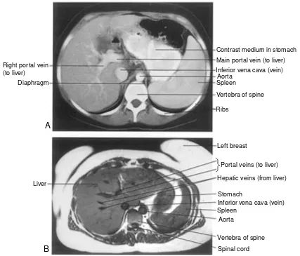
hree imaging techniques that have revolutionized medi-cine are radiography, computed tomography, and mag-netic resonance imaging. With them, physicians today can “see” inside the body without making a single cut. Each tech-nique is so important that its inventor received a Nobel Prize. The oldest is radiography (ra-de-OG-rah-fe), in which a ma-chine beams x-rays (a form of radiation) through the body onto a piece of film. Like other forms of radiation, x-rays dam-age body tissues, but modern equipment uses extremely low doses. The resulting picture is called a radiograph. Dark areas indicate where the beam passed through the body and exposed the film, whereas light areas show where the beam did not pass through. Dense tissues (bone, teeth) absorb most of the x-rays, preventing them from exposing the film. For this reason, radi-ography is commonly used to visualize bone fractures and tooth decay as well as abnormally dense tissues like tumors. Radiography does not provide clear pictures of soft tissues be-cause most of the beam passes through and exposes the film, but contrast media can help make structures like blood vessels and hollow organs more visible. For example, barium sulfate (which absorbs x-rays) coats the digestive tract when ingested.Computed tomography (CT) is based on radiography and also uses very low doses of radiation. During a CT scan, a ma-chine revolves around the patient, beaming x-rays through the body onto a detector. The detector takes numerous pictures of the beam and a computer assembles them into transverse sec-tions, or “slices.” Unlike conventional radiography, CT pro-duces clear images of soft structures such as the brain, liver, and lungs. It is commonly used to visualize brain injuries and tu-mors, and even blood vessels when used with contrast media.
Magnetic resonance imaging uses a strong magnetic field and radiowaves. So far, there is no evidence to suggest that MRI causes tissue damage. The MRI patient lies inside a cham-ber within a very powerful magnet. The molecules in the pa-tient’s soft tissues align with the magnetic field inside the chamber. When radiowaves beamed at the region to be imaged hit the soft tissue, the aligned molecules emit energy that the MRI machine detects, and a computer converts these signals into a picture. MRI produces even clearer images of soft tissue than does computed tomography and can create detailed pic-tures of blood vessels without contrast media. MRI can visual-ize brain injuries and tumors that might be missed using CT.
Medical Imaging: Seeing Without Making a Cut
Medical Imaging: Seeing Without Making a Cut
Contrast medium in stomach Main portal vein (to liver) Inferior vena cava (vein)
Aorta Spleen
Vertebra of spine
Ribs
Left breast
Portal veins (to liver) Hepatic veins (from liver)
Stomach
Inferior vena cava (vein) Spleen
Aorta
Vertebra of spine Spinal cord Right portal vein
(to liver) Diaphragm
Liver
A
B
is made at an angle. The type of section used will
deter-mine what is seen under the microscope, as shown with a
blood vessel in Figure 1-9.
These same terms are used for
im-ages taken by techniques such as
com-puted tomography (CT) or magnetic
resonance imaging (MRI). (See Box
1-2, Medical Imaging: Seeing Without
Making a Cut). In imaging studies, the
term cross section is used more
gener-ally to mean any two-dimensional
view of an internal structure obtained
by imaging, as shown in Figure 1-10.
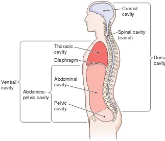
◗
Body Cavities
Internally, the body is divided into a
few large spaces, or cavities,
which
contain the organs. The two main
cav-ities are the dorsal cavity
and ventral
cavity (Fig. 1-11).
Dorsal Cavity
The dorsal body cavity has two
subdivi-sions: the cranial cavity,
containing the
Checkpoint 1-4What are the three planes in which the body can be cut? What kind of a plane divides the body into two equal halves?
brain, and the spinal cavity (canal),
enclosing the spinal cord. These two
areas form one continuous space.
Ventral Cavity
The ventral cavity is much larger than
the dorsal cavity. It has two main
sub-divisions, which are separated by the
di-aphragm
(DI-ah-fram), a muscle used
in breathing. The thoracic
(tho-RAS-ik)
cavity
is located superior to (above) the
diaphragm. Its contents include the
heart, the lungs, and the large blood
vessels that join the heart. The heart is
contained in the pericardial cavity,
formed by the pericardial sac; the lungs
are in the pleural cavity, formed by the
pleurae, the membranes that enclose
the lungs (Fig. 1-12). The mediastinum
(me-de-as-TI-num) is the space
be-tween the lungs, including the organs
and vessels contained in that space.
The
abdominopelvic
(ab-dom-ih-no-PEL-vik) cavity
(see Fig. 1-11)
is
lo-cated inferior to (below) the
di-aphragm. This space is further subdivided into two regions.
The superior portion, the abdominal cavity, contains the
stomach, most of the intestine, the liver, the gallbladder, the
pancreas, and the spleen. The inferior portion, set off by an
imaginary line across the top of the hip bones, is the pelvic
cavity.
This cavity contains the urinary bladder, the rectum,
and the internal parts of the reproductive system.
Thoracic cavity
Mediastinum
Pleural cavity
Pericardial cavity
Diaphragm
Figure 1-12 The thoracic cavity. Shown are the pericardial cavity, which contains the heart, and the pleural cavity, which contains the lungs.
Cranial cavity
Spinal cavity (canal) Thoracic
cavity Diaphragm
Abdominal cavity
Pelvic cavity
Abdomino-pelvic cavity Ventral
cavity
Dorsal cavity
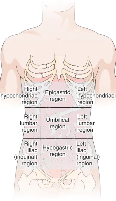
Regions of the Abdomen
It is helpful to divide the
abdomen for examination and reference into nine regions
(Fig. 1-13).
Figure 1-13 The nine regions of the abdomen. Figure 1-14 Quadrants of the abdomen. The organs within each quadrant are shown.
E
very time a patient receives medical treatment, informa-tion is added to the patient’s medical record, which in-cludes data about symptoms, medical history, test results, di-agnoses, and treatment. Health information technicians organize and manage these records, working closely with physicians, nurses, and other health professionals to ensure that medical records provide a complete, accurate basis for quality patient care.Accurate medical records are also essential for administra-tive purposes. Health information technicians assign a codeto each diagnosis and procedure a patient receives, and this in-formation is used for accurate patient billing. In addition, health information technicians analyze medical records to dis-cover trends in health and disease. This research can be used
to improve patient care, manage costs, and help establish new medical treatments.
Health information technicians need a strong clinical knowledge base. A thorough background in medical termi-nology is essential when reading and interpreting medical records. Anatomy and physiology are definitely required!
Most health information technologists work in hospitals and long-term care facilities. Others work in medical clinics, government agencies, insurance companies, and consulting firms. Job prospects are promising because of the growing need for healthcare. In fact, health information technology is projected to be one of the fastest growing careers in the United States. For more information about this profession, contact the American Health Information Management Association.
Health Information Technicians
Box 1-3 • Health Professions
Health Information Technicians
Checkpoint 1-5 There are two main body cavities, oneposte-rior and one anteposte-rior. Name these two cavities.
The three central regions, from superior to inferior are:
◗the epigastric
(ep-ih-GAS-trik) region,
located just
in-ferior to the breastbone
scales. In this text, equivalents in the more familiar units
of inches and feet are included along with the metric
units for comparison. There are 2.5 centimeters (cm) or
25 millimeters (mm) in 1 inch, as shown in Figure 1-15.
Some equivalents that may help you to appreciate the size
of various body parts are as follows:
1 mm
⫽
0.04 inch, or 1 inch
⫽
25 mm
1 cm
⫽
0.4 inch, or 1 inch
⫽
2.5 cm
1 m
⫽
3.3 feet, or 1 foot
⫽
30 cm
Units of Weight
The same prefixes used for linear measurements are used
for weights and volumes. The gram
(g) is the basic unit of
weight. Thirty grams are about equal to 1 ounce, and 1
kilogram to 2.2 pounds. Drug dosages are usually stated
in grams or milligrams. One thousand milligrams equal 1
gram; a 500-milligram (mg) dose would be the equivalent
of 0.5 gram (g), and 250 mg is equal to 0.25 g.
Units of Volume
The dosages of liquid medications are given in units of
volume. The basic metric measurement for volume is the
liter
(L) (LE-ter). There are 1000 milliliters (mL) in a
liter. A liter is slightly greater than a quart, a liter being
equal to 1.06 quarts. For smaller quantities, the milliliter
is used most of the time. There are 5 ml in a teaspoon and
15 mL in a tablespoon. A fluid ounce contains 30 mL.
Temperature
The Celsius (centigrade) temperature scale, now in use
by most countries and by scientists in this country, is
dis-cussed in Chapter 20.
A chart of all the common metric measurements and
their equivalents is shown in Appendix 1. A
Celsius-Fahrenheit temperature conversion scale appears in
Ap-pendix 2.
Checkpoint 1-6Name the three central regions and the three left and right lateral regions of the abdomen.
Checkpoint 1-7Name the basic units of length, weight, and vol-ume in the metric system.
0 Centimeters
2 1
0
1
2
3 4 5
Inches
Figure 1-15 Comparison of centimeters and inches. ◗
the hypogastric
(hi-po-GAS-trik) region,
the most
infe-rior of all the midline regions
The regions on the right and left, from superior to
in-ferior, are:
◗
the
hypochondriac
(hi-po-KON-dre-ak)
regions,
just
inferior to the ribs
◗
the lumbar regions, which are on a level with the
lum-bar regions of the spine
◗
the iliac,
or inguinal
(IN-gwih-nal), regions, named for
the upper crest of the hipbone and the groin region,
re-spectively
A simpler but less precise division into four quadrants
is sometimes used. These regions are the right upper
quadrant (RUQ), left upper quadrant (LUQ), right lower
quadrant (RLQ), and left lower quadrant (LLQ) (Fig.
1-14). (See Box 1-3, Health Information Technicians, for
description of a profession that uses anatomical,
physio-logical, and medical terms)
◗
The Metric System
Now that we have set the stage for further study of the
body’s structure and function, we should take a look at
the metric system, because this system is used for all
sci-entific measurements. The drug industry and the
health-care industry already have converted to the metric
sys-tem, so anyone who plans a career in healthcare should
be acquainted with metrics.
The metric system is like the monetary system in the
United States. Both are decimal systems based on
multi-ples of the number 10. One hundred cents equal one
dol-lar; one hundred centimeters equal one meter. Each
mul-tiple in the decimal system is indicated by a prefix:
kilo
⫽
1000
centi
⫽
1/100
milli
⫽
1/1000
micro
⫽
1/1,000,000
Units of Length
Medical terms are built from standardized word parts (prefixes, roots, and suffixes). Learning the meanings of these parts can help you remember words and interpret unfamiliar terms.
WORD PART MEANING EXAMPLE
Studies of the Human Body
-tomy cutting, incision of Anatomycan be revealed by making incisions in the body.
dis- apart, away from To dissectis to cut apart.
physi/o nature, physical Physiologyis the study of how the body functions.
path/o disease Pathologyis the study of disease.
Body Processes
cata- down Catabolismis the breakdown of complex substances into simpler
ones.
ana- upward, again, back Anabolismis the building up of simple compounds into more complex substances.
home/o- same Homeostasisis the steady state (sameness) within an organism.
stat stand, stoppage, constancy In homeostasis, “-stasis” refers to constancy.
Word Anatomy
Summary
I. Studies of the human body
1. Anatomy—study of structure 2. Physiology—study of function 3. Pathology—study of diseaseA. Levels of organization—chemicals, cell, tissue, organ, organ system, whole organism
II. Body systems
1. Integumentary system—skin and associated structures 2. Skeletal system—support
3. Muscular system—movement
4. Nervous system—reception of stimuli and control of responses
5. Endocrine system—production of hormones for regu-lation of growth, metabolism, reproduction
6. Cardiovascular system—movement of blood for transport 7. Lymphatic system—aids in circulation, immunity, and
absorption of digested fats
8. Respiratory system—intake of oxygen and release of carbon dioxide
9. Digestive system—intake, breakdown, and absorption of nutrients
10. Urinary system—elimination of waste and water 11. Reproductive system—production of offspring
III. Metabolism and its regulation
1. Metabolism—all the chemical reactions needed to sus-tain life
2. Catabolism—breakdown of complex substances into simpler substances; release of energy from nutrients a. ATP (adenosine triphosphate)—energy compound of
cells
3. Anabolism—building of body materials
A. Homeostasis—steady state of body conditions 1. Fluid balance
a. Extracellular fluid—outside the cells b. Intracellular fluid—inside the cells
2. Feedback—regulation by return of information within a system
a. Negative feedback—reverses an action
b. Positive feedback—promotes continued activity B. Effects of aging—changes in all systems
IV. Directions in the body
1. Anatomical position—upright, palms forward, face front, feet parallel
A. Directional terms
1. Superior—above or higher; inferior—below or lower
2. Ventral (anterior)—toward belly or front surface; dorsal (posterior)—nearer to back surface
3. Cranial—nearer to head; caudal—nearer to sacrum 4. Medial—toward midline; lateral—toward side 5. Proximal—nearer to point of origin; distal—farther
from point of origin B. Planes of division
1. Body divisions
a. Sagittal—from front to back, dividing the body into left and right parts
(1) Midsagittal—exactly down the midline b. Frontal (coronal)—from left to right, dividing the
body into anterior and posterior parts
c. Transverse—horizontally, dividing the body into su-perior and inferior parts
2. Tissue sections
V. Body cavities
A. Dorsal cavity—contains cranial and spinal cavities for brain and spinal cord
B. Ventral cavity
1. Thoracic—chest cavity
a. Divided from abdominal cavity by diaphragm b. Contains heart and lungs
c. Mediastinum—space between lungs and the organs contained in that space
2. Abdominopelvic
a. Abdominal—upper region containing stomach, most of intestine, pancreas, liver, spleen, and others b. Pelvic—lower region containing reproductive
or-gans, urinary bladder, rectum c. Nine regions of the abdomen
(1) Central—epigastric, umbilical, hypogastric (2) Lateral (right and left)—hypochondriac,
lum-bar, iliac (inguinal)
d. Quadrants—abdomen divided into four regions
VI. The metric system—based on multiples
of 10
1. Basic units a. Meter—length b. Gram—weight c. Liter—volume
2. Prefixes—indicate multiples of 10 a. Kilo—1000 times
b. Centi—1/100th (0.01) c. Milli—1/1000th (0.001) d. Micro—1/1,000,000 (0.000001) A. Units of length
B. Units of weight C. Units of volume
D. Temperature—measured in Celsius (centigrade) scale
Questions for Study and Review
Building Understanding
Fill in the blanks
1. Tissues may function together as ______.
2. Glands that produce hormones belong to the ______
system.
3. The eyes are located ______ to the nose.
4. Normal body function maintains a state of internal
balance called ______.
5. The basic unit of volume in the metric system is the
______.
Matching
Match each numbered item with the most closely related lettered item.
___ 6. One of two systems that control and coordinate other systems
___ 7. The system that brings needed substances to the body tissues
___ 8. The system that converts foods into a form that body cells can use
___ 9. The cavity that contains the liver
___ 10. The cavity that contains the urinary bladder
a. nervous system
b. abdominal cavity
c. cardiovascular system
d. pelvic cavity
e. digestive system
Multiple choice
___ 11. The study of normal body structure is
a. homeostasis
b. anatomy
c. physiology
d. pathology
___ 12. Fluids contained within cells are described as
a. intracellular
b. ventral
c. extracellular
d. dorsal
___ 13. A type of feedback in which a given action
promotes more of the same is called
a. homeostasis
b. biofeedback
c. positive feedback
d. negative feedback
___ 14. The cavity that contains the mediastinum is the
a. dorsal
b. ventral
c. abdominal
d. pelvic
___ 15. The foot is located ______ to the knee.
a. superior
b. inferior
c. proximal
d. distal
Understanding Concepts
why a disease at the chemical level can have an effect on
organ system function.
24. When glucose levels in the blood drop below
nor-mal the pancreas releases a hormone called glucagon.
Using your understanding of negative feedback, discuss
the possible role of glucagon in blood glucose
home-ostasis.
25. Your patient’s chart reads: “Patient reports pain in
right lower quadrant of abdomen. X-ray reveals mass in
right iliac region.” Locate this region on yourself and
ex-plain why it is important for health professionals to use
anatomical terminology when describing the human
body.
18. Compare and contrast the anatomy and physiology of
the nervous system with that of the endocrine system.
19. What is ATP? What type of metabolic activity
re-leases the energy used to make ATP?
20. Compare and contrast intracellular and extracellular
fluids.
21. Explain how an internal state of balance is
main-tained in the body.
22. List the subdivisions of the dorsal and ventral
cavi-ties. Name some organs found in each subdivision.
Conceptual Thinking
called “lead” in a pencil), coal, charcoal, and diamonds are
different forms of the element carbon.
Elements can be identified by their names or their
chemical symbols, which are abbreviations of the modern
or Latin names of the elements. Each element is also
iden-tified by its own number, which is based on the structure
of its subunits, or atoms. The periodic table is a chart
used by chemists to organize and describe the elements.
Appendix 3 shows the periodic table and gives some
in-formation about how it is used.
Table 2-1
lists some
ele-ments found in the human body along with their
functions.
Atoms
The subunits of elements are
atoms.
These are the
smallest complete units of matter. They cannot be broken
down or changed into another form by ordinary chemical
and physical means. These subunits are so small that
millions of them could fit on the sharpened end of a
pencil.
Atomic Structure
Despite the fact that the atom is
such a tiny particle, it has been carefully studied and has
been found to have a definite structure. At the center of
the atom is a nucleus, which contains positively charged
electrical particles called
protons
(PRO-tonz)
and
non-charged particles called
neutrons
(NU-tronz).
Together,
the protons and neutrons contribute nearly all of the
atom’s weight.
In orbit outside the nucleus are
electrons
(e-LEK-tronz)
(Fig. 2-1)
. These nearly
weight-less particles are negatively charged. It
is the electrons that determine how the
atom will react chemically. The protons
and electrons of an atom always are
equal in number, so that the atom as a
whole is electrically neutral.
The
atomic number
of an element
is equal to the number of protons that
are present in the nucleus of each of its
atoms. Because the number of protons
is equal to the number of electrons, the
atomic number also represents the
number of electrons whirling around
the nucleus. Each element has a
spe-cific atomic number. No two elements
share the same number. In the Periodic
Table of the Elements (see Appendix 3)
the atomic number is located at the top
of the box for each element.
The positively charged protons
keep the negatively charged electrons
in orbit around the nucleus by means
of the opposite charges on the particles.
Positively (
⫹
) charged protons attract
negatively (
⫺
) charged electrons.
G
reater understanding of living organisms has come to
us through
chemistry,
the science that deals with the
composition and properties of matter. Knowledge of
chemistry and chemical changes helps us understand the
normal and abnormal functioning of the body. The
diges-tion of food in the intestinal tract, the producdiges-tion of urine
by the kidneys, the regulation of breathing, and all other
body activities involve the principles of chemistry. The
many drugs used to treat diseases are chemicals.
Chem-istry is used for the development of drugs and for an
un-derstanding of their actions in the body.
To provide some insights into the importance of
chem-istry in the life sciences, this chapter briefly describes
ele-ments, atoms and molecules, compounds, and mixtures
,
which are fundamental forms of matter.
◗
Elements
Matter is anything that takes up space, that is, the
materi-als from which all of the universe is made. Elements are
the substances that make up all matter. The food we eat,
the atmosphere, water—everything around us, everything
we can see and touch, is made of elements. There are 92
naturally occurring elements. (Twenty additional elements
have been created in the laboratory.) Examples of elements
include various gases, such as hydrogen, oxygen, and
ni-trogen; liquids, such as mercury used in barometers and
other scientific instruments; and many solids, such as
iron, aluminum, gold, silver, and zinc. Graphite (the
so-Some Common Chemical Elements*
Table 2•1NAME
SYMBOL
FUNCTION
*The elements are listed in decreasing order by weight in the body. Oxygen
Part of water; needed to metabolize nutrients for energy
Basis of all organic compounds; in carbon dioxide, the waste gas of metabolism Part of water; participates in energy
metabo-lism, acid–base balance
Present in all proteins, ATP (the energy com-pound), and nucleic acids (DNA and RNA) Builds bones and teeth; needed for muscle
contraction, nerve impulse conduction, and blood clotting
Active ingredient in the energy-storing compound ATP; builds bones and teeth; in cell membranes and nucleic acids Nerve impulse conduction; muscle
contrac-tion; water balance and acid–base balance Part of many proteins
Active in water balance, nerve impulse conduction, and muscle contraction Active in water balance and acid–base
balance; found in stomach acid Part of hemoglobin, the compound that
Energy Levels
The electrons of an atom orbit at
spe-cific distances from the nucleus in regions called energy
levels. The first energy level, the one closest to the
nu-cleus, can hold only two electrons. The second energy
level, the next in distance away from the nucleus, can hold
eight electrons.
More distant energy levels can hold more than eight
electrons, but they are stable (nonreactive) when they
have eight.
The electrons in the energy level farthest away from
the nucleus give the atom its chemical characteristics. If
the outermost energy level has more than four electrons
but less than its capacity of eight, the atom normally
com-pletes this level by gaining electrons. In the process, it
be-comes negatively charged, because it has more electrons
than protons. The oxygen atom illustrated in
Figure 2-1
has six electrons in its second, or outermost, level. When
oxygen enters into chemical reactions, it gains two
elec-trons, as when it reacts with hydrogen to form water
(Fig.
2-2)
. The oxygen atom then has two more electrons than
protons.
If the outermost energy level has fewer than four
elec-trons, the atom normally loses those electrons. In so
doing, it becomes positively charged, because it now has
more protons than electrons.
The number of electrons lost or gained by atoms of an
element in chemical reactions is known as the
valence
of
that element (from a Latin word that means “strength”).
The outermost energy level, which determines the
com-bining properties of the element, is the valence level.
Va-lence is reported as a number with a
⫹
or – to indicate
whether electrons are lost or gained in chemical reactions.
Remember that electrons carry a negative charge, so when
an atom gains electrons it becomes negatively charged and
when an atom loses electrons it becomes positively
charged. For example, the valence of oxygen, which gains
two electrons in chemical reactions, is shown as O
2⫺.
◗
Molecules and Compounds
A
molecule
(MOL-eh-kule) is formed when two or more
atoms unite on the basis of their electron structures. A
mol-ecule can be made of like atoms—the oxygen molmol-ecule is
made of two identical atoms—but more often a molecule is
made of atoms of two or more different elements. For
ex-ample, a molecule of water (H2O) contains one atom of
oxygen (O) and two atoms of hydrogen (H)
(see Fig. 2-2).
Substances composed of two or more different
ele-ments are called
compounds.
Molecules are the smallest
subunits of a compound. Each molecule of a compound
contains the elements that make up that compound in the
proper ratio. Some compounds are made of a few elements
in a simple combination. For example, the gas carbon
Checkpoint 2-1 What are atoms?Checkpoint 2-2 What are three types of particles found in atoms?
Electron
Central nucleus 8 protons (red) 8 neutrons (green)
First energy level
Second energy level
Figure 2-1 Representation of the oxygen atom.Eight pro-tons and eight neutrons are tightly bound in the central nucleus. The eight electrons are in orbit around the nucleus, two at the first energy level and six at the second. ZOOMING IN ✦How does the number of protons in this atom compare with the num-ber of electrons?
Oxygen atom
Hydrogen atom Hydrogen atom
Mixtures: Solutions and
Suspensions
Not all elements or compounds combine chemically
when brought together. The air we breathe every day is a
mixture of gases, largely nitrogen, oxygen, and carbon
dioxide, along with smaller percentages of other
sub-stances. The constituents in the air maintain their
iden-tity, although the proportions of each may vary. Blood
plasma is also a mixture in which the various
compo-nents maintain their identity. The many valuable
com-pounds in the plasma remain separate entities with their
own properties. Such combinations are called
mixtures
—blends of two or more substances
(Table 2-2)
.
A mixture formed when one substance dissolves in
another is called a
solution.
One example is salt water. In
a solution, the component substances cannot be
distin-guished from each other and they remain evenly
distrib-uted throughout; that is, the mixture is homogeneous
(ho-mo-JE-ne-us). The dissolving substance, which in
the body is water, is the
solvent.
The substance
dis-solved, salt in the case of salt water, is the
solute.
An
aqueous
(A-kwe-us)
solution
is one in which water is
the solvent. Aqueous solutions of glucose, salts, or both
of these together are used for intravenous fluid
treat-ments.
In some mixtures, the substance distributed in the
background material is not dissolved and will settle out
unless the mixture is constantly shaken. This type of
non-uniform, or heterogeneous (het-er-o-JE-ne-us),
mix-ture is called a
suspension.
The particles in a suspension
are separate from the material in which they are
dis-persed, and they settle out because they are large and
heavy. Examples of suspensions are milk of magnesia,
finger paints, and, in the body, red blood cells suspended
in blood plasma.
The Importance of Water
Water is the most abundant compound in the body. No plant
or animal, including the human, can live very long without
it. Water is of critical importance in all physiological
processes in body tissues. A deficiency of water, or
dehydra-tion (de-hi-DRA-shun), can be a serious threat to health.
Water carries substances to and from the cells and makes
possible the essential processes of absorption, exchange,
se-cretion, and excretion. What are some of the properties of
water that make it such an ideal medium for living cells?
◗
Water can dissolve many different substances in large
amounts. For this reason, it is called the
universal
sol-vent.
Many of the materials needed by the body, such as
gases, minerals, and nutrients, dissolve in water to be
carried from place to place. Substances, such as salts,
that mix with or dissolve in water are described as
hy-drophilic (“water-loving”); those, such as fats, that repel
and do not dissolve in water are described as
hydropho-bic
(“water-fearing”).
◗
Water is stable as a liquid at ordinary
temperatures. Water does not freeze
until the temperature drops to 0
⬚
C
(32
⬚
F) and does not boil until the
temperature reaches 100
⬚
C (212
⬚
F).
This stability provides a constant
en-vironment for body cells. Water can
also be used to distribute heat
throughout the body and to cool the
body by evaporation of sweat from
the body surface.
◗
Water participates in chemical
reac-tions in the body. It is needed
di-rectly in the process of digestion and
in many of the metabolic reactions
that occur in the cells.
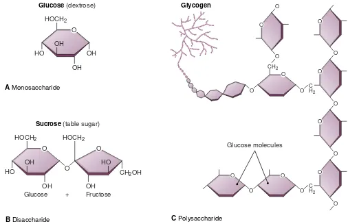
monoxide (CO) contains 1 atom of carbon (C) and 1 atom
of oxygen (O). Other compounds are very large and
plex. Such complexity characterizes many of the
com-pounds found in living organisms. Some proteins, for
ex-ample, have thousands of atoms.
It is interesting to observe how different a compound
is from any of its constituents. For example, a molecule of
liquid water is formed from oxygen and hydrogen, both of
which are gases. Another example is a crystal sugar,
glu-cose (C6H12O6). Its constituents include 12 atoms of the
gas hydrogen, 6 atoms of the gas oxygen, and 6 atoms of
the solid element carbon. The component gases and the
solid carbon do not in any way resemble the glucose.
Checkpoint 2-3 What are molecules?
Checkpoint 2-4 What is the most abun-dant compound in the body?
Mixtures
solves in another (solvent) Heterogeneous mixture inwhich one substance is dispersed in another, but will settle out unless constantly mixed Heterogeneous mixture
in which the suspended material remains evenly distributed based on the small size and opposing charges of the particles
Table salt (NaCl) dissolved in water; table sugar (sucrose) dissolved in water
Red blood cells in blood plasma; milk of magnesia
One other type of mixture is of importance in the
body. Some organic compounds form
colloids,
in which
the molecules do not dissolve yet remain evenly
distrib-uted in the suspending material. The particles have
elec-trical charges that repel each other, and the molecules are
small enough to stay in suspension. The fluid that fills the
cells (cytosol) is a colloidal suspension, as is blood
plasma.
Many mixtures are complex, with properties of
solu-tions, suspensions, and colloidal suspensions. Blood
plasma has dissolved compounds, making it a solution.
The red blood cells and other formed elements give blood
the property of a suspension. The proteins in the plasma
give it the property of a colloidal suspension. Chocolate
milk also has all three properties.
the sodium atom, forming an ionic bond. The two newly
formed ions (Na
⫹and Cl
⫺), because of their opposite
charges, attract each other to produce the compound
sodium chloride, ordinary table salt
(Fig. 2-3 C)
.
Electrolytes
Ionically bonded substances, when they
go into solution, separate into charged particles.
Com-pounds formed by ionic bonds that release ions when
they are in solution are called
electrolytes
(e-LEK-tro-lites). Note that in practice, the term electrolytes
is also
used to refer to the ions themselves in body fluids.
Elec-Electron
Sodium atom Chlorine atom
Sodium ion (Na+)
Na+ Cl–
Sodium chloride (table salt)
Chloride ion (Cl–)
B A
C
11P
17P
17P 17P 11P
11P
Electron
Figure 2-3 Ionic bonding. (A)A sodium atom has 11 pro-tons and 11 electrons. A chlorine atom has 17 propro-tons and 17 electrons. (B)A sodium atom gives up one electron to a chlo-rine atom in forming an ionic bond. The sodium atom now has 11 protons and 10 electrons, resulting in a positive charge of one. The chlorine becomes negatively charged by one, with 17 protons and 18 electrons. (C) The sodium ion (Na⫹) is at-tracted to the chloride ion (Cl-) in forming the compound sodium chloride (table salt).
Checkpoint 2-5 Both solutions and suspensions are types of mixtures. What is the difference between them?
◗
Chemical Bonds
When discussing the structure of the atom, we mentioned
the positively charged (
⫹
) protons that are located in the
nucleus and the equal number of orbiting negatively
charged (
⫺
) electrons that neutralize the protons
(Fig.
2-3 A)
. Atoms interact, however, to reach a stable number of
electrons in the outermost energy level. These chemical
interactions alter the neutrality of the atoms and also form
a bond between them. In chemical reactions, electrons
may be transferred from one atom to another or may be
shared between atoms.
Ionic Bonds
When electrons are transferred from one atom to another,
the type of bond formed is called an
ionic
(i-ON-ik)
bond.
The sodium atom, for example, tends to lose the single
electron in its outermost shell
(Fig. 2-3 B)
, leaving an
out-ermost shell with a stable number of electrons (8).
Re-moval of a single electron from the sodium atom leaves
one more proton than electrons, and the atom then has a
single net positive charge. The sodium atom in this form
is symbolized as Na
⫹. An atom or group of atoms with a
positive or negative charge is called an
ion
(I-on). Any ion
that is positively charged is a
cation
(CAT-i-on).
Alternately, atoms can gain electrons so that there
are more electrons than protons. Chlorine, which has
seven electrons in its outermost energy level, tends to
gain one electron to fill the level to its capacity. Such an
atom of chlorine is negatively charged (Cl
⫺)
(see Fig.
2-3 B)
. (Chemists refer to this charged form of chlorine as
chloride.) Any negatively charged ion is an
anion
(AN-i-on).
trolytes include a variety of salts, such as sodium chloride
and potassium chloride. They also include acids and
bases, which are responsible for the acidity or alkalinity of
body fluids, as described later in this chapter. Electrolytes
must be present in exactly the right quantities in the fluid
within the cell (intracellular fluid) and the fluid outside
the cell (extracellular fluid), or very damaging effects will
result, preventing the cells in the body from functioning
properly.
Ions in the Body
Many different ions are found in
body fluids. Calcium ions (Ca
2⫹) are necessary for the
clotting of blood, the contraction of muscle, and the
health of bone tissue. Bicarbonate ions (HCO3
⫺) are
re-quired for the regulation of acidity and alkalinity of body
fluids. The stable condition of the normal organism,
homeostasis, is influenced by ions.
Because ions are charged particles, electrolyte
solu-tions can conduct an electric current. Records of electric
currents in tissues are valuable indications of the
func-tioning or malfuncfunc-tioning of tissues and organs. The
elec-trocardiogram
(e-lek-tro-KAR-de-o-gram) and the
elec-troencephalogram
(e-lek-tro-en-SEF-ah-lo-gram) are
graphic tracings of the electric currents generated by the
heart muscle and the brain, respectively (see Chapters 10
and 14).
Covalent Bonds
Although ionic bonds form many chemical compounds, a
much larger number of compounds are formed by another
type of chemical bond. This bond involves not the exchange
of electrons but a sharing of electrons between the atoms in
the molecule and is called a
covalent bond.
This name
comes from the prefix co-, meaning “together,” and valence,
referring to the electrons involved in chemical reactions
be-tween atoms. In a covalently bonded molecule, the valence
electrons orbit around both of the atoms, making both of
them stable. Covalent bonds may involve the sharing of one,
two, or three pairs of electrons between atoms.
In some covalently bonded molecules, the electrons are
equally shared, as in the case of a hydrogen molecule (H2)
and other molecules composed of atoms of the same element
(Fig. 2-4)
. Electrons may also be shared equally in some
I
n contrast to ionic and covalent bonds, which hold atoms together, hydrogen bonds hold molecules together. Hydro-gen bonds are much weaker than ionic or covalent bonds—in fact, they are more like “attractions” between molecules. While ionic and covalent bonds rely on electron transfer or sharing, hydrogen bonds form bridges between two mole-cules. A hydrogen bond forms when a slightly positive hydro-gen atom in one molecule is attracted to a slightly negative atom in another molecule. Even though a single hydrogen bond is weak, many hydrogen bonds between two molecules can be strong.Hydrogen bonds hold water molecules together, with the slightly positive hydrogen atom in one molecule attracted to a slightly negative oxygen atom in another. Many of water’s unique properties come from its ability to form hydrogen bonds. For example, hydrogen bonds keep water liquid over a wide range of temperatures, which provides a constant envi-ronment for body cells.
Hydrogen bonds form not only between molecules but also within large molecules. Hydrogen bonds between regions of the same molecule cause it to fold and coil into a specific shape, as in the process that creates the precise
three-dimen-sional structure of proteins. Because a protein’s structure de-termines its function in the body, hydrogen bonds are essen-tial to protein activity.
Box 2-1 A Closer Look
Hydrogen bonds
Water molecules
H
H
O
-+
Hydrogen Bonds: Strength in Numbers
Hydrogen Bonds: Strength in Numbers
Hydrogen bonds. The bonds shown here are holding water mole-cules together.
Checkpoint 2-6 What happens when an electrolyte goes into solution?
+ +
Hydrogen molecule (H2)
molecules composed of different atoms, methane (CH4), for
example. If electrons are equally shared in forming a
mole-cule, the electrical charges are evenly distributed around the
atoms and the bond is described as a nonpolar covalent bond.
That is, no part of the molecule is more negative or positive
than any other part of the molecule. More commonly, the
electrons are held closer to one atom than the other, as in the
case of water (H2O), shown in
Figure 2-2
. In a water
mole-cule, the shared electrons are actually closer to the oxygen at
any one time making that region of the molecule more
neg-ative. Such bonds are called polar covalent bonds, because
one part of the molecule is more negative and one part is
more positive at any one time. Anyone studying biological
chemistry (biochemistry) is interested in covalent bonding
because carbon, the element that is the basis of organic
chemistry, forms covalent bonds with a wide variety of
dif-ferent elements. Thus, the compounds that are characteristic
of living things are covalently bonded compounds. For a
de-scription of another type of bond, see Box 2-1, Hydrogen
Bonds: Strength in Numbers.
ions increases, the concentration of hydrogen ions
de-creases. Acidity and alkalinity are indicated by
pH
units,
which represent the relative concentrations of hydrogen
and hydroxide ions in a solution. The pH units are listed
on a scale from 0 to 14, with 0 being the most acidic and
14 being the most basic
(Fig. 2-5)
. A pH of 7.0 is neutral.
At pH 7.0 the solution has an equal number of hydrogen
and hydroxide ions. Pure water has a pH of 7.0. Solutions
that measure less than 7.0 are acidic; those that measure
above 7.0 are alkaline (basic).
Because the pH scale is based on multiples of 10, each
pH unit on the scale represents a 10-fold change in the
number of hydrogen and hydroxide ions present. A
solu-tion registering 5.0 on the scale has 10 times the number
of hydrogen ions as a solution that registers 6.0. The pH
5.0 solution also has one tenth the number of hydroxide
ions as the solution of pH 6.0. A solution registering 9.0
has one tenth the number of hydrogen ions and 10 times
the number of hydroxide ions as one registering 8.0. Thus,
the lower the pH reading, the greater is the acidity, and the
higher the pH, the greater is the alkalinity.
Blood and other body fluids are close to neutral but
are slightly on the alkaline side, with a pH range of
Increasing acidity
Milk of magnesia (10.5)
Household ammonia (11.5)
Lye (13)
Figure 2-5 The pH scale.Degree of acidity or alkalinity is shown in pH units. This scale also shows the pH of some com-mon substances. ZOOMING IN ✦What happens to the amount of hydroxide ion (OH⫺) present in a solution when the amount of
hydrogen ion (H⫹) increases? Checkpoint 2-7 How is a covalent bond formed?
◗
Compounds: Acids, Bases, and
Salts
An
acid
is a chemical substance capable of donating a
hy-drogen ion (H
⫹) to another substance. A common
exam-ple is hydrochloric acid, the acid found in stomach juices:
HCl
→
H
⫹⫹
Cl
⫺(hydrochloric (hydrogen ion) (chloride ion) acid)
A
base
is a chemical substance, usually containing a
hydroxide ion (OH
⫺), that can accept a hydrogen ion. A
base is also called an
alkali
(AL-kah-li).
Sodium
hydrox-ide, which releases hydroxide ion in solution, is an
exam-ple of a base:
NaOH
→
Na
⫹⫹
OH
⫺(sodium (sodium ion) (hydroxide ion) hydroxide)