BURKITT LYMPHOMA IN GASTROINTESTINAL TRACT:
A REPORT OF TWO CASES
Aleksandar Čubranić1, Marin Golčić2, Dora Fučkar-Čupić3, Boris Brozović1, Domagoj Gajski4 and Ivan Brumini5
1Department of Gastroenterology, Rijeka University Hospital Centre, Rijeka, Croatia; 2Department of Radiotherapy and Oncology, Rijeka University Hospital Centre, Rijeka, Croatia;
3Department of Pathology, Rijeka University Hospital Centre, Rijeka, Croatia; 4Department of Neurosurgery, Sestre milosrdnice University Hospital Centre, Zagreb, Croatia;
5University of Applied Health Sciences, Zagreb, Croatia
SUMMARY – Burkitt lymphoma, a type of non-Hodgkin B-cell lymphoma, is the fastest grow-ing human cancer, presentgrow-ing pathologically with a ‘starry sky’ pattern. It is most often found in the abdomen and the jaw, however, localization in the abdomen other than the ileocecal area is very rare and described only in a handful of cases. Standard treatment consists of initial tumor cytoreduction followed by intense chemotherapy. Most of the relapses occur within one year of the diagnosis, while the 5-year survival is around 80%. We present two cases which are specific for unusual location of Burkitt lymphoma in the colon and stomach, in immunocompetent patients with negative Epstein-Barr virus tests. Also, one of the patients presented is one of the oldest ever reported with abdominal Burkitt lymphoma, while the other patient is an example of diagnostic difficulties in distinguishing Burkitt lymphoma from similar lymphomas. Due to the rapidly growing tumors and urgent need for cytoreductive surgery, it is crucial to consider the diagnosis of Burkitt lymphoma even in atypical lo-calizations or absence of the common risk factors associated with Burkitt lymphoma.
Key words: Burkitt lymphoma; Case reports; Colon; Cytoreduction surgical procedures
Correspondence to: Aleksandar Čubranić, MD, Department of Gas-troenterology, Rijeka University Hospital Centre, Krešimirova 42, HR-51000 Rijeka, Croatia
E-mail: [email protected]
Received March 7, 2018, accepted October 30, 2018
Introduction
Burkitt lymphoma (BL) is a type of non-Hodgkin B-cell lymphoma, found endemically in equatorial Africa and New Guinea in children infected with ma-laria and Epstein Barr virus (EBV), in immunocom-promised patients, or as a sporadic form in Western countries1. BL consists of uniform medium-sized B-lymphocytes with basophilic cytoplasm, numerous mitoses, and apoptosis. Macrophages are scattered among tumor cells, giving BL a distinctive histologic appearance called “starry sky pattern”2.
Tumor cells express membrane immunoglobulin (Ig) M, Ig light chain, B-cellular antigen, B-cell lymphoma (BCL) protein 6, cluster of differentiation (CD) 10, 19, 20 and 22, while expressing negative re-sults for CD 5, 23, and BCL 2. The most common cytogenetic change is t(8;14)(q24;q32), t(q11;q24) or t(8,22)(q24;q11) translocation. The proliferation index (Ki67) of BL is very high, usually over 95%, so it is not surprising that BL is the fastest growing human cancer3-5.
In adults, BL is most often found in the abdomen, followed by the jaw, while central nervous system dis-ease is reported in up to 40% of patients. Localization other than the ileocecal area is very rare. BL can pres-ent as abdominal or jaw pain, melena, nausea, or even acute abdomen6,7, although the symptoms depend on the primary localization and spread of the tumor.
Standard treatment consists of initial tumor cyto-reduction followed by several cycles of intense chemo-therapy based on high doses of cyclophosphamide, methotrexate, rituximab, and intrathecal application of cytostatics along with other necessary supportive med-ications8,9.
Relapses mostly occur within one year of the diag-nosis, while the 5-year survival is around 80%. Around 10%-15% of patients die due to complications during treatment, while 5%-10% die from BL. Prognosis de-pends on patient age and spread of the disease. Adult patients with localized disease have a better prognosis than those with metastatic disease. On the other hand, children have a much better prognosis than adults, even in case of low stage disease10.
In the cases presented, appropriate informed con-sent was obtained, and we ensured everything to pro-tect the patients’ identity, respecting the highest ethical standards.
Case 1
A 73-year-old male Caucasian complained of he-matochezia and symptoms of severe anemia. He had been operated for laryngeal cancer seven years before, without other oncologic treatment, and had a perfo-rated gastric ulcer 19 years before; his previous medical history also included diabetes mellitus.
Esophagogastroduodenoscopy (EGDS) was per-formed following the work-up in the Emergency De-partment, but no recent signs of bleeding were found.
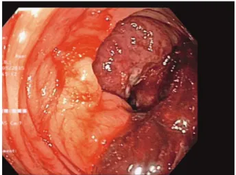
Fig. 1. Colonoscopy view of the tumor in the hepatic flexure.
Fig. 2. Colonoscopy view of the tumor in the hepatic flexure.
Fig. 3. Histology of Burkitt lymphoma found in the colon (HE, magnification X200). Histopathologic appearance of starry sky pattern: solid sheets of middle-sized atypical lymphocytes, with scattered macrophages.
Colonoscopy followed, displaying a tumorous mass in the area of hepatic flexure of the colon (Figs. 1 and 2). The histopathologic diagnosis was BL (Fig. 3). Immu-nohistochemically, tumor cells were negative for panCK, chromogranin, synaptophysin and CD5, and positive for LCA and CD20, with Ki67 over 95%.
For staging purposes, multi-slice computed to-mography (MSCT) of the abdomen was performed, showing a sizeable neoplastic process of hepatic flex-ure with regional lymphadenopathy. No other radio-logical abnormality was described. The case was pre-sented to a multidisciplinary team for colorectal can-cer, and a surgical procedure of right-sided
hemi-colectomy with latero-lateral (LL) anastomosis was suggested and later performed.
Adjuvant therapy consisted of EPOCH protocol along with palonosetron and other necessary support-ive treatment.
Case 2
A 39-year-old male Caucasian started diagnostic procedure due to pain in the upper abdomen without any other symptoms. EGDS was performed to show a stomach mass and biopsy was done. There were seven biopsy specimens of 3 mm in size; histopathologically, these were gastric mucosal fragments infiltrated with dense atypical lymphocytes, with no starry sky
mor-phology. On immunohistochemical analysis, tumor cells were CD20+, CD10+, Bcl6+, cyclinD1-, and nearly 90% Ki67 positive. The histopathologic diagno-sis was non-Hodgkin lymphoma, diffuse large B-cell lymphoma, not otherwise specified (DLBCL NOS), with the origin most likely from the germinative cen-ter. No further cytogenetic analysis was performed, and an R-CHOP chemotherapy protocol was applied.
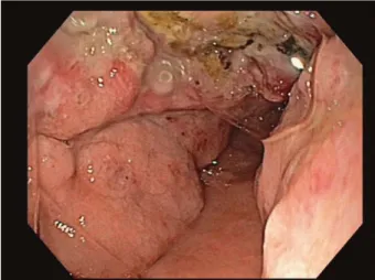
Two months later, the patient was hospitalized as an emergency due to melena. EGDS was performed again to show an exulcerated tumor mass (Figs. 4 and 5). Thorax CT scan confirmed disease progression with enlarged regional lymph nodes and intratumoral necrosis despite therapy. On multidisciplinary team consultation, operative treatment was suggested. The
Fig. 7. Ki67 positivity of 100% tumor cells (magnification X200).
Fig. 4. Tumorous formation in the stomach as displayed on EGDS.
Fig. 5. Tumorous formation in the stomach as displayed on EGDS.
Fig. 6. Histology of Burkitt lymphoma found in the stomach (HE, magnification X200): starry sky pattern.
preoperative process was complicated with secondary obstructive cholangitis due to tumor compression.
Finally, gastrectomy with regional lymphadenec-tomy, omentectomy and cholecystectomy was per-formed. On gross pathologic examination, the stom-ach, the surrounding fat tissue with lymph nodes and the gallbladder wall were infiltrated by tumor tissue. Morphologically, the tumor consisted of monomor-phic medium-sized atypical lymphatic cells with scant basophilic cytoplasm, round nuclei with multiple nu-cleoli, with numerous mitoses and apoptotic cells and with starry sky appearance (Fig. 6). Immunohisto-chemically, tumor cells were positive for CD20, CD10 and Bcl6, and almost negative for Bcl2. Ki67 was 100% (Fig. 7).
Since the morphology and proliferation rate were not the same as on the small endoscopic biopsy, and there was rapid tumor progression despite the therapy administered, the diagnosis was changed to BL.
After the patient had stabilized, a prephase with GMALL protocol was suggested. Unfortunately, the patient died in another hospital before starting the treatment.
Discussion
Adult sporadic BL is commonly a disease of young male adults in their twenties and thirties. Both of our patients were male, but the first patient was much old-er than expected for BL. Both patients presented with sporadic BL, not associated with immunocompro-mised status (HIV negative) and EBV infection, which was also unexpected. Tumor localization in the hepatic flexure (case 1) and stomach (case 2) is not a typical manifestation of the disease either, described only in a handful of cases11,12. Hence, both of our cases serve to emphasize the fact that old age, along with gastroin-testinal localization, does not exclude the possibility of BL and should be a viable differential diagnosis for high-grade non-Hodgkin lymphoma even in patients older than 70 years.
The main challenge in diagnosing BL is distinction between BL and diffuse large B-cell lymphoma, which is difficult. Distinguishing between these two lympho-mas, however, is critical, especially in adults, as the two diseases are treated differently3,13,14. Our second case report serves to stress the problem with distinction be-tween the two types of lymphomas. When atypical age
or localization might point to diffuse large B-cell lym-phoma, we still need to be wary of the possibility of BL, and dubious cases should undergo genetic testing and additional analysis.
Early diagnosis of BL is also essential to prevent life-threatening complications that occur due to high tumor burden and fast increase in size. The second case is a blatant example of the former, with two life-threatening gastrointestinal complications occurring in quick succession, gastrointestinal bleeding and sec-ondary cholangitis after lymph node induced bile duct compression.
With the advancement of medicine and longer survival, it is necessary to consider that BL will appear more often in atypical localization or age group, even without typical risk factors associated with the disease. These case reports emphasize the importance of clini-copathologic correlation in the diagnosis of high-grade lymphomas in everyday practice.
References
1. Burkitt D. A “tumour safari” in east and central Africa. Br J Cancer. 1962;16:379-86. PMID 14017063
2. Fujita S, Buziba N, Kumatori A, Senba M, Yamaguchi A, Tori-yama K. Early stage of Epstein-Barr virus lytic infection lead-ing to the “starry sky” pattern formation in endemic Burkitt lymphoma. Arch Pathol Lab Med. 2004;128:549-52. Doi: 10.1043/1543-2165(2004)128<549:ESOEVL>2.0.CO;2 3. Nakamura N, Nakamine H, Tamaru J, Nakamura S, Yoshino T,
Ohshima K, et al. The distinction between Burkitt lymphoma and diffuse large B-cell lymphoma with c-myc rearrangement. Mod Pathol. 2002 Jul;15(7):771-6.
Doi: 10.1097/01.MP.0000019577.73786.64
4. Chuang SS, Ye H, Du MQ, Lu CL, Dogan A, Hsieh PP, et al. Histopathology and immunohistochemistry in distinguishing Burkitt lymphoma from diffuse large B-cell lymphoma with very high proliferation index and with or without a starry-sky pattern: a comparative study with EBER and FISH. Am J Clin Pathol. 2007 Oct;128(4):558-64. Doi: 10.1309/EQJR3D-3V0CCQGP04
5. Bertrand S, Berger R, Philip T, Bernheim A, Bryon PA, Berto-glio J, et al. Variant translocation in a non-endemic case of Burkitt’s lymphoma: t (8;22) in an Epstein-Barr virus-negative tumour and in a derived cell line. Eur J Cancer. 1981;17: 577-84. PMID: 6271555
6. Mwanda OW. Clinical characteristics of Burkitt’s lymphoma seen in Kenyan patients. East Afr Med J. 2004 Aug;(8 Suppl): S78-89. PMID: 15622606
7. Dunning K, Mattei P. Burkitt lymphoma presenting as colonic ischemia and overwhelming sepsis. J Pediatr Surg. 2007 Aug; 42(8):E15-7. Doi: 10.1016/j.jpedsurg.2007.05.002
8. Molyneux EM, Rochford R, Griffin B, Newton R, Jackson G, Menon G, et al. Burkitt’s lymphoma. Lancet. 2012 Mar 31; 379(9822):1234-44. doi: 10.1016/S0140-6736(11)61177-X. Epub 2012 Feb 13. Doi: 10.1016/S0140-6736(11)61177-X 9. Wildes TM, Farrington L, Yeung C, Harrington AM, Foyil
KV, Liu J, et al. Rituximab is associated with improved survival in Burkitt lymphoma: a retrospective analysis from two US academic medical centers. Ther Adv Hematol. 2014 Feb;5(1): 3-12. Doi: 10.1177/2040620713514682
10. Kenkre VP, Stock W. Burkitt lymphoma/leukemia: improving prognosis. Clin Lymphoma Myeloma. 2009;9 Suppl 3:S231-8. Doi: 10.3816/CLM.2009.s.017
11. Ahluwalia M, Gotlieb V, Damerla V, Saif MW. Aggressive Burkitt-like lymphoma of colon in a patient with prior celiac
disease. Yale J Biol Med. 2006 Dec;79(3-4):173-5. PMID: 17940628
12. Khaled M Musallam KM, Taher AT, Shamseddine AI. Bur-kitt’s lymphoma of the colon and bronchi: three case reports. Cases J. 2008;1:15. Doi: 10.1186/1757-1626-1-15
13. Bellan C, Stefano L, Giulia de F, Rogena EA, Lorenzo L. Burkitt lymphoma versus diffuse large B-cell lymphoma: a practical approach. Hematol Oncol. 2009 Dec;27(4):182-5. Doi: 10.1002/hon.914
14. Shahini L, Gašparov S, Petruševska G, Manxhuka Kerliu S, Veselaj F, Kurshumliu F, et al. Clinical significance of VEGF-A and microvessel density in diffuse large B-cell lymphoma and low-grade follicular lymphoma. Acta Clin Croat. 2017 Dec;56 (4):588-93. Doi: 10.20471/acc.2017.56.04.02
Sažetak
BURKITTOV LIMFOM U PROBAVNOM TRAKTU: PRIKAZ 2 SLUČAJA
A. Čubranić, M. Golčić, D. Fučkar-Čupić, B. Brozović, D. Gajski i I. Brumini
Burkittov limfom spada u non-Hodgkin B-stanične limfome te je najbrže rastući tumor u ljudi, a patološki se prezentira uzorkom ‘zvjezdanog neba’. Najčešće je lokaliziran u trbuhu i u čeljusti, međutim, lokalizacija u trbuhu izvan ileocekalnog područja je izrazito rijetka i opisana samo u nekoliko slučajeva. Uobičajeno liječenje se sastoji od inicijalne citoredukcije tumora nakon čega slijedi intenzivna kemoterapija. Većina recidiva javi se unutar jedne godine od dijagnoze, dok je petogo-dišnje preživljenje oko 80%. U članku prikazujemo dva slučaja koja su specifična zbog rijetke lokalizacije Burkittova limfoma u debelom crijevu i želudcu, u imunokompetentnih bolesnika s negativnim testom na Epstein-Barrov virus. Također, jedan od opisanih bolesnika je jedan od najstarijih slučajeva abdominalnog Burkittova limfoma opisanih u literaturi, dok slučaj drugog bolesnika naglašava izazove u dijagnostici Burkittova limfoma, osobito u bolesnika kod kojih se isti ne očekuje. S obzirom na brz rast tumora i hitnu potrebu citoreduktivne operacije važno je uzeti u obzir dijagnozu Burkittova limfoma čak i u bolesnika s netipičnom lokalizacijom tumora i bez uobičajnih rizičnih čimbenika povezanih s Burkittovim limfomom.