THESIS
THE EFFECT ORAL ADMINISTRATION OF
L-ARGININE ON SPERMATOGENESIS AND SERTOLI
CELLS OF RABBITS TESTES (Oryctolagus cuniculus)
By:
HENING TYAS PITALOKA SIN 061111122
FACULTY OF VETERINARY MEDICINE AIRLANGGA UNIVERSITY
ii
ENDORSEMENT FORM
THE EFFECT ORAL ADMINISTRATION OF
L-ARGININE ON SPERMATOGENESIS AND SERTOLI
CELLS OF RABBITS TESTES (Oryctolagus cuniculus)
Research Result
Submitted in partial fulfillment of the requirement for the degree of Bachelor of Veterinary Medicine
at
Faculty of Veterinary Medicine, Airlangga University, Surabaya
By:
HENING TYAS PITALOKA SIN 061111122
Supervising Committee
Prof. Mas’ud Hariadi, M.Phil, Ph.D., drh Dr. Nove Hidajati, drh., M.Kes.
Supervisor Co-Supervisor
DECLARATION Hereby, I declare that in this thesis entitled:
THE EFFECT ORAL ADMINISTRATION OF
L-ARGININE ON SPERMATOGENESIS AND SERTOLI
CELLS OF RABBITS TESTES (Oryctolagus cuniculus)
There is no work ever published to obtain a college degree in a certain college and to my knowledge there is also no work or opinion ever written or published by others, except those in writing referred to in this paper and mentioned in the references.Surabaya, 10 August 2016
iv
Has been assessed in Result Seminar Date : 1st August 2016
RESULT SEMINAR ASSESSEMENT COMMITTEE Head : Suzanita Utama, drh.,M.Phil., Ph.D. Secretary : Dr. Kadek Rachmawati,drh., M.Kes. Member : Djoko Legowo, drh., M.Si.
Supervisor : Prof. Mas’ud Hariadi, M.Phil, Ph.D., drh Co-Supervisor : Dr. Nove Hidajati, drh., M.Kes.
Has been examined Date : 10th August 2016
THESIS DEFENSE ASSESSMENT COMMITTEE Head : Suzanita Utama, drh.,M.Phil., Ph.D. Members : Dr. Kadek Rachmawati,drh., M.Kes.
Djoko Legowo, drh., M.Si..
Prof. Mas’ud Hariadi, M.Phil, Ph.D., drh Dr. Nove Hidajati, drh., M.Kes.
Surabaya, 10th August 2016 Faculty of Veterinary Medicine
Universitas Airlangga Dean,
vi
THE EFFECT ORAL ADMINISTRATION OF
L-ARGININE ON SPERMATOGENESIS AND SERTOLI
CELLS OF RABBITS TESTES (
Oryctolagus cuniculus
)
Hening Tyas Pitaloka
ABSTRACT
L-Arginine is the precursor of nitric oxide (NO). The aim of this research was to know the effect oral administration of L-arginine on spermatogenesis and the number of Sertoli cell of seminiferous tubules of rabbit (Oryctolagus cuniculus). This researh used sixteen rabbits (6 months old and ± 3 kg of body weight) were devided into two groups, P0 and P1. P0 is a control group and P1 was given L-arginine 6g/300ml/day for 30 consecutive days. After 30 days treatment, their testicles were collected and processed for histological examination. The data were analyzed by Kruskall Wallis Test for Johnsen’s Score and ANOVA for Sertoli cells counting at the significancy level of 5%. The result from statistical analysis showed that treatment with L-arginine did not increase spermatogenesis compared with control group, while the Sertoli cells of rabbit testicular showed that L-arginine increased the number of Sertoli cells.
Keywords : spermatogenesis, Sertoli cells, L-Arginine, nitric oxide, testis
ACKNOWLEDGMENT
First and most of all, praise and thank to ALLAH SWT because of His mercy and guidance this thesis entitled The Effect Oral Administration of L-Arginine on Spermatogenesis and Sertoli Cells of The Testes of Rabbits (Oryctolagus cuniculus), have finished.
On this occasion the author would like to give thanks to :
Prof. Dr. Pudji Srianto, drh.m M.Kes, the dean of Faculty of Veterinary Medicine, Universitas Airlangga, for the occasion to study in Faculty of Veterinary Medicine, Universitas Airlangga.
The supervisor committee, Prof. Mas’ud Hariadi, M.Phil, Ph.D., drh, and Dr. Nove Hidajati, drh., M.Kes., for all the time, patience, advice and support that given to the author.
The examiner committee, Suzanita Utama, drh.,M.Phil., Ph.D., Dr. Kadek Rachmawati,drh., M.Kes., Djoko Legowo, drh., M.Si., for all the advices, knowledge and guidance in the completion of my study.
Dr. Agnes Theresia Soelih Estoepangestie, Drh., as my trustee lecturer for her kindness, help, support and had being my second parent during my study in Faculty of Veterinary Medicine, Universitas Airlangga.
viii
My research and thesis partners, Arif Prasetyo, dr., Syahroni Ibnu,dr., Fachruddin Aziz, Firdausy Kurnia Maulana, Gavrila Amadea, for great teamwork and motivation.
My father Asmuin, S.Pd., my mother Zuliana Surani for your endless affection, education, and motivation for the success of my study all this time. And my beloved twin sister Wening Tyas Pitaloka thank for always beside me, accompany me to finish this thesis.
My dearest best friends Wulan Arfina Ryani, Putri Agnia Kuswandi dan Eka Ardianta for support, prayed, love, advices, patience and motivation all this time.
And all of my vacationeer friends, for the experiences and stories that we have created. All the member of IC 32, for being my second family along this years, and the joyful day we spent together.
The author acknowledges that this writing is still lacking and far from perfect. Therefore, the author expects critics and recommendations that will help in the improvement of this thesis. With a humble heart, the author wishes that this research will be useful for the advancement of science and may give a contribution to the veterinary medicine field and all the people who needs it.
Surabaya, 10 August 2016
Author
LIST OF TABLES
Page Table 4.1. The mean and deviation standard of spermatogenesis and
xii
LIST OF FIGURES
Page
Figure 2.1. Scheme of testis (Trainer, 2007) ... 7
Figure 2.2. Diagram of spermatogenesis (O’Day, 2010) ... 10
Figure 2.3. Histology of testis stains H&E with 1000x magnification (Junqueira, 2007). ... 12
Figure 2.4. Schematic presentation of the presumed NO systems in Leydig cells, testicular blood vessels, and seminiferous tubules (Middendorff et al. 1997) ... 14
Figure 2.5. NO formation in endothelial cells of blood vessel (Koolman and Roehm, 2005) ... 15
Figure 2.6. New Zealand White rabbit ... 19
Figure 4.1. Histological features of spermatogenesis of rabbit testis... 29
Figure 4.2 Histological features of Sertoli cells.. ... 30
APPENDICES
Page
Appendix 1. Figures of Seminiferous Tubules ... 47
Appendix 2. Testes Histology Preparation ... 49
Appendix 3. Histology Observation Result of Seminiferous Tubules ... 50
Appendix 4. Results of Data Analysis of Spermatogenesis ... 54
Appendix 5. Results of Data Analysis of Sertoli Cells Number... ... 56
xiv
ABBREVIATIONS AND SYMBOLICS MEANING
et al : et alii
NO : Nitric Oxide
e.g. : exempli gratia
% : percent
g : gram
NOS : Nitric Oxide Synthase
nNOS : Neural Nitric Oxide Synthase eNOS : Endothelial Nitric Oxide Synthase iNOS : Inducible Nitric Oxide Synthase cGMP : cyclic Guanosine Monophosphate H&E : Hematoxylin and Eosin
FSH : Follicle-stimulating hormone cNOS : Constitutive Nitric Oxide Synthase
NADPH : Nicotinamide Adenine Dinucleotide Phosphate
O2 : Oxygen
ml : mililiter
mg : miligrams
kg : kilograms
h : hours
: Registered
IM : Intra Muscular
CHAPTER 1 INTRODUCTION
1.1 Background of Research
Humans at this time almost all use supplements to improve performance. The management of unusual stress therefore has acquired enormous significance in day-to-day life. It is possible to support the body’s adaptation by using food supplements, dietary elements, herbs and minerals for increasing physical and mental performance (Gupta et al., 2004).
Dietary supplements are intended to provide nutrients that may otherwise not be consumed in sufficient quantities. The most supplement common consumed are purposed to improve the energy, to increase the immunity, to maintain the fitness of body condition or even for sexual necessity.
temperature were not significantly different from those of the controls (Ratnasooriya and Dharmasiri, 2001).
L-Arginine known as semi-essential amino acid, because even though the body normally makes enough of it, supplementation is sometimes needed. L-arginine plays a key role in modulating host defences and cellular immunity. Enviromental factors, such as pesticides, exogenous estrogens, and heavy metals may negatively impact spermatogenesis. Arginine therapies have been shown to improve sperm counts and sperm motility (Husien et al., 2011).
L-Arginine is mainly as the precursor of Nitric Oxide (NO). NO is now known to be produced by various cells in different organs, including smooth muscle cells, mesangial cells, neurons, platelets, hepatocytes, macrophages, fibroblasts and epithelial cells. NO regulates smooth muscle cell tone, platelet aggregation and adhesion, cell growth, apoptosis, neurotransmission and injury as well as infection-induced immune reactions. Because these neurons, blood vessels and cells of the immune system are integral part of reproductive organs, and in view of the important functional role that NO plays in those system, it is likely that NO is an important regulator of the biology and physiology of the reproductive system (Rosselli et al., 1998).
Arginine supplementation significantly improved sperm motility and abnormality without any side effect (Scibona et al., 1994). Researcher found out that feeding arginine-deficient diet to adult men for 9 days decreased sperm counts by ~90% and increased the presentage of non-motile sperm approximately 10 fold (Gad, 2010). A study by Chen et al (1999) revealed a significant
subjective improvement in sexual function in men with organic erectile dysfunction after oral intake of 5 g L-Arginine for 6 weeks, but only if they had decreased NO excretion or production. Long term oral administration of Arginine to diabetic rabbits increased endothelium-dependent relaxation of rabbit corpus cavernosum (Hupertan et al., 2012)
Supplementation of L-Arginine is expected to affect the regulation of the entire part in the seminiferous tubules. The seminiferous tubules are the site of the germination, maturation and transportation of the sperm cells within the male testes. Spermatogenesis through the process of meiosis takes place within seminiferous tubules. Along the way, the maturing sperm cells recieve nutrients and raw materials from Sertoli cells which are located within seminiferous tubules. These component in seminiferous tubules are important roles in reproductive system.
Based on the background above the research untitled the effect of oral L-Arginine administration on spermatogenesis and Sertoli cells of the testes rabbits (Oryctolagus cuniculus) has been conducted.
1.2 Problem Statement
1. Does oral administration of L-arginine increase spermatogenesis of rabbit? 2. Does oral administration of L-arginine increase Sertoli cells of rabbit?
1.3 Theoritical Base
NO receptors present in peritubular lamina propria, Sertoli, and blood vessel cells, suggesting production and activity of NO in these structures (Middendorff et al., 1997).
The endothelium (inner lining) of blood vessels uses NO to signal the surrounding smooth muscle to relax, thus resulting in vasodilation and increasing blood flow. Nitric oxide is highly reactive, yet diffuses freely across membranes (Stryer, 1995). Nitric Oxide has been shown to influence permeability of vascular endothelial cells (Noel et al., 1995; Rumbaut et al., 1995).
In seminiferous tubules, NO-induced cGMP production may mediate relaxation of myofibroblasts. Whereas endothelin, for example, has been shown to be involved in peritubular cell contraction in the rat (Tripiciano et al., 1996). In the peritubular lamina propria, NO may participate in the regulation of the peristaltic activity of the tubules, which in turn, is necessary for sperm transport (Setchell et al., 1994). Furthermore, NO may influence the permeability of the lamina propria and, by this, the transport of nutrients into the tubular lumen for sprematogenesis process (Holstein et al., 1996).
NOS is present and functionally active in testicular blood vessels and seminiferous tubules refers to a local production of NO in these structures, the NO-induced increases in cGMP production observed in isolated tubules may, in part, be caused by NO produced by Sertoli cells. Hence, testicular vasculature and seminiferous tubules are sites of NO production and activity. It is an attractive idea that NO acts locally to regulate the distribution of oxygen, nutrients, and
hormones by testicular vessels, as well as the peristaltic activity of tubules in context of sperm transport (Middendorff et al., 1997).
Because NOS is found in testicular endothelial cells, Sertoli cells, and regulation of eNOS by germ cells, this seemingly suggests that NO is involved in regulating spermatogenesis.
1.4 Aim of Research
1. To know the effect of oral administration of L-arginine on increasing the spermatogenesis in rabbit.
2. To know the effect of oral administration of L-arginine on increasing Sertoli cell number in rabbits.
1.5 Benefit of Research
The benefits of this research are:
1. Providing knowledge and information for researchers and public about the effect of L-Arginine as supplementation on male fertility.
2. Leading the therapy development as therapies to address some types of infertility in male.
1.6 Hypothesis
The hypothesis of this research are:
6
CHAPTER 2 LITERATURE REVIEW
2.1 Reproduction of Male
2.1.1 Testes
The testes are a pair of ovoid glands organs are essential for the function of male reproductive system. Testicular cells responsible for the production of sperm and male sex hormone testosterone
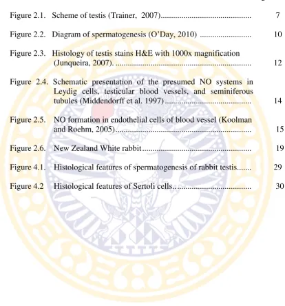
Embryological testes develop in the dorsal part of the abdominal cavity in a retroperitoneal position. As the fetus develops, the testicles migrate the abdomen and descend through the inguinal canal into the scrotum where they are found in adults. Because the testicles develop in the lumbar position, they took the blood and nerve supply from this area. The blood supply and the inervation follow their testicles descend. (Breazile et al., 1971).
Testis consists of two networks that include, vaginal tunic viceral and tunica albuginea. Internal, seminiferous tubules, interstitial cells, nerves, blood vessels, rete testis and efferent ductules found. Each testis is covered with glossy hard connective tissue called visceral vaginal tunic (tunica vaginalis propria). It comes from the peritoneum as the testes descend. It serves to support the testis. Directly below the tunica albuginea testes, which also supports the testes and is the connective tissue capsule that surrounds the blood vessels that meander near the surface of the testicle. Septula testis are strands of connective tissue which is a branch of this layer and connected to the mediastinum testis, which is the core of connective tissue of the testes. This network holds the connective tissue of the
seminiferous tubules and interstitial cells in place and give shape and to support the testicles (Sorensen, 1979).
The mediastinum testis contains blood vessels, nerves, lymphatics, and the rete testis which is a portion of duct system of the testis. Rete testis is the mounth of production per lobule before exit from testis through efferen ducts to the epididymis (Figure 2.1). A vascular layer in the tunica albuginea assists in regulating the temperature of the testis. The bulk of the testis consists of seminiferous tubules and interstitial cells. The seminiferous tubules produce the sperm (Breazile et al., 1971).
Figure 2.1. Scheme of testis (Trainer, 2007)
2.1.2 Seminiferous Tubules
which develop into spermatozoa and Sertoli cells that have the function to support and nutrition.
The epithelium which lining the seminiferous tubules contains two cell types, sustentacular, or Sertoli cells and spermatozoa and their developing germ cells percursor. Sustentacular large cells extend from the base of the epithelium into the lumen of the seminiferous tubules. The shape is irregular because they surround the developing germ cells. Sustentacula cells secrete fluids that bathe the developing germ cells and help with transport spermatozoa out of the tubules to the rete testis and they are required for maturation of spermatozoa (Frandson et al., 2003). Along the way, the maturing sperm cells recieve nutrients and raw materials from the vascular Sertoli cells located in the tubule walls until they become mature primary sperm cells ( spermatozoa).
Line of tubular basement membrane, and the rest in this is the beginning
of spermatogenic cells, the spermatogonia. Supporting the basement membrane are collagen fibers (Constantinides, 1974). There is a developemental progression of cells from the periphery of the tubule to the internal lumen called spermatogenesis (Sorensen, 1979).
2.1.3 Spermatogenesis
Spermatogenesis is the term for the processes involved in the formation of a mature male gametes mostly undifferentiated germ cells. Spermatogenesis takes place in the seminiferous tubules. Rounded immature sperm cells undergo mitosis in a row and meiotic division (spermatocytogenesis) and metamorphic changes (spermiogenesis) to produce spermatozoa.
During spermatocytogenesis, spermatogonia proliferate by mitosis. This is the division of cells from the beginning of sperm formation until a change in shape occurs. The original cells in the process of spermatogenesis are the Type A spermatogonia, which may require testosteron for development from embryonic gonocyte (Steinberger and Duckett, 1967). The A type spermatogonia are containing two or more nucleoli and lie dormant on the basement membrane until they divide mitotically, to be other A type cell in dorman form A cells and active B cells that contain only one nucleolus (Sorensen, 1979). Mitosis ends when a B spermatogonium yields two primary spermatocytes.
The diploid number of primary spermatocytes is halved during meiosis. A primary spermatocyte is transformed into two secondary spermatocytes during meiosis I, these cells then in turn are converted into (1N) spermatids during meiosis II. The second meiotic division is rapid. Spermatocytes and spermatids tend to be larger than their ancestral spermatogonia.
The mature spermatozoa are released from the protective Sertoli cells into the lumen of the seminiferous tubule. The resulting spermatozoa are now mature but lack motility. The non-motile spermatozoa are transported to the epididymis in testicular fluid secreted by Sertoli cells with the aid of peristaltic contraction.
Figure 2.2. Diagram of spermatogenesis (O’Day, 2010)
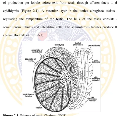
2.1.4 Sertoli Cell
Testicular function is under the control of expression and repression of several gene and gene products, and many of these work through Sertoli cells (Johnson et al., 2008). Sertoli cells are tall simple columnar cells, which span from the basement membrane to the lumen. They surround the proliferating and differentiating germ cells forming pockets around these (Figure 2.3). Sertoli cell functions include: support and nutrition of germ cells; release of mature germ
Primordial Germ Cells
Spermatogonia
Primary Spermatocytes Secondary
Spermatocytes
Spermatids
Spermatozoa
cells into the lumen; translocation of developing germ cells in an adluminal direction; secretion of androgen binding protein, transferrin, inhibin; cell-cell communication via gap junctions to coordinate spermatogenesis; blood-testis barrier.
In the process of spermatogenesis, Sertoli cell provide structural support and cytokines for sperm cells, regulate nutrition development, and play a crucial role in maintaining a stable micro-enviroment for spermatogenesis (Lee and Cheng, 2004). The microtubules of Sertoli cells are involved in spermatogenesis through the regulation of cell morphology, intracellular transport, organelle positioning, cell motility, cell division and other physiological processes (Li et al., 2009)
Sertoli cells are the somatic cells of the testis that are essential for testis formation and spermatogenesis. Sertoli cells facilitate the progression of germ cells to spermatozoa via direct contact and by controlling the enviroment within seminiferous tubules. The regulation of spermatogenesis by FSH and testosterone occurs by the action of these hormones on the Sertoli cells. While the action of testosterone is necessary for spermatogenesis, the action of FSH minimally serves to promote spermatogenic output by increasing the number of Sertoli cells (Griswold, 1998).
isolates the developing spermatogonia, spermatocytes, spermatids and mature spermatozoa from blood. Sertoli cells also produce testicular fluid, including a protein that binds to and concentrates testosterone, which is essential for the development of the spermatozoa
The number of germ cells is supported by a single Sertoli cells is the best reflection of the functional efficiency of the cells and are usually highly correlated with spermatogenic efficiency (daily sperm production per gram testis) (Russell and Peterson, 1984; França and Russell, 1998).
Figure 2.3. Histology of testis stains H&E with 1000x magnification (Junqueira, 2007).
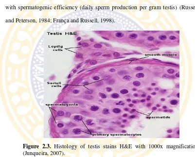
2.2 Nitric Oxide
Nitric oxide (NO) is synthesized from L-arginine by the action of NO synthase (NOS), an enzyme existing in three isoforms. Brain NOS (bNOS) or neuronal NOS (nNOS or NOS1) and endothelial NOS (eNOS or NOS3), also referred to as constitutive NOS (cNOS), are responsible for the continuous basal
release of NO and both require calcium/calmodulin for activation (Griffith and Stuehr, 1995; Snyder, 1995). A third isoform is an inducible calcium-independent form (iNOS or NOS2) that is expressed only in response to inflammatory cytokines and lipopolysaccharides (Nussler and Billiar, 1993; Morris and Biliar, 1994).
The varied expression and activity patterns of the NOS enzymes uniquely suit the different functions of the NO generated from each isoform in normal physiologic functions and in disease states (Lei et al., 2013). The neuronal NOS (nNOS) and endothelial NOS (eNOS) are calcium-dependent and produce low level of NO. The inducible NOS (iNOS) is calcium independent and produce large amount of NO. Under normal condition, the activity of iNOS is very low, but it is stimulated during inflammation by bacterial endotoxins (Braga, 2012).
the Leydig cells, peritubular myofibroblasts relaxation and dilation of blood vessels testis. Because NO can freely diffuse across the membrane, three different systems can affect one another (Middendorff et al. 1997) (Figure 2.4).
The processes are known to be associated with biology, physiology and pathophysiology of various reproductive processes and NO has been recognized as a molecule that importantly regulates the biology and physiology of reproductive function (VidyaGarg and Garg, 2011). The nitrogen derivate free radical nitric oxide also appear to play a significant role in reproduction and fertilisation (Rosselli et al., 1998).
Figure 2.4. Schematic presentation of the presumed NO systems in Leydig cells, testicular blood vessels, and seminiferous tubules (Middendorff et al. 1997).
2.3 L-Arginine
L-Arginine (2-amino-5-guanidino-pentanoic acid) is a conditionally
essential amino acid that is a natural constituent of dietary proteins. Arginine is a
stable nutrient in an aqueous solution, and is not destroyed by serilization conditions (e.g. high temperature and high pressure). Arginine is not toxic and its administration is generally safe for human and animals (Flynn et al., 2002).
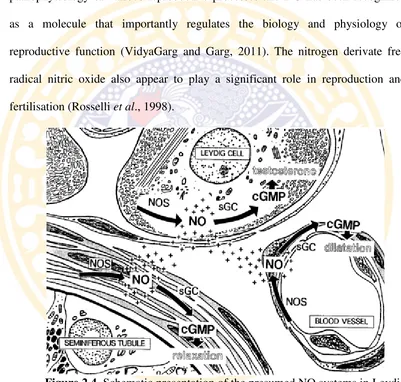
L-arginine is the biological precursor of nitric oxide, an endogenous gaseous messenger molecule involved in a variety of endothelium-depedent physiological functions (Wu and Meininger, 2000), including its critical role in cardiovascular protection and immune support (Duru et al, 2011; Sunita et al., 2000).
Figure 2.5. NO formation in the endothelial cells of blood vessel (Koolman and Roehm, 2005).
arginosuccinate synthetase and arginosuccinatelyase (Morris, 1992) and catabolized by arginase (Albina et al., 1988). NO arises from arginine in the endothelial cells of blood vessels triggered by Ca2+-calmodulin. NO diffuses from endothelial cells into vascular muscle cells where it leads to the formation of the cGMP, which is the trigger of relaxation of the smooth muscle and vessels dilatation (Koolman and Roehm, 2005) (Figure 2.5).
Arginine rapidly enters the circulation reaches a plasma peak 40-60 minutes after intake (pharmacokinetics are similar in the rat, the pig, the rabbit and human) and slowly decreases during the next 24 hour (Boger et al.,1998). It is thus clear that upon administration Arginine has prolonged contact with the vascular endothelium prior to any other tissue. Arginine is also very well tolerated and can be administrated at high dosages (Barbul, 1986).
It has been shown that endogenous arginine synthesis is couples to NO synthesis (Morris et al., 1994; Nussler et al., 1994). Low concetrations of arginine caused by release of arginase in wound are responsible for reduced NO synthesis (Albina et al., 1988). Apart from synthesis, transport of arginine into the cells can also regulate NOS activity. In this regard, it has been shown that hepatic L-arginine transport, which is normally low, is enhanced during sepsis (Sax et al., 1988; Inoue et al.,1993)
Dietary L-arginine supplementation attenuates the oxidative stress induced by burn injury with a better macrophage response (Tsai et al., 2002). These beneficial effect of L-arginine have been attributed to its dependent formation of NO within the endothelial lining ( Brandes et al., 2000).
2.3.1 L-Arginine in Male Reproductive System
L-arginine is a dietary supplement often used to improve the performance of sexual health, for example, erectile dysfunction. L-arginine is also known as sexual nutrients that safe and effective. L-arginine plays an important role in regulating the body's defenses, as well as play an active role in the formation of sperm. Arginine amino acid deficiency can cause a metabolic disorder that causes a decrease in sperm motility and disruption of spermatogenesis process.
Effect of amino acid arginine improves the quality of sperm occurs through multiple mechanisms. Arginine amino acids can protect the sperm plasma membrane from damage caused by lipid peroxidation that is by increasing the production of nitric oxide. The mechanism is similar to the mechanism of antioxidant to protect cells from free radicals (Adams et al., 1994). A study of nitric oxide (NO) have been carried out both in vitro and in vivo and the results showed that nitric oxide (NO) has multiple biological activities and pharmacological among others are increasing fertility through increased testosterone levels and activates the release of luteinizing hormone - releasing hormone (LHRH), which is a hormone stimulator for secretion of FSH and LH hormones that affect the improvement of the process of spermatogenesis ( Rosselli et al., 1998; Elgohary et al., 1999).
important role in stimulating sperm motility in humans, rabbits, and goats under in vitro conditions (Radany et al., 1981).
L-Arginine is capable of regulating penile erection. Because NO acts as a mediator of penile erection and expressed in the cavernosal smooth muscle cells of the penis (Garban et al., 1997). Within the testis, NO has also been shown to regulate blood flow, cell permeability and contractile function of myofibroblasts, which in turn regulate steroid synthesis and transport (Rosselli et al., 1998).
2.4 Rabbit (Oryctolagus cuniculus)
Rabbit race classification according to binominal system is as follows: (Linnaeus, 1758)
Kingdom : Animalia
Phylum : Chordata Sub phylum : Vertebrata Class : Mamalia
Ordo : Lagomorpha
Family : Leporidae Sub family : Leporinae Genus : Oryctolagus
Species : Oryctolaguscuniculus
Figure 2.6. New Zealand White rabbit.
Rabbits have a thin skin and dense fur that consists of a soft undercoat and stiff guard hairs. They do not have footpad; instead the feet are covered with thick fur, the skin on the neck is loose and pendulous and forms a pronounced dewlap in females of some breed. The nostrils are sensitive with large number of tactile vibrissae. Scent glands are situated in the deep inguinal spaces that are found on either side of the anus immediately dorsal to urogenital opening. In the male rabbit, the testicles are found in hairless scrotal sacs on either side of the penis. The inguinal canal remains open and the testicles can be retracted into the abdomen. Retraction occurs during periods of sexual inactivity or during periods of insufficient food. Male rabbits have rudimentary nipples (Harcourt-Brown, 2002)
of the male and the inguinal spaces in both sexes (Weisbroth et al., 1974). New Zealand white rabbits have a life span of over 6-13 years. The urine volume is usually about 130ml/kg BW/24 h and water intake in rabbits is 50-100 ml/kgBW/24 h (Harcourt-Brown, 2002).
Rabbit has served as a good and practical model in studies of basic spermatogenesis as well as for investigating effects of potential toxins or other agents on that process (Castro et al., 2002).
2.4.1 Characteristic of Male Rabbits Reproduction.
In the male, the oval-shaped testes within the scrotum remain in communication with the abdominal cavity, where they were at birth. The testicles descend at about two months and can move freely from the scrotum to the abdomen through an opening in the inguinal canal (Campos et al, 2014). The short, back-slanting penis points forward when erect.The male rabbit tends to mature slower than the female. Spermatogenesis begins between days 40 and 50. The testicular tubes become active at about 84 days. The first spermatozoa are present in the ejaculate at about 110 days. Sexual maturity, defined as the moment when daily sperm production ceases to increase, is reached at 32 weeks by New Zealand White rabbits in temperate climates. However, a young buck in these same conditions can be used for reproduction from the age of 20 weeks. Coitus may occur for the first time at about 100 days, but the viability of the sperm cells is very weak or nil in the first ejaculates. So first mating should be timed for age 135 to 140 days (Lebas et al, 1997).
.
CHAPTER 3 MATERIALS AND METHODS
3.1 Time and Location of Research
This research was held at non-infectious animal laboratory, the Institute of Tropical Disease, Universitas Airlangga, Surabaya, as place for treatment, termination and collecting testes. Preparation and staining of seminiferous tubules histological slides were done at The Laboratory of Pathology, Faculty of Veterinary Medicine, Universitas Airlangga, Surabaya. This research was held on February-June 2015.
3.2 Material of Research
3.2.1 Expeerimental Animals
This research used 16 healthy male New Zealand White rabbit (Oryctolagus cuniculus) with average 6-12 months of ages, 3 kg of body weight collected from rabbit farmers located in Malang, East Java.
3.2.2 Research Materials
Materials used in this research were mineral water, Susu PAP feed
concentrate (PT. Japfa Comfeed Indonesia, Tbk), GNC L-Arginine 500 (Nutra
Manufacturing USA) for maintenance and treatment.
Absolute alcohol and 70%, 80%, 90%, 95% alcohol concentration, xylol, paraffin, Hematoxylin-eosin staining used for staining and preparation of histological materials.
3.2.3 Research Equipments
Equipments used for maintenance in this research were rabbit cages and bottles for drink.
Mask, gloves, syringe 1ml, small plastic containers, forceps, scalpel, blades, surgical scissors were used for preparation of the organ samples.
Object glass, cover glass, pipette, tissue processor automatic, water bath, hot plate, microtome, light microscope (Olympus® CX-21). and camera were used for histological observation.
3.3 Research Methods
3.3.1 Animal Preparation
Rabbits were divided into two groups, then adapted to their new enviroment for one week. The rabbits were given feed and drunk ad libitum and placed in cages made of steel with size 90x60x40 cm, in the room with normal temperature, humidity, and good air ventilation.
3.3.2 Treatment
Rabbits were divided into two groups. First group was used as control, while rabbits in the other group recieved L-arginine in their drinking water.
arginine dosage is determined using dose 2g/kg of body weight/day. arginine dissolved in drinking water, 6 gram of arginine (12 capsules of L-arginine). The treatment was given for 4 weeks (Okazaki et al., 1997).
Treatment type in this study:
P0(control group) : The rabbits were given drink water only for four weeks. P1(treatment group) : The rabbits were given treatment with L-arginine
dissolved in drinking water for four weeks. 3.3.3 Organ Collection
At the end of the period of treatment, rabbits were sacreficed instead of castration, since it was a research groups. All groups of rabbits were injected with a combination of ketamine-xylazine with dose of 25 mg/kg/IM for ketamine and 3mg/kg/IM for xylazine.
After the rabbit has reached a stable level of anaesthesia at about 10 minutes after the injection, and positioned in dorsal recumbence, the fur around the scrotum was shaved. The incision was made through the skin, at the cranial end of the scrotum. The fibrous tunic was incised then the vaginal tunic was exposed. The testis, epididymis and the deferent duct were pushed and advanced through the incision. Removed the testis by cutting through the spermatic cord and cut the ligament of the epididymis and the testis.
3.3.4 Histological Preparation of Testes
Preparation process of testis and Hematoxylin- Eosin staining can be seen in (Appendix 2).
3.3.5 Seminiferous Tubules Histological Examination
Data is determined by calculating the average score of spermatogenesis in 20 visual field of seminiferous tubules of each slide preparation in a light microscope with 400 times magnification. For standardization, it used scoring system for testicular biopsies (Johnsen score) (Yama et al., 2013). For the figures of
Johnsen’s score can be seen in the Appendix 1.
1 = no seminiferous epithelium
2 = no germinal cells, Sertoli cells only 3 = spermatogonia only
4 = no spermatozoa or spermatids, few spematocytes 5 = no spermatozoa or spermatids, many spematocytes 6 = no spermatozoa, no late spermatids, few early spermatids 7 = no spermatozoa, no late spermatids, many early spermatids 8 = less than five spermatozoa per tubule, few late spermatids
9 = slightly impaired spermatogenesis, many late spermatids, disorganized epithelium
10 = full spermatogenesis
Counting the amount of Sertoli cells can be done by observing 20 of view on 20 seminiferous tubules on each slide preparation. Calculation results of each field
of view in preparation are summed and then calculate the average. Observation are done using a light microscope with magnification of 400 times.
3.4 Research Design
This research is an experimental study with Completely Randomized Design (CRD), with two treatments. The number of repetitions in each treatment is determined by formulation t (n-1) ≥ 15, whereas n for quantity of sample for each treatment and t is number of experiment (Kusriningrum, 2008).
t (n-1) ≥ 15
2 (n –1) ≥ 15
2n – 2 ≥ 15
n ≥ 8.5 (8)
Based on the calculation above, this research used eight rabbits repetition of each group.
3.5 Reasearch Variables
3.5.1 Dependent Variable
The histology score of spermatogenesis and the amount of Sertoli cells.
3.5.2 Independent Variable
Oral administration of L-Arginine 3.5.3 Controlled Variables
3.6 Operational Definition of Variables
3.6.1 L-Arginine is an amino acid supplement that formed into capsule.
3.6.2 Spermatogenesis is the process in male gametes are produced from male primordial germ cells and transform into spermatozoa. With hematoxylin and eosin staining, nucleus of spermoatogonium, primary and secondary spermatocytes are stained in blue or purple, while the cytoplasm are pink.
3.6.3. Sertoli cells extend from the basement membrane to the luminal surface of the seminiferous epithelium. With hematoxylin and eosin staining, Sertoli cells are recognized in the seminifereous tubules by their pale invaginated irregular nuclei. Nuclei should have a slightly wrinkled nuclear membrane and have prominent nucleoli.
3.7 Data Analysis
This research is an experimental study with Completely Randomized Design (CRD). The data were obtained in the form of semi-quantitative data of spermatogenesis and quantitative of Sertoli cells of rabbits testes. Data were
presented as mean standard deviation.. To determine treatment differences was
analyzed by Kruskall Wallis Test for Johnsen’s Score and ANOVA for Sertoli cells counting at the significancy level of 5% (Kusriningrum, 2008). The data was processed using the program Statistical Package for the Social Sciences (SPSS) 21.0 for Windows.
3.8 Research Framework
16 male rabbits
P0
8 Rabbits given mineral water
P1
8 Rabbits given L-arginine 6gram/300cc of mineral water for each
Treatment for 4 weeks
Sacrificed, testes collection
Preparation and HE staining
Microscopic observation (spermatogenesis and number of
Sertoli cells)
CHAPTER 4 RESULT
Microscopic observation was conducted using histological preparations with
H.E staining of the rabbit testicles to observed the spermatogenesis and the
number of Sertoli cells after given treatment for 4 weeks. There were two groups,
in control group (P0) and group with L-arginine administration orally (P1).
Observation was done by using a microscope magnification of 400 times on
spermatogenesis of 20 seminiferous tubules of each slide preparation then
averaged and Sertoli cells were observed in 20 different field of view of each slide
preparation then averaged.
4.1. Spermatogenesis
The data result of observation and assessment was obtained by using Johnsen’s Score method. Data of Johnsen’s score for spermatogenesis in each treatment are shown in Table 4.1 (Appendix 3).
Table 4.1 The mean and deviation standard of spermatogenesis and Sertoli cells number. P0 = for spermatogenesis and number of Sertoli cells without L-Arginine administration and P1 = with L-L-Arginine administration.
Group Johnsen’s Score for
Kruskall-Wallis test result (Appendix 4), shows the score of spermatogenesis in the control treatment without the administration of L-arginine not significant different (p>0.05) from the treatment of l-arginine administration of a dose of 6g / 300ml / day.
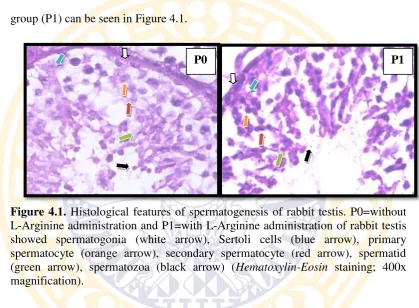
Spermatogenesis of rabbit testis both in control group (P0) and treatment group (P1) can be seen in Figure 4.1.
Figure 4.1. Histological features of spermatogenesis of rabbit testis. P0=without L-Arginine administration and P1=with L-Arginine administration of rabbit testis showed spermatogonia (white arrow), Sertoli cells (blue arrow), primary spermatocyte (orange arrow), secondary spermatocyte (red arrow), spermatid (green arrow), spermatozoa (black arrow) (Hematoxylin-Eosin staining; 400x magnification).
4.2. Sertoli Cells
Histological observation of Sertoli cells in rabbit testis control and treatment group data are showed in Appendix 3. The means and deviation standards of Sertoli cells in rabbit testis both in the control group and the treatment group are presented in Table 4.1.
ANOVA on the number of Sertoli cells (Appendix 5), shows the number of Sertoli cells in the control without the administration of L-arginine different
P0 P1
significantly (p<0.05) from the treatment of L-arginine administration of a dose of 6g / 300ml / day.
The number of Sertoli cells of rabbit testis both in control group (P0) and treatment group (P1) can be seen in Figure 4.2.
Figure 4.2. Histological features of Sertoli cells. P0=without L-Arginine administration P1=with Arginine administration . Seminiferous tubule with L-Arginine administration shown by higher amount of Sertoli cells (white arrow) (Hematoxylin-Eosin staining; 400x magnification).
CHAPTER 5 DISCUSSION
In recent time, the attention is to the use of L-Arginine supplementation used widely to enhance tissue growth and general performance, in treatment of men sterility and pevention of male impotance. Nitric oxide (NO) is synthesized from L-arginine by the action of NO synthase (NOS). Nitric oxide serves as a neurotransmitter in the nervous system and as a mediator of endothelium-dependent relaxation of blood vessels (Moncada et al., 1991; Schmidt and Walter, 1994). Research on nitric oxide (NO) have been carried both in vitro and in vivo and the results showed that nitric oxide (NO) has multiple biological and pharmacological activity, among others, are increasing fertility.
In the testis, NOS has been shown to regulate an array of functions, including sperm motility and maturation, as well as germ cell apoptosis in the testis. NO production and NOS expression was detected in testes (Lee and Cheng, 2004). Peritubular cells, Sertoli cells and testicular blood vessels appear to be sites of NO production and activity (Fujisawa et al., 2001).
This study obtained results that L-arginine may affect the amount of the Sertoli cells and increase spermatogenesis of the testes of rabbits (Orygtolagus cuniculus).
5.1 Spermatogenesis
The lack of statistical significance might be due to that spermatogenesis of experimental animals could have been normal in this study. This result similar to recent studies, the absence of L-arginine effect in healthy young men on vessel size or endothelium-dependent or smooth muscle-dependent vasodilatation given oral L-arginine 21 g daily for three days (Adams et al., 1995) and in experimental conducted by Duru et al (2011) in adult Sprague-Dawley rats administrated 15 mg/100 mg body weight L-arginine orally for 8 weeks that showed the normal histological architecture of the seminiferous epithelium and interstitial spaces were essentially normal. Husein et al (2011) investigated the effects of L-arginine orally administrated with 0,1mg/100ml water on sperm morphology in albino male mice for 30 days and considered that not showed histological changes compared with control. Same results of another research conducted by Jarad et al (2011) in healthy male rats were administrated L-Arginine intraperitonicaly 200mg/kg body weight for 60 days did not showed histological changes in spermatogenesis. Another similar result obtained from healthy crossbred albino rats were orally administrated 100 mg/kg/day and 200 mg/kg/day for 7 days and had no significant effect on sexual competence (in term of sexual arousal, libido, sexual vigour and sexual performance) (Ratnasooriya and Dharmasiri, 2001).
The most likely explanation is that normal endothelium, which already secretes nitric oxide in the basal state, does not normally produce significantly more nitric oxide even if extra substrate is available (Adams et al., 1995). Another possibly explanation is that supplementation of L-arginine directed for treatment of men sterility, prevention of male impotence (Kocic et al.,
2012) and supporting abnormal condition like in treatment for diabetic rat (Jarad et al., 2011), the treatment of L-arginine deficiency and spermatogenic arrest (Jungling and Bunge, 1976). In this study L-Arginine suggested as supplementation that can contribute to the maintenance of normal the spermatogenesis.
5.2 Sertoli Cells
The administration of L-arginine in rabbits for 30 days with a dose 6gram / 300cc / day showed that it increased the number of the Sertoli cells (Table 4.2).
guanylate cyclase and thus increase the synthesis of cGMP, which is responsible for the release of FSH (Kosior-Korzecka and Bobowiec, 2006).
FSH increases the rate of proliferation of Sertoli cells (Sharpe et al., 2003) FSH minimally serves to promote spermatogenic output by increasing the number of Sertoli cells (Griswold, 1998).FSH has been implicated as a major factor regulating Sertoli cell proliferation. The increase in secretion of Follicle Stimulating Hormone (FSH) increase the number of Sertoli cells through proliferation (Maechem, et al., 1996).
The number of Sertoli cells determines testicular size. Change in diameter of seminiferous tubules is a major determinant of testicular weight. Increases in seminiferous tubule length and in the absolute volume of seminiferous epithelium and lumen. This increase in epithelial volume was associated with proportionate increases in numbers of Sertoli cells (Orth, et al., 1988).
It was concluded that testes containing a larger total number of Sertoli cells were heavier than testes with fewer Sertoli cells. Larger testes would contain a greater volume of seminiferous tubules, characterized by either greater total tubular length and (or) diameter (Berndtson et al., 1987). These statement relates to a previous study conducted by Aziz (2015) that the administration of the same dose of L-arginine in rabbits for four weeks increased the diameter of the seminiferous tubules and thickness of epithelium of seminiferous tubules.
5.3 Relationship between spermatogenesis and Sertoli cells
The function and efficiency of Sertoli cells appear to be limiting to germ cell numbers that the number of germ cells appears to be directly related to the number of Sertoli cells and probably to their synthetic capability (Griswold, 1995).
Because of their intimate physical relationship with the germ cells and the variety of supportive roles they perform, it seemed reasonable to speculate that the absolute numerical size of the Sertoli cell population might establish the upper limit for spermatozoal production in testis. The adult population of Sertoli cells to be stable throughout life and therefore complicated equipped to fully support spermatogenesis (Russell et al., 1990).
36
CHAPTER 6 CONCLUSION AND SUGGESTIONS
6.1 Conclusion
The conclusion of this study is
1. Oral administration of L-arginine can not increase spermatogenesis of
rabbit.
2. Oral administration of L-arginine can increase number of Sertoli cells
of rabbit.
6.2 Suggestions
Based on the result of research, there are some suggestions:
1. While the explanation about the increase number of Sertoli cells do not
associated with spermatogenesis changes in rabbit testes after
supplementation of L-arginine, further research is needed.
2. Need some continuation researches about varying doses, periods of use,
the way of administration and with different spescies of experimental
animals to know about potential of L-arginine against testicular tissue.
SUMMARY
HENING TYAS PITALOKA. The effect oral administration of
L-Arginine to spermatogenesis and sertoli cells of rabbits testes (Oryctolagus
cuniculus). This research was conducted under the supervision of Prof. Mas’ud
Hariadi, M.Phil, Ph.D., drh, as supervisor and Dr. Nove Hidajati, drh., M.Kes., as
co-supervisor.
L-Arginine known as semi-essential amino acid, because even though the
body normally makes enough of it, supplementation is sometimes needed.
L-Arginine mainly as the precursor of Nitric Oxide (NO). NO regulates smooth
muscle cell tone, platelet aggregation and adhesion, cell growth, apoptosis,
neurotransmission and injury as well as infection-induced immune reactions.
Nitric oxide (NO) has multiple biological and pharmacological activity, among
others, are increasing fertility carried both in vitro and in vivo. L-Arginine plays
an important role in improvement of erectile dysfunction and stimulating sperm
motility in humans, rabbits, and goats under in vivo conditions. L-Arginine
supplementation significantly improved sperm motility and abnormality without
any side effects.
This research used 16 healthy male rabbits. It used complete randomized
design with two groups and eight repetitions each. The treatment given by the
criteria of male rabbits, 6 months old, with an average weight of 3 kg type of New
Zealand and the treatments: P0: standard ration, P1: standard ration with
preceded by a period of adaptation for seven days. L-arginine doses used in this
study is a series of research studies main groups entitle "Pengaruh pemberian
l-arginine secara oral pada penggunaan allograft arteri yang dipreservasi dengan
tehnik beku kering (freeze drying) pada arteri carotis kelinci (Oryctolagus
cuniculus) untuk mengurangi stenosis akibat intimal hiperplasia". Observation
was done on the spermatogenesis and Sertoli cell microscopically using
histological preparations. Data were analyzed by Kruskall Wallis Test for
Johnsen’s Score and ANOVA for Sertoli cells counting at the significancy level of
5%. Noted that spermatogenesis after treatment of administration of oral
L-arginine showed not significant different (p>0.05) between group. While on the
observation of the Sertoli cells showed significant differences (P <0.05).
The results of this study provide information that L-Arginine does not
improve spermatogenesis but may increase the number of Sertoli cells of rabbit
testes.
REFERENCES
Adams, M.R., Forsyth, C.J., Jessup, W., Robinson, J., Celermajer, D.S. 1995. Oral L-Arginine Inhibits Platelet Aggregation but Does Not Enhance Endothelium-Dependent Dilatation in Healthy Young Men. Journal of The American College of Cardiology. 26: 1054-1061.
Albina, J.E., Mills, C.D., Barbul, A. 1988. Arginine Metabolism in Wounds. American Journal of Physiology. 254: E459-E467.
Aziz, F. 2015. Pengaruh Pemberian L-Arginine Secara Oral terhadap Gambaran Histologi Tubulus Seminiferus Testis Kelinci (Oryctolagus cuniculus) [Skripsi]. Fakultas Kedokteran Hewan. Universitas Airlangga.
Barbul, A. 1986. Arginine: Biochemistry, Physiology, and Therapeutic Implications. Journal of Parenteral and Enteral Nutrition 10 (2):227-238.
Berndtson, W.E., Igboeli, G., Pickett, B.W. 1987. Relationship of Absolute Numbers of Sertoli Cella to Testicular Size and Spermatogenesis in Youn Beef Bulls. J. Anita. Sci. 64: 241-246.
Braga, M. 2012. Perioperative Immunonutrition and Gut Function. Curr. Opin. Clin. Nutr. Metab. Care. 15 (5): 485-488
Brandes, R.P., Brandes, S., Boger, R.H., Bode-Boger, S.M. and Mugge, A. 2000. L-Arginine Supplementation in Hypercholesterolamic Rabbits Normalizes Leukocyte Adhesion to Non-Endothelial Matrix. Life Sciences. 66: 1519-1524.
Breazile, J.E., Beames, G., Cardielhac, P.T., Newcomer, W.S. 1971. Textbook of Veterinary of Physiology. Lea & Febiger. Philadelphia.
Bode-Boger, S.M., Boger, R.H., Galland, A., Tsikas, D., & Frolich, J.C. 1998. L-Arginine Induced Vasodilation in Healthy Humans, Pharmacokinetic-Pharmacodynamic Relationship. British Journal of Clinical Pharmacology. 46: 489–497.
Campos, A.C.N., Gadelha, C.R.F., Guerreiro, M.E.F., Pereira, E.S., Lima, I.C.S., Linard, M.A.B., Meneses, H.M., Castelo-Branco K.F. and Estevam, F.N.L. 2014. Male Rabbit Reproductive Physiology. Standard Research Journal of Agricultural Sciences. 2(8): 120-128.
Spermatogenic Efficiency of Rabbits. Brazilian Journal of Medical and Biological Research. 35 (4): 493-498.
Chen, J., Wollman, Y., Chernichovsky, T., Iaina, A., Sofer, M., Matzkin, H. 1999. Effect of Oral Administration of High-Dose Nitric Oxide Donor L-Arginine in Men with Organic Erectile Dysfunction: Results of A Double-Blind, Randomized, Placebo-Controlled Study. BJU International. 83(3): 269–73.
Constantinides, P. 1974. Functional Electronic Histology. Elsevier, New York. Duru, F.I.O., Olalekan, O.O., Azu, O.O., Okoko, I.I. 2011. L-Arginine Augments
Oxidative Stress in Cryptorchid Testes of Adult Spague-Dawley Rats. Journal of Medicine and Medical Science. 2 (4): 777-782.
Elgohary, M., W.M. Awara., S. Nassar., S. Hawas. 1999. Deltamethrin-Induced Testicular Apoptosis in Rats: The Protective Effect of Nitric Oxide Synthase Inhibitor. Toxicol 132: 1-8.
Franca, L.R., Russell, L.D. 1998. The Testis of Domestic Animals. Churchill Livingstone, Madrid.
Frandson, R.D., Wilke, W.L., Fails, A.D. 2003. Anatomy and Physiology of Farm Animals, 6th ed. Lippincott Williams & Wilkins, Baltimore.
Flynn, N.E., Meininger, C.J., Haynes, T.E., Wu, G. 2002. The Metabolic Basis of Arginine Nutrition and Pharmacotherapy. Biomedicine and Pharmacotherapy. 56: 427-438.
Fujisawa, M., Yamanaka, K., Tanaka, H., Tanaka, H., Okada, H., Arakawa, S., Kamidono, S. 2001. Expression of Endothelial Nitric Oxide Synthase in Sertoli Cells of Men with Infertility of Various Causes. British Journal of Urology International. 87: 85-88.
Gad, M.Z. 2010. Anti-aging Effects of L-Arginine. Journal of Advanced Research. 1: 169-177.
Garban, H., Marquez, D., Magee, T., et al. 1997. Cloning of Rat and Human Inducible Penile Nitric Oxide Synthase: Application for Gene Therapy of Erectile Dysfunction. Biology of Reproduction. 56: 954-963.
Griffith, O.W., Stuehr, D.J. 1995. Nitric Oxide Synthase Properties and Catalytic Mechanism. Annual Review of Physiology. 57: 707-736
Griswold, M.D. 1995. Interactions Between Germ Ccells and Sertoli Cells in The Testis. Biology of Reproduction. 52: 211-216.
Griswold, M.D. 1998. The Central Role of Sertoli Cells in Spermatogenesis. Seminars in Cell and Developmental Biology. 9 (4): 411-416.
Yao, G.D., Shu, Y.M., Shi, S.L., Peng, Z.F., Song, W.Y., Jin, H.X., Sun, Y.P. 2014. Expression and Potential Roles of HLA-G in Human Spermatogenesis and Early Embryonic Development. Plos One. 9(3): 1-9. Gupta, V., Gupta, A., Saggu, S., Divekar, H.M., Grover, S.K. and Kumar, R. 2004. Anti-stress and Adaptogenic Activity of L-Arginine Supplementation. Oxford University Press. 2 (1): 93-97.
Harcourt-Brown, F. 2002. Textbook of Rabbit Medicine. Butterworth Heinemann, Oxford.
Henwood, A. 2010. Connection Special Stains and H&E. Volume 14. Dako. USA. 115-120.
Hikim, A. P. S., Vera, Y., Vernet, D., Castanares, M., Diaz-Romero, M., Ferrini, M., Swerdloff, R. S., Gonzalez-Cadavid, N. F. and Wang, C. 2005. Involvement of Nitric Oxide-Mediated Intrinsic Pathway Signaling in Age-Related Increase in Germ Cell Apoptosis in Male Brown-Norway Rats. Journal of Gerontology: Biological Sciences. 60A (6): 702–708. Holstein, A.F., Maekawa, M., Nagano, T., Davidoff, M.S. 1996. Myofibroblasts
in The Lamina Propria of Human Seminiferous Tubules are Dynamic Structures of Heterogenous Phenotype. Archives of Histology and Cytology. 59: 109-125.
Holt, L.E., Albanesi, A.A. 1994. Observation of Amino Acids Deficiencies in Man. Transactions of the Association of Americanphysicians Journal. 58: 143-156.
Hupertan, V., Neuzillet, Y., Stȕcker, O., Pons, C., Leammel, E., Lebret, T. 2012. Effects of Nucleotides Adenosine Monophosphate and Adenosine Triphosphate in Combination with L-Arginine on Male Rabbit Corpus Cavernosum Tissue. International Journal of Andrology. 35 (6): 860-866. Husein, R.H., Ahmed, M.O., Muhammed, S.M. 2011. Effect of L-Arginine,
Vitamin E and Their Combinations on Sperms Morphology in Albino Male Mice. Journla of Al-Nahrain University. 14 (2): 137-143.
Jarad, A.S., Al-Samawy, E.R.M., Al-Badran, A.S.H. 2011. Effect of L-Arginine on Spermatogenesis of The Diabetic Rat. Basrah Journal of Veterinary Research. 10 (2): 19-32.
Johnson, L., Thompson, D.L. Jr., Varner, D.D. 2008. Role of Sertoli Cell Number And Function on Regulation of Spermatogenesis. Animal Reproduction Science. 105 (1): 23-51.
Jungling, M.L. and Bunge, R.G. 1976. The Treatment of Spermatogenic Arrest with Arginine. Fertility and Strerility. 27: 282-283.
Junqueira,LC., 2007. Persiapan Jaringan untuk Pemeriksaan Mikroskopik. Histology Dasar: teks dan atlas. Edisi 10. Jakarta : EGC. 3-5.
Jusuf, A. A. 2009. Histoteknik Dasar: Rangkaian Proses Histoteknik Pembuatan Sediaan Histologi. Fakultas Kedokteran Universitas Indonesia. Depok. Kocic,G., Nikolic, J., Jevtovic-Stolmenov, T., Sokolovic, D., Kocic, H.,
Cvetkovic, T., Pavlovic, D., Cencic, A. and Stojanovic, D. 2012. L-Arginine Intake Effect on Adenine Nucleotide Metabolism in Rat Parenchymal and Reproductive Tissues. The Scientific World Journal. 2012: 1-4.
Koolman, J., Roehm, K.H. 2005. Color Atlas of Biochemistry, 2nd edition. Thieme. Stuttgart.
Kosior-Korzecka, U., Bobowiec, R. 2006.Leptin Effect on Nitric Oxide and GnRH-Induced FSH Secretion from Ovine Pituitary Cells in Vitro. Journal of Physiology and Pharmacology. 57 (4): 637-647.
Kusriningrum, R.S. 2008. Perancangan Percobaan. Edisi Ke-2. Airlangga University Press. Surabaya. 15.
Lebas, F., Coudert, P., de Rochambeau, H., Thébault, R.G. 1997. The Rabbit-Husbandry, Health and Production. Food and Agriculture Organization of The United Nations. Rome.
Lee, N.P., Cheng, C.Y. 2004. Adaptors, Junction Dynamics And Spermatogenesis. Biology of Reproduction. 71: 392-404.
Lei, J., Y. Vodovotz, E. Tzeng and T.R. Billiar. 2013. Nitric oxide, A Protective Molecule in The Cardiovascular System. A review. Nitric Oxide. 35: 175– 185.
Li, Y., Xian-zhong, W., Meng-bo, Y., Jia-hua, Z. 2009. Effect of Nitric Oxide on Sertoli Cell Microtubule of Piglets. Chinese Journal of Agricultural Biotechnology. 6 (3): 257-263.
Linnaeus, C. 1758. Tomus I. Systema Naturae Per Regna Tria Naturae, Secundum Classes, Ordines, Genera, Species, Cum Characteribus, Differentiis, Synonymis, Locis. Editio Decima, Reformata. Stockholm. Laurentii Salvii: (1-4), 1-824.
Maechem, S.J., McLachlan, R.I., de Kretser.D.M., Robertson, D.M., Wreford, N.G. 1996. Neonatal Exposure of Rats to Recombinant Follicle Stimulating Hormone Increases Adult Sertoli and Spermatogenic Cell Numbers. Biology of Reproduction. 54: 36-44.
Middendorff, R., Muller, D., Wichers, S., Holstein, A.F. and Davidoff, M.S. 1997. Evidence for Production and Functional Activity of Nitric Oxide in Seminiferous Tubules and Blood Vessels of the Human Testis. Journal of Clinical Endocrinology and Metabolism. 82 (12): 4154-4161.
Moncada, S., Palmer, R.M.J., Higgs, E.A. 1991. Nitric Oxide Physiology, Pathophysiology and Pharmacology. Pharmacological Reviews. 43: 109-142.
Morris, S.M, Jr. 1992. Regulation of Enzymes of Urea and Arginine Synthesis. Annual Review of Nutrition. 12: 81-101.
Morris, S.M., Billiar, T.R. 1994. New Insights into The Regulation of Inducible Nitric Oxide Synthesis. American Journal of Physiology. 266: E829-E839. Muntiha, M. 2001. Teknik Pembuatan Preparat Histopatologi dari Jaringan Hewan
Dengan Pewarnaan Hematoksilin dan Eosin (H&E). Balai penelitian Veteriner. Bogor. 156-163.
Noel, A.A., Fallek, S.R., Hobson, R.W., Duran, W.N. 1995.inhibition of Nitric Oxide Synthase Attenuates Primed Microvascular Permeability in The in vivo Microcirculation. Journal of Vascular Surgery. 22: 661-670.
Nussler, A.K., Billiar, T.R. 1993. Inflammation, Immunoregulation and Inducible Nitric Oxide Synthase. Journal of Leukocyte Biology. 54: 171-178.
O’Day, D.H. 2010. Formation of the Male Sex Cells: Spermiogenesis. Human Development. University of Toronto, Mississauga.
Okazaki, J., Komori, K., Kawasaki, K., Eguchi, D., Ishida, M., Sugimachi, K. 1997. L-Arginine Inhibits Smooth Muscle Cell Proliferation of Vein Graft Intimal Thickness in Hypercholesterolemic Rabbits. Cardiovascular Research. 36: 429-436.
Orth, J.M., Gunsalus, G.L. & Lamperti, A.A. 1988. Evidence from Sertoli Cell-Depleted Rats Indicates That Spermatid Number in Adults Depends on
Numbers of Sertoli Cells Produced During Perinatal
Development. Endocrinology. 122: 787–794.
Radany, E.W., Atherton, R.W., Forrester, I.T. 1981. Arginine Uptake in Rabbit Spermatozoa. Archives of Biochemistry and Biophysics. 210: 770-774. Ratnasooriya, W. D., Dharmasiri, M.G. 2001. L-Arginine, The Substrate of Nitric
Oxide Synthase, Inhibit Fertility of Male Rats. Asian Journal of Andrology. 3 (2): 97-103.
Rodriguez, I., Ody, C., Araki, K., Garcia, I., Vassalli, P. 1997. An Early and Massive Wave of Germinal Cell Apoptosis is Required for The Development of Functional Spermatogenesis. EMBO Journal. 16 :2262-2270.
Rosselli, M., Keller, P.J., Dubey, R.K. 1998. Role of Nitric Oxide in The Biology, Physiology and Pathophysiology of Reproduction. Human Reproduction. 4 (1): 3-24.
Rumbaut, R.E., McKay, M.K.,Huxley, V.H. 1995. Capillary Hydraulic Conductivity Is Decreased by Nitric Oxide Synthase Inhibition. American Journal of Physiology. 268: H1856-H1861.
Russell, L.D., Ettlin, R.A., Sinha-Hikim, A.P. and Clegg, E.D. 1990. Histological and Histopathological Evaluation of the Testis. Cache River Press, Vienna, IL, USA.
Russell, L.D., Peterson, R.N. 1984. Determination of The Elongate Spermatid-Sertoli Cell Ratio in Various Mammals. Journal of Reproduction and Fertility. 70: 635-664.
Saeed, K., Tahir, M., Lone, K.P. 2015. Effect of Phoenix Dactyliferia (Date Palm) Pit Powder on Nicotine Induced Spermatotoxicity in Adult Albino Mice. Journal of Pakistan Medical Association. 65 (1): 43-48.
Sandford, J.C. 1996. The Domestic Rabbit, 5th edn. Blackwell Science.
Sax, H.C., Hasselgren, P.O., Talamini, M.A. 1988. Amino Acid Uptake in Isolated, Perfused Liver: Effect of Trauma and Sepsis. Journal of Surgical Research. 45: 50-55.
Schmidt, H.H.W., Walter, U. 1994. NO at Work. Cell. 78: 919-925.
Scibona, M., Meschini, P., Capparelli, S. 1994. L-Arginine and Male Infertility. Minerva Urologica e Nefrologica. 46: 251-253.
Setchell, B.P., Maddocks, S., Brooks, D.E. 1994. Anatomy, Vasculature, Innervation, and Fluids of The Male Reproductive Tract. Raven Press, New York.
Sharma, R.K., Bhat, R.A., Goyal, A.K., Bhardwaj, J.K. 2015. Germ Cells Apoptosis during Spermatogenesis in Mammals. Journal of Entomology and Zoology Studies. 3(3): 506-515.
Sharpe, R.M., McKinnell, C., Kivlin, C., Fisher, J.S. 2003. Proliferation and Functional Maturation of Sertoli Cells, And Their Relevance to Disorders of Testis Function in Adulthood. Reproduction. 125: 769-784.
Snyder, S.H. 1995. Nitric Oxide: NO Endothelial NO. Nature. 377: 196-197. Sorensen., A.M. 1979. Animal Reproduction. McGraw-Hill. New York.
Steinberger, E., Duckett, G.E. 1967. Hormonal Control of Spermatogenesis. Journal of Reproduction and Fertility Supplement. 2:75-87.
Sunita, R., Goutam, R., Mishra, S.C., Raviprakash, V. 2000. Role of Nitric Oxide in Central Regulation of Humoral Response in Rats. Indian Journal of Pharmacology. 32: 318-320.
Tripiciano, A., Filippini, A., Giustiniani, Q., Palombi, F. 1996. Direct Visualization of Rat Peritubular Myoid Cell Contraction in Response to Endothelin. Biology of Reproduction. 55: 25-31.
Tsai, H.J., Shang, H.F., Yeh, C.L., Yeh, S.L. 2002. Effects of Arginine Supplementation on Antioxidant Enzyme Activity and Macrophage Response in Burned Mice. Burns. 28: 258-263.
VidyaGarg, Garg, S.P. 2011. Role of Nitric Oxide in Male Infertility. Journal of Indian Academy of Forensic Medicine. 33 (1): 65-68.
Trainer TD. 2007. Testis and Excretory Duct System. In: Mills SE, (ed.). Histology for Pathologists. 3rd edition ed. Virginia: Lippincott Williams & Wilkins, p. 944-60.
Wu, G., Meininger, C.J. 2000. Arginine Nutrition and Cardiovascular Function. Journal of Nutrition. 130: 2626-2629.
Yama, O.E., Amah, C.I., Okoko, I.E., Kusemiju, T.O., Ekundayo, O.T. 2013. Fertility Ratio and The Johnsen Histological Scoring of Testicular Tissues in Momordica charantia-Treated Male Sprague-Dawley Rats. Journal of Basic Medical Sciences. 1 (1): 22-26.
Appendix 1. Figures of Seminiferous Tubules
(A) Johnsen Score 10. Spermatogonia close to the basement membrane (dark red arrow) primary spermatocytes (dark blue arrow), round spermatid (brown arrow),elongated spermatid (green arrows) and mature spermatozoa(black arrow) seen. Interstitial cells of Leydig (light blue arrow) conspicuous between
A B
C D