FUNCTIONAL
NEUROANATOMY
A T L A S O F
Walter J. Hendelman, M.D., C.M.
FUNCTIONAL
NEUROANATOMY
A T L A S O F
Library of Congress Card Number 2005049418
This book contains information obtained from authentic and highly regarded sources. Reprinted material is quoted with permission, and sources are indicated. A wide variety of references are listed. Reasonable efforts have been made to publish reliable data and information, but the author and the publisher cannot assume responsibility for the validity of all materials or for the consequences of their use.
No part of this book may be reprinted, reproduced, transmitted, or utilized in any form by any electronic, mechanical, or other means, now known or hereafter invented, including photocopying, microfilming, and recording, or in any information storage or retrieval system, without written permission from the publishers.
For permission to photocopy or use material electronically from this work, please access www.copyright.com (http://www.copyright.com/) or contact the Copyright Clearance Center, Inc. (CCC) 222 Rosewood Drive, Danvers, MA 01923, 978-750-8400. CCC is a not-for-profit organization that provides licenses and registration for a variety of users. For organizations that have been granted a photocopy license by the CCC, a separate system of payment has been arranged.
Trademark Notice: Product or corporate names may be trademarks or registered trademarks, and are used only for identification and explanation without intent to infringe.
Library of Congress Cataloging-in-Publication Data Hendelman, Walter.
Atlas of functional neuroanatomy / Walter Hendelman.-- 2nd ed. p. ; cm.
Includes bibliographical references and index. ISBN 0-8493-3084-X
1. Neuroanatomy--Atlases. I. Title: Functional neuroanatomy. II. Title.
[DNLM: 1. Central Nervous System--anatomy & histology--Atlases. WL 17 H495a 2005]
QM451.H347 2005
611.8'022'2--dc22 2005049418
Visit the Taylor & Francis Web site at
http://www.taylorandfrancis.com
and the CRC Press Web site at
http://www.crcpress.com
DEDICATION
I wish to dedicate this book to people who have made a meaningful impact on my life as a professional, both teacher and scientist, and as a person.
To my wife and life partner, Teena and to our daughter, Lisanne
and sadly now to the memory of our daughter, Devra
To the many teachers and mentors and colleagues in my career as a neuroscientist, and particularly with respect and gratitude to
Dr. Donald Hebb Dr. Richard Bunge Dr. Malcolm Carpenter
PREFACE
This atlas grew out of the seeds of discontent of a teacher attempting to enable medical students to understand the neuroanatomical framework of the human brain, the central nervous system. As a teacher, it is my conviction that each slide or picture that is shown to students should be accompanied by an explanation; these explanations formed the basis of an atlas. Diagrams were created to help students understand the structures and pathways of the nervous system and each illustration was accompanied by explanatory text, so that the student could study both together.
The pedagogical perspective has not changed over the various editions of the atlas as it expanded in content, but the illustrations have evolved markedly. They changed from simple artwork to computer-based graphics, from no color to 2-color, to the present edition in full color. The illustrations now include digital photographs, using carefully selected and dissected specimens.
Most of the diagrams in the atlas were created by medical students, with artistic and/or technological ability, who could visualize the structural aspects of the nervous system. These students, who had completed the basic neuroanatomy course, collaborated with the author to create the diagrams intended to assist the next generation of students to learn the material more easily and with better understanding. I sincerely thank each of them for their effort and dedication and for their frequent, intense discussions about the material (please see the acknowledgements). They helped decide which aspects should be included in an atlas intended for use by students early in their career with limited time allotted for this course of study during their medical studies.
This atlas has benefited from the help of colleagues and staff in the department of which I have been a member for over 30 years, and from professional colleagues who have contributed histological and radiological enhancements, as well as advice. Their assistance is sincerely appreciated.
The previous edition of this atlas included a CD ROM containing all the images in full color. At that time, few texts had such a learning companion. It is to the credit of CRC Press that they were willing to accept the idea of this visual enhancement as an aid to student learning. The CD-ROM accompanying this new edition of the atlas, thanks to another student, employs newer software that allows the creative use of “rollover” labeling, and also adds animation to some of the illustrations (please see the User’s Guide).
A final comment about the word “functional” in the title is appropriate. The central nervous system, the CNS, is a vast, continually active set of connections, ever-changing and capable of alteration throughout life. The orientation of the written text is to describe both the structural aspects of the CNS and the connections between the parts, and to explain the way those structures of the brain operate as a functional unit. In addition, there are clinically relevant comments included in the descriptive text, where there is a clear relation between the structures being described and neurological disease.
No book could be completed without the support and encouragement of the people who are part of the process of transforming a manuscript to a published work, from the publisher and the project editor, to the technical staff that handles the illustrations, to the proofreaders and copyeditors who work to improve and clarify the text. Each individual is an important contributor to the final product, and I wish to thank them all.
I sincerely hope that you, the learner, enjoy studying from the Atlas of Funtional Neuroanatomy and its accompanying CD-ROM, and that the text and illustrations, along with the dynamic images, help you to gain a firm understanding of this fascinating, complex organ—the brain.
AUTHOR BIOGRAPHY
Dr. Walter Hendelman, M.D.,C.M., is a Canadian, born and raised in Montreal. He did his undergraduate studies at McGill University in science with honors in psychology. As part of his courses in physiological psychology, he assisted in an experimental study of rats with lesions of the hippocampus, which was then a little known area of the brain. At that time, Professor Donald Hebb was the chair of the Psychology Department and was gaining prominence for his theory known as “cell assembly,” explaining how the brain functions.
Dr. Hendelman proceeded to do his medical studies at McGill. The medical building is situated in the shadow of the world-famous Montreal Neurological Institute (MNI) where Dr. Wilder Penfield and colleagues were forging a new frontier in the understanding of the brain. Subsequently, Dr. Hendelman completed an internship and a year of pediatric medicine, both in Montreal.
Having chosen the brain as his lifelong field of study and work, the next decision involved the choice of either clinical neurology or brain research—Dr. Hendelman chose the latter, with the help of Dr. Francis McNaughton, a senior neurologist at the MNI. Postgraduate studies continued for 4 years in the United States, in the emerging field of developmental neuroscience, using the “new” techniques of nerve tissue culture and electron microscopy. Dr. Richard Bunge was his research mentor at Columbia University Medical Center in New York City, while his neuroanatomy mentor was Dr. Malcolm Carpenter, author of the well-known textbook Human Neuroanatomy.
Dr. Hendelman returned to Canada and has made Ottawa his home for his academic career at the Faculty of Medicine of the University of Ottawa, in the Department of Anatomy, now merged with Physiology and Pharmacology into the Department of Cellular and Molecular Medicine. He began his teaching in gross anatomy and neuroanatomy, and in recent years has focused on the latter. His research continued, with support from Canadian granting agencies, using nerve tissue culture to examine the development of the cerebellum; more recently he has been involved in studies on the development of the cerebral cortex. Several investigations were carried out in collaboration with summer and graduate students and with other scientists. He has been a member of various neuroscience and anatomy professional organizations, has attended and presented at their meetings, and has numerous publications on his research findings.
In addition to research and teaching and the usual academic “duties,” Dr. Hendelman was involved with the faculty and university community, including a committee on research ethics. He has also been very active in curriculum planning and teaching matters in the faculty. During the 1990s, when digital technology became available, Dr. Hendelman recognized its potential to assist student learning, particularly in the anatomical subjects and helped bring the new technology into the learning environment of the faculty. Recently, he organized a teaching symposium for the Canadian Association of Anatomy, Neurobiology and Cell Biology on the use of technology for learning the anatomical sciences. In 2002, Dr. Hendelman completed a program in medical education and received a Master’s degree in Education from the Ontario Institute of Studies in Education (OISE), affiliated with the University of Toronto. In the same year, following retirement, he began a new stage of his career, with the responsibility for the development of a professionalism program for medical students at the University of Ottawa.
As a student of the brain, Dr. Hendelman has been deeply engaged as a teacher of the subject throughout his career. Dedicated to assisting those who wish to learn functional neuroanatomy, he has produced teaching videotapes and four previous editions of this atlas. As part of this commitment he has collaborated in the creation of two computer-based learning modules, one on the spinal cord based upon the disease syringomyelia and the other on voluntary motor pathways; both contain original graphics to assist in the learning of the challenging and fascinating subject matter, the human brain.
ACKNOWLEDGMENTS
This atlas has been a cumulative “work-in-progress,” adding and altering and deleting material over time. The illustrations have been created by talented and dedicated individuals—artists, photographers, and students, and with the help of staff and colleagues—whom the author has had the pleasure of working with over these many years.
PREVIOUS EDITIONS
The atlas was originally published with the title of Student's Atlas of Neuroanatomy. The diagrams in the first editions were created by Mr. Jean-Pierre Morrissey, a medical student at the time he did the work. To these were added photographs of brain specimens taken by Mr. Stanley Klosevych, who was then the director of the Health Sciences Communication Services, University of Ottawa. Mr. Emil Purgina, a medical artist with the same unit, assisted in these early editions and added his own illustration. Dr. Andrei Rosen subsequently created the airbrush diagrams (note particularly the basal ganglia, thalamus, and limbic system) and expanded the pool of illustrations. For the previous edition of the atlas under its new title The Atlas of Functional Neuroanatomy many of the earlier illustrations were replaced by computer-generated diagrams done by Mr. Gordon Wright, a medical illustrator. Mr. Wright also put together the CD-ROM for the previous edition, which contained all the illustrations in this atlas. The efforts of the staff of the University of Ottawa Press and of W.B. Saunders, who published the previous editions, are very much appreciated and acknowledged.
PRESENT EDITION
ILLUSTRATIONS AND PHOTOGRAPHS
Dr. Tim Willett, a medical student during the preparation of the atlas, created many new illustrations and retouched several others. In addition, all the photographs were redone, using original dissections and digital photography, with the assistance of Dr. Willett.
CD-ROM
Mr. Patrick O’Byrne, a doctoral candidate in the nursing program at the Faculty of Health Sciences, University of Ottawa, has put together the present CD-ROM, using Macromedia Flash software to create “rollover” labeling and animated illustrations.
MEDICAL ARTIST
Mr. Mohammad Dayfallah created the overview diagrams and those of the ventricular system.
RADIOGRAPHS
With thanks to all
CONTENTS
List of Illustrations User’s Guide Foreword
Section A: Orientation
OverviewSpinal Cord Brainstem
Cranial Nerve Nuclei Diencephalon
Thalamus
Cerebral Hemispheres Cortex
Corpus Callosum White Matter Ventricles
Basal Ganglia
Section B: Funtional Systems
Part I: Sensory SystemsSpinal Cord Dorsal Column Anterolateral System Trigeminal Pathways Audition
Vision
Part II: Reticular Formation Part III: Motor Systems
Spinal Cord Spinal Tract Vestibular System
Medial Longitudinal Fasciculus Motor Regulatory System Cerebellum
Section C: Neurological Neuroanatomy
Blood SupplyThalamus
Annotated Bibliography
LIST OF ILLUSTRATIONS
Section A: Orientation
FIGURE OA: Overview Diagram — Anterior View FIGURE OL: Overview Diagrsm — Lateral View
FIGURE 1: Spinal Cord 1 — Longitudinal (Vertebral) View FIGURE 2A: Spinal Cord 2 — Longitudinal View (photograph) FIGURE 2B: Spinal Cord 3 — Cervical Region (photograph) FIGURE 2C: Spinal Cord 4 — Cauda Equina (photograph) FIGURE 3: Spinal Cord 5 — MRI: Longitudinal View (radiograph) FIGURE 4: Spinal Cord 6 — Cross-Sectional Views
FIGURE 5: Spinal Cord 7 — MRI: Axial View (radiograph) FIGURE 6: Brainstem 1 — Ventral View with Cranial Nerves FIGURE 7: Brainstem 2 — Ventral View (photograph)
FIGURE 8A: Brainstem 3 — Cranial Nerves Nuclei — Motor FIGURE 8B: Brainstem 4 — Cranial Nerves Nuclei — Sensory
FIGURE 9A: Brainstem 5 — Dorsal View with Cerebellum (photograph)
FIGURE 9B: Brainstem 6 — Dorsal Inferior View with Cerebellum (photograph) FIGURE 10: Brainstem 7 — Dorsal View — Cerebellum Removed
FIGURE 11: Thalamus 1 — Orientation FIGURE 12: Thalamus 2 — Nuclei
FIGURE 13: Cerebral Hemispheres 1 — Dorsal View (photograph) FIGURE 14A: Cerebral Hemispheres 2 — Dorsolateral View (photograph) FIGURE 14B: Cerebral Hemispheres 3 — The Insula (photograph)
FIGURE 15A: Cerebral Hemispheres 4 — Inferior View with Brainstem (photograph) FIGURE 15B: Cerebral Hemispheres 5 — Inferior View with Midbrain (photograph) FIGURE 16: Cerebral Hemispheres 6 — Superior View (photograph)
FIGURE 17: Cerebral Hemispheres 7 — Medial View (photograph) FIGURE 18: Cerebral Hemispheres 8 — MRI: Sagittal View (radiograph)
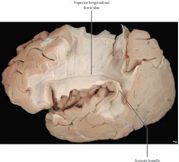
FIGURE 19A: Cerebral Hemispheres 9 — Medial Dissected View: Corpus Callosum (photograph) FIGURE 19B: Cerebral Hemispheres 10 — Lateral Dissected View: Association Bundles (photograph) FIGURE 20A: Ventricles 1 — Lateral View
FIGURE 20B: Ventricles 2 — Anterior View FIGURE 21: Ventricles 3 — CSF Circulation FIGURE 22: Basal Ganglia 1 — Orientation
FIGURE 41B: Visual System 2 — Visual Pathway 2 and Visual Cortex (photograph) FIGURE 41C: Visual System 3 — Visual Reflexes
Part II: Reticular Formation
FIGURE 42A: Reticular Formation 1 — Organization FIGURE 42B: Reticular Formation 2 — Nuclei
FIGURE 43: Reticular Formation 3 — Pain Modulation System
Part III: Motor Systems
FIGURE 44: Spinal Cord Nuclei — Motor
FIGURE 45: Cortico-Spinal Tract — Pyramidal System
FIGURE 46: Cortico-Bulbar Tracts — Nuclei of the Brainstem FIGURE 47: Rubro-Spinal Tract
FIGURE 48: Descending Tracts and Cortico-Pontine Fibers FIGURE 49A: Pontine (Medial) Reticulo-Spinal Tract FIGURE 49B: Medullary (Lateral) Reticulo-Spinal Tract FIGURE 50: Lateral Vestibulo-Spinal Tract
FIGURE 51A: Vestibular Nuclei and Eye Movements FIGURE 51B: Medial Longitudinal Fasciculus (MLF) FIGURE 52: Basal Ganglia Circuitry
FIGURE 53: Thalamus — Motor Circuits FIGURE 54: Cerebellum 1 — Functional Lobes FIGURE 55: Cerebellum 2 — Cerebellar Afferents
FIGURE 56A: Cerebellum 3 — Intracerebellar (Deep Cerebellar) Nuclei FIGURE 56B: Cerebellum 4 — Intracerebellar Circuitry
FIGURE 57: Cerebellum 5 — Cerebellar Efferents
Section C: Neurological Neuroanatomy
FIGURE 58: Blood Supply 1 — Arterial Circle of Willis (photograph with overlay) FIGURE 59A: Blood Supply 2 — MR Angiogram: MRA (radiograph)
FIGURE 59B: Blood Supply 3 — Cerebral Angiogram (radiograph)
FIGURE 60: Blood Supply 4 — Cortical Dorsolateral Surface (photograph with overlay) FIGURE 61: Blood Supply 5 — Cortical Medial Surface (photograph with overlay) FIGURE 62: Blood Supply 6 — Internal Capsule (photograph with overlay)
FIGURE 63: Thalamus: Nuclei and Connections FIGURE 64A: Brainstem Histology: Ventral View FIGURE 64B: Brainstem Histology: Sagittal View
FIGURE 65: Brainstem Histology — Midbrain (upper — photograph) FIGURE 65A: Brainstem Histology — Upper Midbrain
FIGURE 65B: Brainstem Histology — Lower Midbrain
FIGURE 66: Brainstem Histology — Pons (upper — photograph) FIGURE 66A: Brainstem Histology — Upper Pons
FIGURE 66B: Brainstem Histology — Mid-Pons FIGURE 66C: Brainstem Histology — Lower Pons
FIGURE 67: Brainstem Histology — Medulla (mid — photograph) FIGURE 67A: Brainstem Histology — Upper Medulla
FIGURE 68: Spinal Cord — Nuclei and Tracts FIGURE 69: Spinl Cord Histology — Cross Sections
Section D: The Limbic System
FIGURE 70A: Limbic Lobe 1 — CorticalFIGURE 70B: Limbic Lobe 2 — Cingulum Bundle (photograph) FIGURE 71: Limbic System — Noncortical
FIGURE 72A: Hippocampus 1 — Hippocampal Formation
FIGURE 72B: Hippocampus 2 — Hippocampal Formation (3 parts) FIGURE 73: Hippocampus 3 — The Hippocampus (photograph) FIGURE 74: Hippocampus 4 — Coronal View (photograph) FIGURE 75A: Amygdala 1 — Location
FIGURE 75B: Amygdala 2 — Connections
FIGURE 76: Limbic Structures and Lateral Ventricle FIGURE 77A: Limbic Diencephalon 1 — Anterior Nucleus FIGURE 77B: Limbic Diencephalon 2 — Dorsomedial Nucleus FIGURE 78A: Hypothalamus
FIGURE 78B: Medial Forebrain Bundle — Septal Region and Limbic Midbrain FIGURE 79: Olfactory System
(pain, temperature & crude touch from the body)
Trigeminal system
(touch, pain, temperature & proprioception from the head)
Special senses
(vision, audition & taste)
Reticular Formation
(arousal & regulation of muscle t one and reflexes)
Motor:
Voluntary
(movement of body and face)
Parasympathetic
(“rest & digest”)
Other
(non-voluntary motor & visual coordination)
Vestibular nuclei & tracts
(balance & gravity adjustments)
Cerebellum & associated tracts
(motor coordination)
Substantia nigra
(motor initiation)
Red nucleus & tract
(non-voluntary motor)
Other
(miscellaneous)
USER’S GUIDE
COLOR CODING
Color adds a significant beneficial dimension to the learn-ing of neuroanatomy. The colors have a functional role in this atlas, in that they are used consistently for the pre-sentation of sensory, motor, and other components. The following is the color coding used in this atlas, as shown on the opposite page:
For students who enjoy a different learning approach, a black and white photocopy of the illustration can be made and then the color added, promoting active learning. Some students may wish to add color to some of the airbrush diagrams, including the basal ganglia, thalamus, and limbic system.
REFERENCE TO OTHER FIGURES
CLINICAL ASPECT
Various clinical entities are mentioned where there is a clear connection between the structures being discussed and a clinical disease, for example, Parkinson’s disease and the substantia nigra. In Section C, the vascular ter-ritories are discussed and the deficits associated with occlusion of these vessels is reviewed. Textbooks of neurology should be consulted for a detailed review of clinical diseases (see the Annotated Bibliography). Man-agement of the disease and specific drug therapies are not part of the subject matter of this atlas.
ADDITIONAL DETAIL
On occasion, a structure is described that has some importance but may be beyond what is necessary, at this stage, for an understanding of the system or pathway under discussion. In other cases, a structure is labeled in an illustration but is discussed at another point in the atlas.
DEVELOPMENTAL ASPECT
For certain parts of the nervous system, knowledge of the development contributes to an understanding of the structure seen in the adult. This is particularly so for the spinal cord, as well as for the ventricular system. Knowl-edge of development is also relevant for the cerebral hemispheres, and for the limbic system (i.e., the hippoc-ampal formation).
NOTE TO THE LEARNER
This notation is added at certain points in the text when, in the author's experience, it might be beneficial for a student learning the matter to review a certain topic; in other cases there is a recommendation to return to the section at a later stage. Sometimes, consulting other texts is suggested. Of course, this is advice only, and each student will approach the learning task in his or her own
Sensory (nuclei and tracts) Dorsal Column – Medial
Lemniscus
Cobalt Blue
Anterolateral System (Pain and Temperature)
Deep Blue
Trigeminal Pathways Purple Special Senses (Audition,
Vision, Taste)
Violet
Reticular Formation Yellow
Motor (nuclei and tracts)
Voluntary Cadmium Orange
Parasympathetic Orange
Other Motor (e.g. visual motor) Light Red Vestibular (nuclei and tracts) Lime Green Cerebellum (nuclei and tracts) Turquoise
Special Nuclei:
FOREWORD
We are about to embark on an amazing and challenging journey — an exploration of the human brain. The com-plexity of the brain has not yet been adequately described in words. The analogies to switchboards or computers, although in some ways appropriate to describe some aspect of brain function, do not do the least bit of justice to the totality. The brain functioning as a whole is infinitely more than its parts. Our brains encompass and create a vast universe.
In the past decade we have come to appreciate that our brains are in a dynamic state of change in all stages of life. We knew that brain function was developing throughout childhood and this has been extended into the teen years, and even into early adulthood. We now are beginning to understand that the brain has the potential to change throughout life, in reaction to the way we live and our personal experiences in this world. The generic term for this is plasticity, and the changes may significantly alter the connections of the brain and its pattern of “pro-cessing” information, whether from the external world, from our internal environment, or from the brain itself as it generates thoughts and feelings.
ORGANIZATION
The Atlas is divided into four sections, each with an intro-ductory text. The focus is on the illustrations, photographs, diagrams, radiographs, and histological material, accom-panied by explanatory text on the opposite page.
Section A:The Atlas starts with an Overview of the var-ious parts of the central nervous system, the CNS. Then we embark on an Orientation to the structural compo-nents of the CNS, and this is presented from the spinal cord upward to “the brain”; additional material on the spinal cord is added in other parts of the Atlas. Radio-graphic images have been included, because that is how the CNS will be viewed and investigated in the clinical setting.
(Part II), which has both sensory and motor aspects. Included as part of the motor systems are the major con-tributors to motor function, the basal ganglia and the cer-ebellum.
Section C: The third section, Neurological Neuroanat-omy, includes a neurological orientation and detailed neu-roanatomical information, to allow the student to work through the neurological question: Where is the disease process occurring (i.e., neurological localization)? Because vascular lesions are still most common and relate closely to the functional neuroanatomy, the blood supply to the brain is presented in some detail, using photographs with overlays. The emphasis in this section is on the brain-stem, including a select series of histological cross-sec-tions of the human brainstem. In addition, there is a sum-mary of the spinal cord nuclei and tracts, along with a histological view of levels of the human cord.
Section D: The section on the Limbic System has once again been revised. New photographs of limbic structures enhance the presentation. This material is sometimes taught within the context of other systems in the curricu-lum.
ANNOTATED BIBLIOGRAPHY
Students may wish to consult more complete texts on the anatomy and physiology of the nervous system, and cer-tainly some neurology books concerning diseases of the nervous system. A guide to this reference material is included, with commentary, as an annotated bibliography, with an emphasis on recent publications. Added are sug-gestions for material available on CD-ROM, as well as the Internet. Students are encouraged to search out addi-tional (reliable) resources of this nature.
GLOSSARY
inconsis-1
INTRODUCTION
An understanding of the central nervous system — the CNS — and how it functions requires knowing its com-ponent parts and their specialized operations, and the con-tribution of each of the parts to the function of the whole. The first section of this atlas introduces the student to the CNS from an anatomical and functional viewpoint. The subsequent section (Section B) will use these components to build the various systems, such as the sensory and motor systems. The blood supply and the detailed anatomical organization are found in Section C. Emotional behavior is discussed in Section D.
FUNCTIONAL NEUROHISTOLOGY
The major cell of the CNS is the neuron. Human brains have billions of neurons. A neuron has a cell body (also called soma, or perikaryon); dendrites, which extend a short distance from the soma; and an axon, which con-nects one neuron with others. Neuronal membranes are specialized for electro-chemical events, which allow these cells to receive and transmit messages to other neurons. The dendrites and cell bodies of the neurons receive infor-mation, and the axons transmit the firing pattern of the cell to the next neuron. Generally, each neuron receives synaptic input from hundreds or perhaps thousands of neurons, and its axon distributes this information via col-laterals (branches) to hundreds of neurons.
Within the CNS, neurons that share a common func-tion are usually grouped together; such groupings are called nuclei (singular nucleus, which is somewhat con-fusing as it does not refer to the part of a cell). In other parts of the brain, the neurons are grouped at the surface, forming a cortex. In a cortical organization, neurons are arranged in layers and the neurons in each layer are func-tionally alike and different from those in other layers. Older cortical areas have three layers (e.g., the cerebel-lum); more recently evolved cortices have six layers (the cerebral cortex) and sometimes sublayers.
Some neurons in the nervous system are directly linked to sensory (afferent) or motor (efferent) functions. In the CNS, the overwhelming majority of neurons inter-connect, that is, form circuits that participate in the
pro-cessing of information. These neurons are called inter-neurons, and more complex information processing, such as occurs in the human brain, is correlated with the dra-matic increase in the number of interneurons in our brains. Communication between neurons occurs almost exclu-sively at specialized junctions called synapses, using bio-logical molecules called neurotransmitters. These modify ion movements across the neuronal membranes of the syn-apse and alter neurotransmission — they can be excitatory or inhibitory in their action, or modulate synaptic excitabil-ity. The post-synaptic neuron will modify its firing pattern depending on the summative effect of all the synapses act-ing upon it at any moment in time. The action of neurotrans-mitters depends also on the specific receptor type; there is an ever increasing number of receptor subtypes allowing for even more complexity of information processing within the CNS. Drugs are being designed to act on those receptors for therapeutic purposes.
Much of the substance of the brain consists of axons, also called fibers, which connect one part of the brain with other areas. These fibers function so that the various parts of the brain communicate with each other, some going a short distance linking neurons locally and others traveling a long distance connecting different areas of the brain and spinal cord. Many of the axons are myelinated, an “insula-tion,” which serves to increase the speed of axonal conduc-tion; the thicker themyelin sheath, the faster the conduc-tion. Axons originating from one area (cortex or nucleus) and destined for another area usually group together and form a tract, also called a pathway (or fasciculus).
The other major cells of the CNS are glia; there are more glia than neurons. There are two types of glial cells:
• Astrocytes, which are involved in supportive structural and metabolic events
• Oligodendrocytes, which are responsible for the formation and maintenance of the myelin that ensheaths the axons
Some of the early maturation that we see in infants and children can be accounted for by the progressive myelination of the various pathways within the CNS throughout childhood.
2 Atlas of Functional Neutoanatomy
FUNCTIONAL NEUROANATOMY OF THE CNS
One approach to an understanding of the nervous system is to conceptualize that it is composed of a number of functional modules, starting with simpler ones and evolv-ing in higher primates and humans to a more complex organizational network of cells and connections. The func-tion of each part is dependent upon and linked to the function of all the modules acting in concert.
The basic unit of the CNS is the spinal cord (see Figure 1 and Figure 2), which connects the CNS with the skin and muscles of the body. Simple and complex reflex circuits are located within the spinal cord. It receives sen-sory information (afferents) from the skin and body wall, which are then transmitted to higher centers of the brain. The spinal cord receives movement instructions from the higher centers and sends motor commands (efferents) to the muscles. Certain motor patterns are organized in the spinal cord, and these are under the influence of motor areas in other parts of the brain. The autonomic nervous system, which supplies the internal organs and the glands, is also found within the spinal cord.
As the functional systems of the brain become more complex, new control “centers” have evolved. These are often spoken of as higher centers. The first set of these is located in the brainstem, which is situated above the spinal cord and within the skull (in humans). The brain-stem includes three distinct areas — the medulla, pons, and midbrain (see Figure OA,Figure OL,Figure 6, and Figure 7). Some nuclei within the brainstem are concerned with essential functions such as pulse, respiration, and the regulation of blood pressure. Other nuclei within the brainstem are involved in setting our level of arousal and play an important role in maintaining our state of con-sciousness. Special nuclei in the brainstem are responsible for some basic types of movements in response to gravity or sound. In addition, most of the cranial nerves and their nuclei, which supply the structures of the head, are anchored in the brainstem (see Figure 8A and Figure 8B). Many nuclei in the brainstem are related to the cerebellum. The cerebellum has strong connections with the brainstem and is situated behind the brainstem (inside the skull) in humans (see Figure OA, Figure OL, and Figure
spheres and acts as the gateway to the cerebral cortex. The thalamus consists of several nuclei, each of which projects to a part of the cerebral cortex and receives reciprocal connections from the cortex. The hypothalamus, a much smaller part of the diencephalon, serves mostly to control the neuroendocrine system via the pituitary gland, and also organizes the activity of the autonomic nervous sys-tem. Parts of the hypothalamus are intimately connected with the expression of basic drives (e.g., hunger and thirst), with the regulation of water in our bodies, and with the manifestations of “emotional” behavior as part of the limbic system (see below).
With the continued evolution of the brain, the part of the brain called the forebrain undergoes increased devel-opment, a process called encephalization. This has culmi-nated in the development of the cerebral hemispheres, which dominate the brains of higher mammals, reaching its zenith (so we think) in humans. The neurons of the cerebral hemispheres are found at the surface, the cerebral cortex (see Figure 13 and Figure 14A), most of which is six-layered (also called the neocortex). In humans, the cerebral cortex is thrown into ridges (gyri, singular gyrus) and valleys (sulci, singular sulcus). The enormous expan-sion of the cerebral cortex in the human, both in terms of size and complexity, has resulted in this part of the brain becoming the dominant controller of the CNS, capable, so it seems, of overriding most of the other regulatory systems. We need our cerebral cortex for almost all inter-pretations and actions related to the functioning of the sensory and motor systems, for consciousness, language, and thinking.
Buried within the cerebral hemispheres are the basal ganglia, large collections of neurons (see Figure OA, Fig-ure OL, and Figure 22) that are involved mainly in the initiation and organization of motor movements. These neurons affect motor activity through their influence on the cerebral cortex.
The CNS is laced with blood vessels as neurons depend upon a continuous supply of oxygen and glucose. This aspect will be discussed further with the section on vasculature (e.g., see Figure 58).
STUDYOFTHE CNS
Early studies of the normal brain were generally descrip-tive. Brain tissue does not have a firm consistency, and the brain needs to be fixed for gross and microscopic examination. One of the most common fixatives used to preserve the brain for study is formalin, after which it can be handled and sectioned. Areas containing predominantly neuronal cell bodies (and their dendrites and synapses) become grayish in appearance after formalin fixation, and this is traditionally called gray matter. Tracts containing myelinated axons become white in color with formalin fixation, and such areas are likewise simply called the white matter (see Figure 27 and Figure 29).
We have learned much about the normal function of the human CNS through diseases and injuries to the
disrupting neurotransmission. Biochemical disturbances may disrupt the balance of neurotransmitters and cause functional disease states.
The recent introduction of functional imaging of the nervous system is revealing fascinating information about the functional organization of the CNS. We are slowly beginning to piece together an understanding of what is considered by many as the last and most impor-tant frontier of human knowledge, an understanding of the brain.
CLINICAL ASPECT
4 Atlas of Functional Neutoanatomy
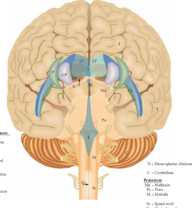
FIGURE OA
OVERVIEW — ANTERIOR VIEW
Constructing a three-dimensional visualization of the brain and its various parts is a challenging task for most people, and this diagram and its companion (the next illustration) are designed to assist the learner in this task. This is a semi-anatomic representation of the brain and the parts of the CNS. This general diagrammatic view should be consulted as the learner is orienting to the place-ment of the structures within the brain. These same struc-tures are viewed from the lateral perspective with the next illustration.
The cerebral hemispheres: The large cerebral hemi-spheres, with its extensive cerebral cortex, is by far the most impressive structure of the CNS and the one that most are referring to when speaking about “the brain.” In fact there are two cerebral hemispheres that are connected across the midline by a massive communication link called the corpus callosum (see Figure 16 and Figure 19A). The hemispheres are discussed with Figure 13–Figure 19 of the Orientation section.
Many parts of the brain are found deep inside the hemispheres. This illustration is done so that these struc-tures should be visualized “within” the hemispheres. Included are:
• Basal ganglia: These large neuronal areas are found within the brain; its three parts are shown — the caudate nucleus (head and tail), the putamen, and the globus pallidus. The basal ganglia are discussed with Figure 22–Figure 30 of the Orientation section.
• Ventricles of the brain: Each hemisphere has within it a space remaining from the neural tube, from which the brain developed, called a ventricle — the lateral ventricle (also called ventricles 1 and 2). The ventricles are presented in this anterior perspective with Figure 20B.
The massive cerebral hemispheres hide the other parts of the brain from view, when looking from the anterior perspective, although some of these parts can be seen if the brain is viewed from below (see Figure 15A and Figure 15B). These structures include:
• Diencephalon: The largest part of the dien-cephalon is the thalamus; in fact, this is a paired structure. The unpaired third ventricle should be noted between the thalamus of each side. The thalamus is discussed with Figure 11 and Figure 12 of the Orientation section. • Brainstem: By definition, the brainstem
con-sists of the midbrain, pons, and medulla; the cranial nerves are attached to the brainstem. The brainstem and cranial nerves are consid-ered in Figure 6–Figure 10 of the Orientation section. The ventricular space within the brain-stem is the fourth ventricle.
• Cerebellum: Part of the cerebellum can be seen from this perspective. This “little brain” is usually considered with the brainstem and is discussed with Figure 9A and Figure 9B of the Orientation section.
• Spinal cord: This long extension of the CNS continues from the medulla and is found in the vertebral canal. The spinal cord is discussed with Figure 1–Figure 5 of the Orientation sec-tion.
FIGURE OA: Overview Diagram — Anterior View
F
3 LV LV
Ch P
GP
D Aq
Md Ct
Po
T
4
M
Cc Sc
C Cerebral hemispheres
F = Frontal lobe T = Temporal lobe
Basal Ganglla Ch = Caudate head Ct = Caudate tail P = Putamen GP = Globus pallidus
Ventricles
LV = Lateral ventricle 3 = 3rd ventricle Aq = Aqueduct 4 = 4th ventricle
D = Diencephalon (thalamus)
C = Cerebellum
Sc = Spinal cord Cc = Central canal Brainstem Md = Midbrain
6 Atlas of Functional Neutoanatomy
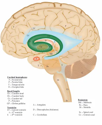
FIGURE OL
OVERVIEW — LATERAL VIEW
This is the companion diagram to the previous illustration, created to assist the learner in placing the brain and its various divisions in a three-dimensional construct.
This is a semi-anatomic view of the brain from the lateral perspective. The front pole of the brain is on the left side of this illustration; the posterior pole is on the right side. The structures included are:
• Cerebral hemispheres: The extensive cerebral hemisphere of one side is seen, with the top edge of the other hemisphere in view (this same view is presented in Figure 14). The lower part of the hemisphere seen on this view is the tem-poral lobe.
• Lateral ventricles: The shape of the ventricles within the hemispheres is now clearly seen (like a reversed letter C), with its continuation into the temporal lobe. The ventricle of the other hemisphere is seen as a “shadow.” (A similar view is presented in Figure 20B.)
• Basal ganglia: The three parts of the basal ganglia are represented in this view. The cau-date (head, body, and tail) follows the ventri-cle. The putamen can be seen from the lateral perspective, but the globus pallidus is hidden from view because it lies medial to the puta-men; its position is indicated by the dashed ellipse. (A similar view is presented in Figure 25.) The two nuclei together are called the lentiform or lenticular nucleus.
One additional nucleus belonging, by definition, with the basal ganglia is seen within the temporal lobe — the amygdala. It will be discussed with the limbic system (in Section D).
• Diencephalon: The thalamus of one side can be visualized from this perspective, almost completely hidden from view by the putamen and the globus pallidus, the lentiform nucleus. The third ventricle is seen just behind it, occu-pying the midline (see Figure 25).
• Brainstem: The upper parts of the brainstem, namely the midbrain and upper pons, cannot be seen from this view of the brain, but their posi-tion is shown as if one could “see through” the temporal lobe. The lower part of the pons and the medulla may be seen. The shape of the fourth ventricle within the brainstem should also be noted.
• Cerebellum: Only the lower portion of one of the hemispheres of the cerebellum can be seen from this lateral perspective, below the cerebral hemispheres.
The brainstem and cerebellum occupy the posterior cranial fossa of the skull.
• Spinal cord: The spinal cord continues from the bottom of the medulla. A view similar to this is seen in a neuroradiologic image in Figure 3.
FIGURE OL: Overview Diagram — Lateral View
LV
LV
Ch
Gp
D 3
P
Cb
Md
A
T Po
Ct
M
Cc
Sc
C 4
O F
Cerebral hemispheres F = Frontal lobe P = Parietal lobe T = Temporal lobe O = Occipital lobe
Basal Ganglia Ch = Caudate head Cb = Caudate body Ct = Caudate tail P = Putamen GP = Globus pallidus
Brainstem Md = Midbrain Po = Pons M = Medulla
Sc = Spinal cord Cc = Central canal Ventricles
LV = Lateral ventricle 3 = 3rd ventricle
4 = 4th ventricle
A = Amygdata
D = Diencephalon (thalamus)
8 Atlas of Functional Neutoanatomy
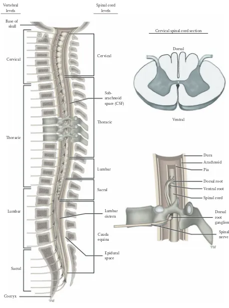
FIGURE 1
SPINAL CORD 1
SPINAL CORD: LONGITUDINAL VIEW
The spinal cord is the extension of the CNS below the level of the skull. It is an elongated structure that is located in the vertebral canal, covered with the meninges — dura, arachnoid, and pia — and surrounded by the subarach-noid space containing cerebrospinal fluid (CSF) (see Fig-ure 21). There is also a space between the dura and ver-tebra, known as the epidural space. Both of these spaces have important clinical implications (see Figure 2C and Figure 3).
The spinal cord, notwithstanding its relatively small size compared with the rest of the brain, is absolutely essen-tial for our normal function. It is the connector between the central nervous system and our body (other than the head). On the sensory (afferent) side, the information arriving from the skin, muscles, and viscera informs the CNS about what is occurring in the periphery; this information then “ascends” to higher centers in the brain.
On the motor (efferent) side, the nerves leave the spinal cord to control our muscles. Although the spinal cord has a functional organization within itself, these neu-rons of the spinal cord receive their “instructions” from higher centers, including the cerebral cortex, via several descending tracts. This enables us to carry out normal movements, including normal walking and voluntary activities. The spinal cord also has a motor output to the viscera and glands, part of the autonomic nervous system (see Figure 4).
UPPER INSET: CERVICAL SPINAL CORD CROSS-SECTION
The neurons of the spinal cord are organized as nuclei, the gray matter, and the various pathways are known as white matter. In the spinal cord, the gray matter is found on the inside, with the white matter all around. The divi-sions of the gray matter are introduced with Figure 4; the functional aspects will be described with the sensory (see Figure 32) and motor (see Figure 44) systems. The tracts
68). Histological cross-sections of the spinal cord are also presented (see Figure 69).
LOWER INSET: NERVE ROOTS
The dorsal root (sensory) and ventral root (motor) unite within the intervertebral foramina to form the (mixed) spinal nerve (see also Figure 5). The nerve cell bodies for the dorsal root are located in the dorsal root ganglion (DRG). Both the roots and the dorsal root ganglion belong to the peripheral nervous system (PNS) (where the Schwann cell forms and maintains the myelin).
DEVELOPMENTAL PERSPECTIVE
During early development, the spinal cord is the same length as the vertebral canal and the entering/exiting nerve roots correspond to the spinal cord vertebral levels. During the second part of fetal development, the body and the bony spine continue to grow, but the spinal cord does not. After birth, the spinal cord only fills the ver-tebral canal to the level of L2, the second lumbar vertebra (see also Figure 3). The space below the termination of the spinal cord is the lumbar cistern, filled with cere-brospinal fluid.
Therefore, as the spinal cord segments do not corre-spond to the vertebral segments, the nerve roots must travel in a downward direction to reach their proper entry/exit level between the vertebra, more so for the lower spinal cord roots (see the photographic view in Figure 2A and Figure 2C). These nerve roots are collectively called the cauda equina, and they are found in the lumbar cistern (see Figure 2A, Figure 2C, and Figure 3).
CLINICAL ASPECT
The four vertebral levels — cervical, thoracic, lumbar, and sacral — are indicated on the left side of the illustration. The spinal cord levels are indicated on the right side. One must be very aware of which reference point — the ver-tebral or spinal — is being used when discussing spinal cord injuries.
FIGURE 1: Spinal Cord 1 — Longitudinal (Vertebral) View
Cervical
Thoracic
Lumbar
Sacral
Coccyx
Epidural space Lumbar cistern
Cauda equina Sacral Lumbar
Sub-arachnoid space (CSF)
Thoracic Ventral
Dura
Arachnoid
Pia
Dorsal root
Ventral root
Spinal cord
Dorsal root ganglion
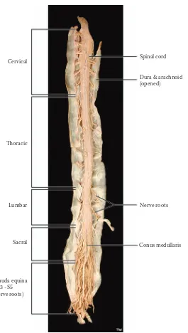
FIGURE 2A
SPINAL CORD 2
SPINAL CORD: LONGITUDINAL VIEW (PHOTOGRAPH)
This is a photographic image of the spinal cord removed from the vertebral canal. The dura-arachnoid has been opened and the anterior aspect of the cord is seen, with the attached spinal roots; from this anterior perspective, most of the roots seen are the ventral (i.e., motor) roots.
The spinal cord is divided into parts according to the region innervated: cervical (8 spinal roots), thoracic (12 spinal roots), lumbar (5 spinal roots), sacral (5 spinal roots), and coccygeal (1 root).
The nerve roots attached to the spinal cord, connecting the spinal cord with the skin and muscles of the body, give the cord a segmented appearance. This segmental organi-zation is reflected onto the body in accordance with embryological development. Areas of skin are supplied by certain nerve segments — each area is called a der-matome (e.g., inner aspect of the arm and hand = C8; umbilical region = T10), with overlap from adjacent seg-ments. The muscles are supplied usually by two adjacent segments, called myotomes (e.g., biceps of the upper limb = C5 and C6; quadriceps of the lower limb = L3 and L4). This known pattern is very important in the clinical setting (see below).
There are two enlargements of the cord: at the cervical level for the upper limb (seen at greater magnification in Figure 2B), the roots of which will form the brachial plexus, and at the lumbosacral level for the lower limb, the roots of which form the lumbar and sacral plexuses. The cord tapers at its ending, and this lowermost portion is called the conus medullaris. Below the vertebral level of L2 in the adult, inside the vertebral canal, are numerous
nerve roots, both ventral and dorsal, collectively called the cauda equina; these are found within the lumbar cistern, an expansion of the subarachnoid space, a space contain-ing CSF (see Figure 1, and shown at a greater magnifica-tion and discussed in Figure 2C; also shown in the MRI in Figure 3).
CLINICAL ASPECT
The segmental organization of the spinal cord and the known pattern of innervation to areas of skin and to mus-cles allows a knowledgeable practitioner, after performing a detailed neurological examination, to develop an accu-rate localization of the injury or disease (called the lesion) at the spinal cord (segmental) level.
The spinal cord can be affected by tumors, either within the cord (intramedullary), or outside the cord (extramedullary). There is a large plexus of veins on the outside of the dura of the spinal cord (see Figure 1), and this is a site for metastases from pelvic (including prostate) tumors. These press upon the spinal cord as they grow and cause symptoms as they compress and interfere with the various pathways (see Section B).
FIGURE 2A: Spinal Cord 2 — Longitudinal View (photograph)
Thoracic
Lumbar
Sacral
Cauda equina (L3 - S5 nerve roots)
Dura & arachnoid (opened)
Nerve roots
FIGURE 2B
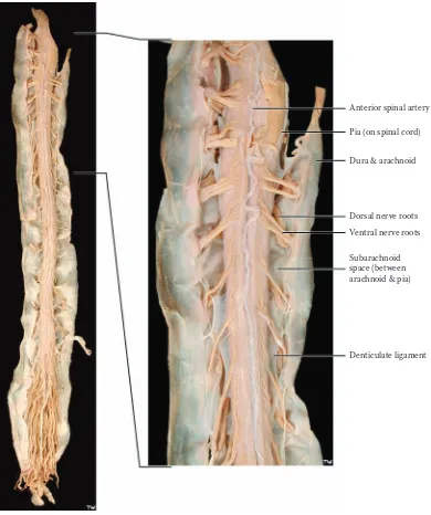
SPINAL CORD 3
SPINAL CORD: CERVICAL REGION (PHOTOGRAPH)
This is a higher magnification photographic image of the cervical region of the spinal cord. Most of the attached roots are the motor/ventral roots, coming from the ventral horn of the spinal cord (discussed with Figure 4); a few of the dorsal/sensory roots can be seen, which enter the cord in the dorsal horn. These roots exit the vertebral canal and carry a sleeve of arachnoid-dura with them for a very short distance, as they head for the intervertebral spaces (see Figure 1).
The somewhat tortuous artery running down the mid-line of the cord is the anterior spinal artery. This artery, which is the major blood supply to the ventral portion of the upper part of the cord, is formed by a branch from each of the vertebral arteries (see Figure 58). This artery receives supplementary branches from the aorta along its way, called radicular arteries, which follow the nerve roots. There are two very small posterior spinal arteries. The most vulnerable area of the spinal cord blood supply is around the mid-thoracic level. There is a particularly important branch off the aorta that supplies this critical region of the spinal cord. This is important clinically (see below).
The pia is attached directly to the spinal cord. Sheets of pia are found in the subarachnoid space, between the ventral and dorsal roots, and can be seen attaching to the inner aspect of the arachnoid — these pial extensionsare called denticulate ligaments. These ligaments, which are
located at intervals along the cord, are thought to tether the cord, perhaps to minimize movement of the cord.
CLINICAL ASPECT
Because of its tenuous blood supply, the spinal cord is most vulnerable in the mid-thoracic portion. A dramatic drop in blood pressure, such as occurs with a cardiac arrest or excessive blood loss, may lead to an infarction of the spinal cord. The result can be just as severe as if the spinal cord was severed by a knife. The most serious consequence of this would be the loss of voluntary motor control of the lower limbs, known as paraplegia. The clinical picture will be understood once the sensory and motor tracts of the spinal cord have been explained (in Section B).
Surgeons who operate on the abdominal aorta, for example, for aortic aneurysm, must make every effort to preserve the small branches coming off the aorta as these are critical for the vascular supply of the spinal cord. One would not want the end result of an aneurysmal repair to be a paraplegic patient.
DEVELOPMENTAL ASPECT
FIGURE 2B: Spinal Cord 3 — Cervical Region (photograph)
Anterior spinal artery
Pia (on spinal cord)
Dura & arachnoid
Dorsal nerve roots
Ventral nerve roots
FIGURE 2C
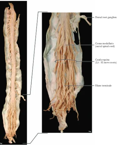
SPINAL CORD 4
SPINAL CORD: CAUDA EQUINA (PHOTOGRAPH)
This is a higher magnification photographic image of the lowermost region of the spinal cord, the sacral region. The tapered end of the spinal cord is called the conus med-ullaris, and this lower portion of the cord corresponds approximately to the sacral segments.
The collection of dorsal and ventral nerve roots, below the level of the termination of the cord, is collectively called the cauda equina. These roots, which belong to the lumbar and sacral segments of the spinal cord, fill the expanded subarachnoid space in this region, known as the lumbar cistern (see Figure 3). The roots are traveling from the spinal cord levels to exit at their appropriate (embryological) intervertebral level (see Figure 1). The roots are floating in the CSF of the lumbar cistern.
The pia mater of the cord gathers at the tip of the conus medullaris into a ligament-like structure, the filum terminale, which attaches to the dura-arachnoid at the termination of the vertebral canal, at the level of (verte-bral) S2. The three meningeal layers then continue and attach to the coccyx as the coccygeal ligament.
CLINICAL ASPECT
Sampling of CSF for the diagnosis of meningitis, an inflammation of the meninges, or for other neurological diseases, is done in the lumbar cistern. This procedure is called a lumbar puncture and must be performed using sterile technique. A trochar (which is a large needle with a smaller needle inside) is inserted below the termination of the spinal cord at L2, in the space between the vertebra, usually between the vertebra L4–L5 (see Figure 1). The trochar must pierce the very tough ligamentum flavum (shown in the next illustration), then the dura-arachnoid, and then “suddenly” enters into the lumbar cistern; the (inner) needle is withdrawn and CSF drips out to be col-lected in sterile vials. This is not a pleasant procedure for a patient and is especially unpleasant, if not frightening, when performed on children.
The nerve roots exit the spinal cord at the appropriate intervertebral level. The roots to the lower extremity, those exiting between L4–L5 and L5–S1, are the ones most commonly involved in the everyday back injuries that affect many adults. The student should be familiar with the signs and symptoms that accompany degenerative disc disease in the lumbar region (see also Figure 1).
FIGURE 2C: Spinal Cord 4 — Cauda Equina (photograph)
Dorsal root ganglion
Conus medullaris (sacral spinal cord)
Cauda equina (L3 - S5 nerve roots)
FIGURE 3
SPINAL CORD 5
SPINAL CORD MRI – T1: LONGITUDINAL VIEW (RADIOGRAPH)
This is a magnetic resonance image (MRI) of the verte-bral column and spinal cord, viewed in a midsagittal plane. This is called a TI-weighted image, in which the cere-brospinal fluid (CSF) is dark. (The various radiological techniques used to image the nervous system are discussed below.) This image is from an adult, in which no pathology was found in the spinal cord radiological examination.
Because of the length of the spinal cord, it is being shown in two parts — upper and lower. The vertebral bodies, the intervertebral discs and the spinous processes posteriorly have been labeled, as well as the ligamentum flavum (discussed with the previous illustration). The ver-tebral bodies have been numbered at various levels — C2, T1, L1, and S1.
The UPPER portion shows the spinal cord to be a continuation of the medulla of the brainstem, at the lower-most border of the skull, the foramen magnum. The pons, medulla, and cerebellum are seen above foramen magnum occupying the posterior cranial fossa.
The spinal cord tissue is located in the middle of the vertebral column, surrounded by the meninges (which can dimly be visualized), with the dura-arachnoid separating the subarachnoid space containing CSF from the space outside the meninges, the epidural space, between the meninges and vertebra (see Figure 1). The epidural space in the lower thoracic region and in the lumbar and sacral regions often contains fat (epidural fat), which is seen as bright on this image.
The LOWER portion of the spinal cord shows the spinal cord itself, tapering as the conus medullaris and terminating around the level of vertebra L1–L2. Below that level is the enlarged subarachnoid space — called a cistern, the lumbar cistern — within which are the nerve roots, dorsal and ventral, for the lower extremity (shown in the previous illustration).
ADDITIONAL DETAIL
RADIOLOGICAL IMAGING
Ordinary x-rays show the skull and its bony structures but not the brain. A remarkable revolution occurred in clinical neurology and our understanding of the brain when imag-ing techniques were developed that allowed for visualiza-tion of the brain. This now includes:
• Computed tomography (CT) (often pro-nounced as a “CAT” scan, meaning computer assisted tomography see Figure 28A). This is done using x-rays, and there is a computer reconstruction of the brain after a series of views are taken from a large number of per-spectives. In this view the bones of the skull are bright and the CSF is dark, with the brain tissue “gray” but not clear. This image can be obtained in several seconds, even with a very sick patient. • Magnetic resonance imaging (MRI) does not use x-rays; the image is created by capturing the energy of the hydrogen ions of water. An extremely strong magnet is used for MRI, and capturing the images requires more time. Again, there is a computer reconstruction of the images. The brain itself looks “anatomic.” This view can be weighted during the acquisition of the image so as to produce a TI image, in which the CSF is dark (this illustration), or a T2 image, in which the CSF is bright (see Figure 28B). With MRI, the bones of the skull are dark, while fatty tissue (including the bone marrow) is bright. Other settings are now available to visualize the brain, such as FLAIR.
As imaging and technology improve, we are able to visualize the brain during functional activity — func-tional MRIs are becoming more widely available; this allows us to “see” which areas of the brain are particularly active during a certain task, based upon the increased blood supply to that area during the active period.
FIGURE 3: Spinal Cord 5 — MRI: Longitudinal View (radiograph)
Subarachnoid space (CSF)
Spinal cord
Subcutaneous fat
Spinous processes
Ligamentum flavum
Conus medullaris
Epidural fat
Lumbar cistern T1
S1 L1 Vertebral bodies
Aorta
FIGURE 4
SPINAL CORD 6
SPINAL CORD: CROSS-SECTIONAL VIEWS
UPPER DIAGRAM
The upper diagram is a cross-section through the spinal cord at the C8 level, the eighth cervical segmental level of the spinal cord (not the vertebral level, see Figure 1). The gray matter is said to be arranged in the shape of a butterfly (or somewhat like the letter H). The gray matter of the spinal cord contains a variety of cell groups (i.e. nuclei), which subserve different functions. Although it is rather difficult to visualize, these groups are continuous longitudinally throughout the length of the spinal cord.
The dorsal region of the gray matter, called the dorsal or posterior horn, is associated with the incoming ( affer-ent) dorsal root, and is thus related to sensory functions. The cell body of these sensory fibers is located in the dorsal root ganglion (see Figure 1). The dorsal horn is quite prominent in this region because of the very large sensory input to this segment of the cord from the upper limb, particularly from the hand. The situation is similar in the lumbar region (as shown in the middle of the three lower illustrations).
The ventral gray matter, called the ventral or anterior horn, is the motor portion of the gray matter. The ventral horn has the large motor neurons, the anterior horn cells, which are efferent to the muscles (see Figure 44). These neurons, because of their location in the spinal cord, which is “below” the brain, are also known as lower motor neurons. (We will learn that the neurons in the cerebral cortex, at the “higher” level, are called upper motor neu-rons — discussed with Figure 45.) The ventral horn is again prominent at this level because of the large number of motor neurons supplying the small muscles of the hand. The situation is similar in the lumbar region, with the motor neurons supplying the large muscles of the thigh (as shown in the illustration below).
The area in between is usually called the intermediate gray and has a variety of cell groups with some associa-tion-type functions (see Figure 32 and Figure 44).
The autonomic nervous system to the organs of the chest, abdomen, and pelvis is controlled by neurons located in the spinal cord.
• Preganglionic sympathetic neurons form a dis-tinctive protrusion of the gray matter, called the lateral horn, which extends throughout the tho-racic region, from spinal cord level T1 to L2 (as shown in the first of the three lower illus-trations). The post-ganglionic nerves supply the organs of the thorax, abdomen, and pelvis. • Parasympathetic preganglionic neurons are
located in the sacral area and do not form a separate horn (as shown in the illustration). This region of the spinal cord in the area of the conus medullaris (the last of the three lower illustra-tions) controls bowel and bladder function, sub-ject to commands from higher centers, including the cerebral cortex.
The parasympathetic control of the organs of the tho-rax and abdomen comes from the vagus nerve, CN X, a cranial nerve (see Figure 6 and Figure 8A).
The central canal of the spinal cord (see Figure 20A, Figure 20B, and Figure 21) is located in the center of the commissural gray matter. This represents the remnant of the neural tube and is filled with CSF. In adults, the central canal of the spinal cord is probably not patent throughout the whole length of the spinal cord. A histological view of these levels of the spinal cord is shown in Figure 69 in Section C.
Note to the Learner: The white matter, which con-tains the ascending sensory and descending motor path-ways, will be described with the pathways in Section B; a summary diagram with all the tracts is shown in Section C (see Figure 68).
ADDITIONAL DETAIL
FIGURE 4: Spinal Cord 6 — Cross-Sectional Views
Dorsal horn
Intermediate gray
Ventral horn
Ventral median fissure
Efferent (motor) neuron Motor nuclei Central canal Sensory nuclei Dorsal root of spinal nerve
Lateral horn Thoracic
Lumbar
FIGURE 5
SPINAL CORD 7
SPINAL CORD MRI – T2: AXIAL VIEWS (RADIOGRAPH)
MRI views of the spinal cord are shown in the axial plane at the C4 (fourth cervical vertebral) level; the orientation should be noted with anterior (ventral) at the top. The CSF is bright in these T2-weighted images. The position of the spinal cord can be easily visualized within the vertebral canal, with the surrounding CSF space. The vertebral bod-ies and lamina are dark; the muscles of the neck can be visualized.
In both images it is possible to see the “butterfly” shape of the gray matter of the spinal cord (see Figure 1 and Figure 4). The orientation of the cord should be noted. In the upper image, the dorsal root and ventral root can be seen, as they head for the intervertebral foramen to form the spinal nerve (see Figure 1); neuroradiologists often call this the neural foramen. In the lower image, taken just a few millimeters below, the spinal nerve can be seen in the intervertebral (neural) foramen.
Note to the Learner: In viewing these radiographs, the left side of the image is in fact the right side of the patient and likewise on the other side — this is the con-vention. The veins, internal jugular and external jugular, appear white with MRI imaging; the common carotid
artery appears dark because of the rapid flow of blood in the arteries; note the presence of the vertebral artery (dark) in the foramen in the transverse process.
CLINICAL ASPECT
FIGURE 5: Spinal Cord 7 — MRI: Axial View (radiograph)
artery
External jugular vein
Internal jugular vein
Spinal roots: Ventral
Dorsal
Vertebral body
Vertebral artery (within transverse foramen)
Spinal nerve
Lamina of vertebra
Subarachnoid space (CSF)
Spinal cord Dorsal
Right Left
Ventral
Dorsal
FIGURE 6
BRAINSTEM 1
BRAINSTEM AND DIENCEPHALON: VENTRAL VIEW
The brainstem is the lowermost part of the brain and is located above the spinal cord. It can be seen by viewing the brain from below (see Figure 15A; also Figure OA and Figure OL). This specimen has been obtained by dissecting out the brainstem, and cerebellum, along with the diencephalon; a photographic view of this specimen is shown in the next illustration (Figure 7). The dienceph-alon will be described subsequently (see Figure 11 and Figure 12).
In the human brain, the brainstem is a relatively small mass of brain tissue compared to the large hemispheres, but it is packed with various nuclei and tracts. Among these nuclei are those of 10 of the cranial nerves (CN III to CN XII). Many basic brain activities are located in the brainstem, including key vital functions (control of blood pressure, pulse, and respiration). Some motor functions are found at various brainstem levels, some as part of the reticular formation; the reticular formation is also part of a system that is responsible for consciousness. Most important, the ascending sensory and descending motor tracts/pathways that connect the spinal cord with “higher” areas of the brain pass through the brainstem (described in Section B). In addition, many of the connections to the cerebellum, including pathways and nuclei, are found in the brainstem. Finally, each part of the brainstem has a part of the ventricular system.
The brainstem is divided anatomically into three parts — the narrow midbrain, which is located under the dien-cephalon; the pons, with its ventral bulge; and the medulla, which connects with the spinal cord. Each of the parts has distinctive features that allow for the identification of the parts, both on the gross brain specimen or a microscopic cross-section.
• The midbrain region (mesencephalon) has two large “pillars” anteriorly called the cerebral peduncles, which consist of millions of axons
tract, is located within the pyramid. Behind each is a prominent bulge, called the olive, the inferior olivary nucleus, which connects with the cerebellum.
CRANIAL NERVES AND THEIR ATTACHMENT
The cranial nerves of the brainstem will be presented in numerical order, starting at the midbrain level.
Midbrain Level
• CN III, the oculomotor nerve, emerges ven-trally between the cerebral peduncles (in the interpeduncular fossa).
• CN IV, the trochlear nerve, which exits pos-teriorly, is a thin nerve that wraps around the lowermost border of the cerebral peduncle.
Pontine Level
• CN V, the trigeminal nerve, is a massive nerve attached along the middle cerebellar peduncle. • CN VI, the abducens nerve, is seen exiting anteriorly at the junction between the pons and medulla.
• CN VII, the facial nerve, and CN VIII (the vestibulocochlear nerve), are both attached to the brainstem at the ponto-cerebellar angle.
Medullary Level
• CN IX, the glossopharyngeal, and CN X, the vagus, are attached to the lateral margin of the medulla, behind the inferior olive.
• CN XI, the spinal accessory nerve, from the uppermost region of the spinal cord, enters the skull and then exits from the skull as if it were a cranial nerve; by convention it is included as a cranial nerve.
FIGURE 6: Brainstem 1 — Ventral View with Cranial Nerves
Thalamus
Midbrain
Pons
Cerebellum
Medulla
Spinal cord
O Py Py O Po
CP M CP
FI FI
Pituitary stalk
Oculomotor nerve (CN III) Trochlear nerve (CN IV)
Trigeminal nerve (CN V)
Abducens nerve (CN VI) Facial nerve (CN VII) Vestibulocochlear nerve (CN VIII)
Glossopharyngeal nerve (CN IX) Vagus nerve (CN X)
Hypoglossal nerve (CN XII)
Spinal accessory nerve (CN XI)
M = Mammillary bodies CP = Cerebral peduncle Po = Pons