123
Nathalia Peixoto
Margarida Silveira
Hesham H. Ali
Carlos Maciel
Egon L. van den Broek (Eds.)
10th International Joint Conference, BIOSTEC 2017
Porto, Portugal, February 21–23, 2017
Revised Selected Papers
in Computer and Information Science
881
Commenced Publication in 2007 Founding and Former Series Editors:
Alfredo Cuzzocrea, Xiaoyong Du, Orhun Kara, Ting Liu, DominikŚlęzak, and Xiaokang Yang
Editorial Board
Simone Diniz Junqueira Barbosa
Pontifical Catholic University of Rio de Janeiro (PUC-Rio), Rio de Janeiro, Brazil
Phoebe Chen
La Trobe University, Melbourne, Australia Joaquim Filipe
Polytechnic Institute of Setúbal, Setúbal, Portugal Igor Kotenko
St. Petersburg Institute for Informatics and Automation of the Russian Academy of Sciences, St. Petersburg, Russia
Krishna M. Sivalingam
Indian Institute of Technology Madras, Chennai, India Takashi Washio
Osaka University, Osaka, Japan Junsong Yuan
University at Buffalo, The State University of New York, Buffalo, USA Lizhu Zhou
Hesham H. Ali
•Carlos Maciel
Egon L. van den Broek (Eds.)
Biomedical Engineering
Systems and Technologies
10th International Joint Conference, BIOSTEC 2017
Porto, Portugal, February 21
–
23, 2017
Revised Selected Papers
George Mason University Communications in Computer and Information Science
ISBN 978-3-319-94805-8 ISBN 978-3-319-94806-5 (eBook) https://doi.org/10.1007/978-3-319-94806-5
Library of Congress Control Number: 2018947372
©Springer International Publishing AG, part of Springer Nature 2018
This work is subject to copyright. All rights are reserved by the Publisher, whether the whole or part of the material is concerned, specifically the rights of translation, reprinting, reuse of illustrations, recitation, broadcasting, reproduction on microfilms or in any other physical way, and transmission or information storage and retrieval, electronic adaptation, computer software, or by similar or dissimilar methodology now known or hereafter developed.
The use of general descriptive names, registered names, trademarks, service marks, etc. in this publication does not imply, even in the absence of a specific statement, that such names are exempt from the relevant protective laws and regulations and therefore free for general use.
The publisher, the authors and the editors are safe to assume that the advice and information in this book are believed to be true and accurate at the date of publication. Neither the publisher nor the authors or the editors give a warranty, express or implied, with respect to the material contained herein or for any errors or omissions that may have been made. The publisher remains neutral with regard to jurisdictional claims in published maps and institutional affiliations.
Printed on acid-free paper
This Springer imprint is published by the registered company Springer International Publishing AG part of Springer Nature
The present book includes extended and revised versions of a set of selected papers from the 10th International Joint Conference on Biomedical Engineering Systems and Technologies (BIOSTEC 2017), held in Porto, Portugal, during February 21–23, 2017. BIOSTEC is composed offive co-located conferences, each specialized in a different knowledge area, namely, BIODEVICES, BIOIMAGING, BIOINFORMATICS, BIO-SIGNALS, and HEALTHINF.
BIOSTEC 2017 received 297 paper submissions from 56 countries, of which only 6% are included in this book. This reflects our care in selecting contributions. These papers were selected by the conference chairs on the basis of a number of criteria that include the classifications and comments provided by the Program Committee mem-bers, the session chairs’assessment, and the program chairs’meta-review of the papers that were included in the technical program. The authors of selected papers were invited to submit a revised, extended, and improved version of their conference paper, including at least 30% new material.
The purpose of the BIOSTEC joint conferences is to bring together researchers and practitioners, including engineers, biologists, health professionals, and informatics/computer scientists. Research presented at BIOSTEC included both theo-retical advances and applications of information systems, artificial intelligence, signal processing, electronics, and other engineering tools in areas related to advancing biomedical research and improving health care.
The papers included in this book contribute to the understanding of relevant research trends in biomedical engineering systems and technologies. As such, they provide an overview of thefield’s current state of the art.
We thank the authors for their contributions and Monica Saramago for process management. In particular, we express our gratitude to the reviewers, who helped to ensure the quality of this publication.
February 2017 Nathalia Peixoto
Conference Co-chairs
Ana Fred Instituto de Telecomunicações/IST, Portugal Hugo Gamboa LIBPHYS-UNL/FCT - New University of Lisbon,
Portugal
Mário Vaz INEGI LOME, FEUP, Portugal
Program Co-chairs
BIODEVICES
Nathalia Peixoto Neural Engineering Lab, George Mason University, USA
BIOIMAGING
Margarida Silveira Instituto Superior Técnico (IST), Portugal BIOINFORMATICS
Hesham Ali University of Nebraska at Omaha, USA BIOSIGNALS
Carlos Maciel University of São Paulo, Brazil HEALTHINF
Egon L. van den Broek Utrecht University, The Netherlands BIODEVICES Program Committee
Azam Ali University of Otago, New Zealand Mohammed Bakr CCIT-AASTMT, Egypt
Steve Beeby University of Southampton, UK
Hadar Ben-Yoav Ben-Gurion University of the Negev, Israel Dinesh Bhatia North Eastern Hill University, India Efrain Zenteno Bolaños Universidad Católica San Pablo, Peru Luciano Boquete Alcala University, Spain
Carlo Capelli Norwegian School of Sport Sciences, Norway Vítor Carvalho IPCA and Algoritmi Research Centre, UM, Portugal Hamid Charkhkar Case Western Reserve University, USA
Wenxi Chen The University of Aizu, Japan
James M. Conrad University of North Carolina at Charlotte, USA Albert Cook University of Alberta, Canada
Maeve Duffy NUI Galway, Ireland
Paddy French Delft University of Technology, The Netherlands Juan Carlos Garcia University of Alcala, Spain
Javier Garcia-Casado Universitat Politècnica de València, Spain Bryon Gomberg Geyra Gassner IP Law, Israel
Miguel Angel García Gonzalez
Universitat Politècnica de Catalunya, Spain Klas Hjort Uppsala University, Sweden
Toshiyuki Horiuchi Tokyo Denki University, Japan Leonid Hrebien Drexel University, USA
Sandeep K. Jha Indian Institute of Technology Delhi, India Eyal Katz Tel Aviv University, Israel
Michael Kraft University of Liege, Belgium
Ondrej Krejcar University of Hradec Kralove, Czech Republic Ning Lan Shanghai Jiao Tong University, China
Jung Chan Lee Seoul National University, South Korea Chwee Teck Lim National University of Singapore, Singapore Mai S. Mabrouk Misr University for Science and Technology, Egypt Jordi Madrenas Universitat Politècnica de Catalunya, Spain
Jarmo Malinen Aalto University, Finland Karen May-Newman San Diego State University, USA
Joseph Mizrahi Technion, Israel Institute of Technology, Israel Raimes Moraes Universidade Federal de Santa Catarina, Brazil Umberto Morbiducci Politecnico di Torino, Italy
Antoni Nowakowski Gdansk University of Technology, Poland Eoin O’Cearbhaill University College Dublin, Ireland Mónica Oliveira University of Strathclyde, UK Abraham Otero Universidad San Pablo CEU, Spain Gonzalo Pajares Universidad Complutense de Madrid, Spain Sofia Panteliou University of Patras, Greece
Nancy Paris British Columbia Institute of Technology, Canada
Lionel Pazart CHU, France
Nathalia Peixoto George Mason University, USA
Marek Penhaker VŠB, Technical University of Ostrava, Czech Republic Dmitry Rogatkin Moscow Regional Research and Clinical
Institute MONIKI, Russian Federation Wim L. C. Rutten University of Twente, The Netherlands Seonghan Ryu Hannam University, South Korea Ashutosh Sabharwal Rice University, USA
V. V. Raghavendra Sai IIT Madras, India
Chutham Sawigun Mahanakorn University of Technology, Thailand Michael J. Schöning FH Aachen, Germany
Alcimar Barbosa Soares Universidade Federal de Uberlândia, Brazil Filomena Soares Algoritmi Research Centre, UM, Portugal Akihiro Takeuchi Kitasato University School of Medicine, Japan
Gil Travish Adaptix Ltd., UK
John Tudor University of Southampton, UK Renato Varoto University of Campinas, Brazil
Pedro Vieira Faculdade de Ciências e Tecnologia, Universidade Nova de Lisboa, Portugal
Bruno Wacogne FEMTO-ST, France Huikai Xie University of Florida, USA Sen Xu Merck & Co., Inc., USA Hakan Yavuz ÇukurovaÜniversity, Turkey BIOIMAGING Program Committee
Sameer K. Antani National Library of Medicine, National Institutes of Health, USA
Peter Balazs University of Szeged, Hungary Grégory Barbillon EPF-Ecole d’Ingénieurs, France
Alpan Bek Middle East Technical University, Turkey Mads Sylvest Bergholt Imperial College London, UK
Obara Boguslaw University of Durham, UK
Alberto Bravin European Synchrotron Radiation Facility, France Tom Brown University of St. Andrews, UK
Enrico G. Caiani Politecnico di Milano, Italy Rita Casadio University of Bologna, Italy
Alessia Cedola CNR, Institute of Nanotechnology, Italy Heang-Ping Chan University of Michigan, USA
James Chan University of California Davis, USA Dean Chapman University of Saskatchewan, Canada
Guanying Chen Harbin Institute of Technology and SUNY Buffalo, China/USA
Jyh-Cheng Chen National Yang-Ming University, Taiwan Christos E. Constantinou Stanford University, USA
Edite Maria Areias Figueiras
National Physical Laboratory, Portugal Dimitrios Fotiadis University of Ioannina, Greece Patricia Haro Gonzalez Universidad Autonoma Madrid, Spain P. Gopinath Indian Institute of Technology Roorkee, India Dimitris Gorpas Technical University of Munich, Germany Alberto Del Guerra University of Pisa, Italy
Tzung-Pei Hong National University of Kaohsiung, Taiwan
Kazuyuki Hyodo High Energy Accelerator Research Organization, Japan
Shu Jia Stony Brook University, USA
Pluim Josien Eindhoven University of Technology, The Netherlands Patrice Koehl University of California, USA
Adriaan A. Lammertsma VU University Medical Center Amsterdam, The Netherlands
Sang-Won Lee Korea Research Institute of Standards and Science, South Korea
Rainer Leitgeb Medical University Vienna, Austria Ivan Lima North Dakota State University, USA
Xiongbiao Luo XMU-TMMU, China
Modat Marc University College London, UK David McGloin University of Dundee, UK
Aidan D. Meade Centre for Radiation and Environmental Science, Dublin Institute of Technology, Ireland
Erik Meijering Erasmus University Medical Center, The Netherlands Israel Rocha Mendoza Centro de Investigación Científica y de Educación
Superior de Ensenada, (CICESE), Mexico Kunal Mitra Florida Institute of Technology, USA Christophoros Nikou University of Ioannina, Greece Joanna Isabelle Olszewska University of West Scotland, UK Kalman Palagyi University of Szeged, Hungary
Joao Papa UNESP, Universidade Estadual Paulista, Brazil Tae Jung Park Chung-Ang University, South Korea
Gennaro Percannella University of Salerno, Italy
Ales Prochazka University of Chemistry and Technology, Czech Republic
Jia Qin University of California/University of Washington, USA
Wan Qin University of Washington, USA
Miroslav Radojevic Erasmus MC, Biomedical Imaging Group Rotterdam, The Netherlands
Joseph Reinhardt University of Iowa, USA
Giovanna Rizzo Consiglio Nazionale delle Ricerche, Italy Bart M. ter Haar Romeny Eindhoven University of Technology (TU/e),
The Netherlands
Emanuele Schiavi Universidad Rey Juan Carlos, Spain
Jan Schier The Institute of Information Theory and Automation of the Czech Academy of Sciences, Czech Republic Leonid Shvartsman Hebrew University, Israel
Chikayoshi Sumi Sophia University, Japan
Chi-Kuang Sun National Taiwan University, Taiwan
Pablo Taboada University of Santiago de Compostela, Spain Xiaodong Tao University of California, Santa Cruz, USA Pécot Thierry Medical University of South Carolina, France Kenneth Tichauer Illinois Institute of Technology, USA
Eric Tkaczyk Vanderbilt University, USA
Carlos M. Travieso University of Las Palmas de Gran Canaria, Spain Benjamin M. W. Tsui Johns Hopkins University, USA
Vladimír Ulman Masaryk University, Czech Republic GözdeÜnal Istanbul Technical University, Turkey
Sandra Rua Ventura School of Allied Health Technologies/Escola Superior de Saúde do Porto, Portugal
Yuanyuan Wang Fudan University, China
Quan Wen University of Electronic Science and Technology of China, China
Hongki Yoo Hanyang University, South Korea Habib Zaidi Geneva University Hospital, Switzerland BIOIMAGING Additional Reviewers
Xiaoli Qi Johns Hopkins University, USA Qinqin Zhang University of Washington, USA BIOINFORMATICS Program Committee
Tatsuya Akutsu Kyoto University, Japan
Jens Allmer Izmir Institute of Technology, Turkey
Sameer K. Antani National Library of Medicine, National Institutes of Health, USA
Marco Antoniotti Universitàdegli Studi di Milano Bicocca, Italy Joel Arrais Universidade de Coimbra, Portugal
Rolf Backofen Albert-Ludwigs-Universität, Germany Lucia Ballerini University of Edinburgh, UK
Ugur Bilge Akdeniz University, Turkey Leonardo Bocchi Universitàdi Firenze, Italy
Ulrich Bodenhofer Johannes Kepler University Linz, Austria Luca Bortolussi University of Trieste, Italy
Andrea Bracciali University of Stirling, UK Giulio Caravagna University of Edinburgh, UK
JoséPedro Cerón Carrasco Universidad Católica San Antonio de Murcia, Spain Claudia Consuelo Rubiano
Castellanos
Universidad Nacional de Colombia, Bogota, Colombia Santa Di Cataldo Politecnico di Torino, Italy
Wai-Ki Ching The University of Hong Kong, SAR China Mark Clement Brigham Young University, USA
Netta Cohen University of Leeds, UK
Federica Conte National Research Council of Rome, Italy
Antoine Danchin Institute of Cardiometabolism and Nutrition, France Sérgio Deusdado Instituto Politecnico de Bragança, Portugal
Richard Edwards University of Southampton, UK Fabrizio Ferre University of Rome Tor Vergata, Italy António Ferreira Universidade de Lisboa, Portugal
Alexandre P. Francisco Instituto Superior Técnico, Universidade de Lisboa, Portugal
Dario Ghersi University of Nebraska at Omaha, USA
Arndt von Haeseler Center of Integrative Bioinformatics Vienna, MFPL, Austria
Christopher E. Hann University of Canterbury, New Zealand Ronaldo Fumio Hashimoto University of São Paulo, Brazil
Shan He University of Birmingham, UK
Volkhard Helms Universität des Saarlandes, Germany Song-Nian Hu Chinese Academy of Sciences, China Jari Hyttinen Tampere University of Technology, Finland Sohei Ito National Fisheries University, Japan
Bo Jin MilliporeSigma, Merck KGaA, USA
Giuseppe Jurman Fondazione Bruno Kessler, Italy
Yannis Kalaidzidis Max Planck Institute Molecular Cell Biology and Genetics, Germany
Michael Kaufmann Witten/Herdecke University, Germany Natalia Khuri Stanford University, USA
Sami Khuri San JoséState University, USA Inyoung Kim Virginia Tech, USA
Toralf Kirsten University of Leipzig, Germany
JiríKléma Czech Technical University in Prague, Czech Republic Peter Kokol University of Maribor, Slovenia
Malgorzata Kotulska Wroclaw University of Technology, Poland Ivan Kulakovskiy Engelhardt Institute of Molecular Biology RAS,
Russian Federation
Yinglei Lai George Washington University, USA Carlile Lavor University of Campinas, Brazil Matej Lexa Masaryk University, Czech Republic Antonios Lontos Frederick University, Cyprus Shuangge Ma Yale University, USA Pawel Mackiewicz Wroclaw University, Poland Thérèse E. Malliavin CNRS/Institut Pasteur, France Elena Marchiori Radboud University, The Netherlands Andrea Marin University of Venice, Italy
Majid Masso George Mason University, USA Petr Matula Masaryk University, Czech Republic
Claudine Médigue CEA/Genomic Institute/Genoscope and CNRS, France
Ivan Merelli ITB CNR, Italy
Armin Mikler University of North Texas, USA Paolo Milazzo University of Pisa, Italy
Saad Mneimneh Hunter College CUNY, USA
Pedro Tiago Monteiro INESC-ID/IST, Universidade de Lisboa, Portugal Vincent Moulton University of East Anglia, UK
JoséLuis Oliveira Universidade de Aveiro, Portugal Hakan S. Orer Koc University, Turkey
Giulia Paciello Politecnico di Torino, Italy
Marco Pellegrini Consiglio Nazionale delle Ricerche, Italy Matteo Pellegrini University of California, Los Angeles, USA Nadia Pisanti Universitádi Pisa, Italy
Olivier Poch Universitéde Strasbourg, France Alberto Policriti Universitàdegli Studi di Udine, Italy Gianfranco Politano Politecnico di Torino, Italy
Giuseppe Profiti University of Bologna, Italy
Mark Ragan The University of Queensland, Australia Jagath Rajapakse Nanyang Technological University, Singapore Javier Reina-Tosina University of Seville, Spain
Laura Roa University of Seville, Spain
David Rocke University of California, Davis, USA Simona E. Rombo Universitàdegli Studi di Palermo, Italy Eric Rouchka University of Louisville, USA
Carolina Ruiz WPI, USA
J. Cristian Salgado University of Chile, Chile Alessandro Savino Politecnico di Torino, Italy
Sophie Schbath French National Institute for Agriculatural Research, France
Noor Akhmad Setiawan Universitas Gadjah Mada, Indonesia Joao C. Setubal Universidade de São Paulo, Brazil Christine Sinoquet University of Nantes, France Neil R. Smalheiser University of Illinois Chicago, USA
Pavel Smrz Brno University of Technology, Czech Republic Gordon Smyth Walter and Eliza Hall Institute of Medical Research,
Australia
Yinglei Song Jiansu University of Science and Technology, China Peter F. Stadler Universität Leipzig, IZBI, Germany
Andrew Sung University of Southern Mississippi, USA David Svoboda Masaryk University, Czech Republic
Peter Sykacek BOKU, University of Natural Resources and Life Sciences, Austria
Jerzy Tiuryn Warsaw University, Poland
Takashi Tomita Japan Advanced Institute of Science and Technology, Japan
Alexander Tsouknidas Aristotle University of Thessaloniki, Greece Gabriel Valiente Technical University of Catalonia, Spain Juris Viksna University of Latvia, Latvia
Thomas Werner University of Michigan, Germany Yanbin Yin Northern Illinois University, USA Malik Yousef Zefat Academic College, Israel
Nazar Zaki United Arab Emirates University, UAE Helen Hao Zhang University of Arizona, USA
Leming Zhou University of Pittsburgh, USA Zexuan Zhu Shenzhen University, China BIOINFORMATICS Additional Reviewers
Roberta Gori Universitádi Pisa, Italy Artem Kasianov VIGG, Russian Federation BIOSIGNALS Program Committee
Marko Ackermann FEI University, Brazil
Jean-Marie Aerts M3-BIORES, Katholieke Universitëit Leuven, Belgium Robert Allen University of Southampton, UK
Sergio Alvarez Boston College, USA Sridhar P. Arjunan RMIT University, Australia Ofer Barnea Tel Aviv University, Israel
Eberhard Beck Brandenburg University of Applied Sciences, Germany Egon L. van den Broek Utrecht University, The Netherlands
Guy Carrault University of Rennes 1, France Maria Claudia F. Castro Centro Universitário da FEI, Brazil Sergio Cerutti Polytechnic University of Milan, Italy Bruno Cornelis Vrije Universiteit Brussel, Belgium Jan Cornelis Vrije Universiteit Brussel, Belgium Adam Czajka University of Notre Dame, USA Bruce Denby UniversitéPierre et Marie Curie, France Gordana Jovanovic Dolecek Institute INAOE, Mexico
Petr Dolezel University of Pardubice, Czech Republic Pier Luigi Emiliani Italian National Research Council, Italy Pedro Encarnação Universidade Católica Portuguesa, Portugal Poree fabienne Universitéde Rennes 1, France
Luca Faes Universitàdegli Studi di Trento, Italy Dimitrios Fotiadis University of Ioannina, Greece Arfan Ghani Coventry University, UK Inan Güler Gazi University, Turkey
Thomas Hinze Friedrich Schiller University Jena, Germany Roberto Hornero University of Valladolid, Spain
Jiri Jan University of Technology Brno, Czech Republic Tzyy-Ping Jung University of California San Diego, USA Theodoros Kostoulas University of Geneva, Switzerland
Dagmar Krefting Berlin University of Applied Sciences, Germany Vaclav Kremen Czech Technical University in Prague, Czech Republic Lenka Lhotska Czech Technical University in Prague, Czech Republic Julián David Arias Londoño Universidad de Antioquia, Colombia
Ana Rita Londral Universidade de Lisboa, Portugal
Carlos Maciel University of São Paulo, Brazil Hari Krishna Maganti Saga, UK
Armando Malanda Universidad Pública de Navarra, Spain Pramod Kumar Meher Nanyang Technological University, Singapore Jirí Mekyska Brno University of Technology, Czech Republic Roberto Merletti Politecnico di Torino, Italy
Mihaela Morega University Politehnica of Bucharest, Romania Percy Nohama Pontifícia Universidade Católica do Paraná, Brazil Andres Orozco-Duque Instituto Tecnológico Metropolitano, Colombia Krzysztof Pancerz University of Rzeszow, Poland
Gennaro Percannella University of Salerno, Italy
Vitor Pires Escola Superior de Tecnologia de Setúbal, Instituto Politécnico de Setúbal, Portugal Ales Prochazka University of Chemistry and Technology,
Czech Republic
Heather Ruskin Dublin City University, Ireland Tomasz Rutkowski The University of Tokyo, Japan
Andres Santos Universidad Politécnica de Madrid, Spain Roberto Sassi Universitàdegli studi di Milano, Italy Edward Sazonov University of Alabama, USA
Gerald Schaefer Loughborough University, UK Christian Schmidt University of Rostock, Germany Reinhard Schneider Fachhochschule Vorarlberg, Austria Lotfi Senhadji University of Rennes 1, France Samuel Silva Universidade de Aveiro, Portugal Alan A Stocker University of Pennyslvania, USA
Nicola Strisciuglio University of Groningen, The Netherlands
Asser Tantawi IBM, USA
António Teixeira University of Aveiro, Portugal
João Paulo Teixeira Polytechnic Institute of Bragança, Portugal Carlos Eduardo Thomaz Centro Universitário da FEI, Brazil
Carlos M. Travieso University of Las Palmas de Gran Canaria, Spain Pedro Gómez Vilda Universidad Politécnica de Madrid, Spain Gert-Jan de Vries Philips Research Healthcare, The Netherlands Yuanyuan Wang Fudan University, China
Quan Wen University of Electronic Science and Technology of China, China
Kerstin Witte Otto von Guericke University Magdeburg, Germany Didier Wolf Research Centre for Automatic Control, CRAN CNRS
UMR 7039, France
Pew-Thian Yap University of North Carolina at Chapel Hill, USA Chia-Hung Yeh National Sun Yat-sen University, Taiwan
HEALTHINF Program Committee
Anurag Agrawal CSIR Institute of Genomics and Integrative Biology, Center for Translational Research in Asthma and Lung, India
Hesham Ali University of Nebraska at Omaha, USA Adil Alpkocak Dokuz Eylul University, Turkey
Flora Amato Universitàdegli Studi di Napoli Federico II, Italy Francois Andry Philips, France
Wassim Ayadi LERIA, University of Angers, France and LaTICE, University of Tunis, Tunisia
Bert-Jan van Beijnum University of Twente, The Netherlands Patrick Boissy Universitéde Sherbrooke and Research Centre
on Aging, Canada
Sorana D. Bolboaca Iuliu Hatieganu University of Medicine and Pharmacy, Cluj-Napoca, Romania
Alessio Bottrighi Universitádel Piemonte Orientale, Italy Andrew D. Boyd University of Illinois at Chicago, USA Egon L. van den Broek Utrecht University, The Netherlands Berry de Bruijn National Research Council, Canada
Federico Cabitza Universitàdegli Studi di Milano-Bicocca, Italy
Eric Campo LAAS CNRS, France
Guillermo Lopez Campos The University of Melbourne, Australia Marc Cavazza University of Kent, UK
Philip K. Chan Florida Institute of Technology, USA
Taridzo Chomutare University Hospital of North Norway, Norway Mihail Cocosila Athabasca University, Canada
Miguel Coimbra IT, University of Porto, Portugal Emmanuel Conchon XLIM, France
Carlos Costa Universidade de Aveiro, Portugal Andre Dekker MAASTRO Clinick, The Netherlands Chrysanne DiMarco University of Waterloo, Canada
Liliana Dobrica University Politehnica of Bucharest, Romania Stephan Dreiseitl Upper Austria University of Applied Sciences
at Hagenberg, Austria JoséFonseca UNINOVA, Portugal Daniel Ford Dell Research, USA
Christoph M. Friedrich University of Applied Sciences and Arts Dortmund, Germany
Ioannis Fudos University of Ioannina, Greece
Henry Gabb University of Illinois at Urbana-Champaign, USA Hugo Gamboa New University of Lisbon, Portugal
Mauro Giacomini University of Genoa, Italy
Alejandro Rodríguez González
Centro de Tecnología Biomédica, Spain Yves Gourinat ISAE-SUPAERO, France
David Greenhalgh University of Strathclyde, UK Jorge Henriques Universidade de Coimbra, Portugal Dragan Jankovic University of Nis, Serbia
Noreen Kamal University of Calgary, Canada Enkelejda Kasneci University of Tübingen, Germany Finn Kensing University of Copenhagen, Denmark Irena Koprinska University of Sydney, Australia Sofia Kyratzi University of the Aegean, Greece
Elyes Lamine University of Toulouse, CUFR J.F. Champollion, France
Gondy Leroy University of Arizona, USA Giuseppe Liotta University of Perugia, Italy Guillaume Lopez Aoyama Gakuin University, Japan Martin Lopez-Nores University of Vigo, Spain
Alda Marques University of Aveiro, Portugal
Paloma Martínez Universidad Carlos III de Madrid, Spain Ken Masters Sultan Qaboos University, Oman Sally Mcclean University of Ulster, UK Gerrit Meixner Heilbronn University, Germany Christo El Morr York University, Canada Hammadi Nait-Charif Bournemouth University, UK Goran Nenadic University of Manchester, UK JoséLuis Oliveira Universidade de Aveiro, Portugal
Calvin Or The University of Hong Kong, SAR China Rui Pedro Paiva University of Coimbra, Portugal
Guy Pare HEC Montréal, Canada
Vimla L. Patel Arizona State University, USA Sotiris Pavlopoulos ICCS, Greece
Göran Petersson eHealth Institute, Linnaeus University, Sweden Enrico Maria Piras Fondazione Bruno Kessler, Trento, Italy Arkalgud Ramaprasad University of Illinois at Chicago, USA Grzegorz Redlarski Gdansk University of Technology, Poland Ita Richardson University of Limerick, Ireland
Valter Roesler Federal University of Rio Grande do Sul, Brazil Elisabetta Ronchieri INFN, Italy
Senjuti Basu Roy The University of Washington, Tacoma, USA
Carolina Ruiz WPI, USA
George Sakellaropoulos University of Patras, Greece
Ovidio Salvetti National Research Council of Italy, Italy
Akio Sashima AIST, Japan
Jacob Scharcanski Universidade Federal do Rio Grande do Sul, Brazil Bettina Schnor Potsdam University, Germany
Jan Sliwa Bern University of Applied Sciences, Switzerland George Spyrou The Cyprus Institute of Neurology and Genetics,
Cyprus
Jan Stage Aalborg University, Denmark
Vicente Traver Universidad Politécnica de Valencia, Spain
Mohy Uddin King Abdullah International Medical Research Center, Saudi Arabia
Gary Ushaw Newcastle University, UK Aristides Vagelatos CTI, Greece
Sumithra Velupillai KTH, Sweden and King’s College, UK Sitalakshmi Venkatraman Melbourne Polytechnic, Australia Francisco Veredas Universidad de Málaga, Spain Justin Wan University of Waterloo, Canada
Szymon Wilk Poznan University of Technology, Poland Janusz Wojtusiak George Mason University, USA
Clement T. Yu University of Illinois at Chicago, USA Xuezhong Zhou Beijing Jiaotong University, China AndréZúquete Universidade de Aveiro, Portugal HEALTHINF Additional Reviewers
Koundinya Desiraju Institute of Genomics and Integrative Biology, Mall Road, Delhi, India
Angela Locoro Universitàdegli Studi di Milano-Bicocca, Italy Imen Megdiche IRIT, France
Sebastian Rauh Heilbronn University of Applied Sciences, Germany Kateryna Sergieieva Heilbronn University of Applied Science, Germany
Invited Speakers
Bart M. ter Haar Romeny Eindhoven University of Technology (TU/e), The Netherlands
Kristina Höök Royal Institute of Technology, Sweden Bethany Bracken Charles River Analytics Inc., USA
Biomedical Electronics and Devices
An Electronic-Engineered Sensory Sternal Retractor Aimed
at Post-sternotomy Pain Reduction . . . 3 Giovanni Saggio, Alessandra Bianco, Giancarlo Orengo,
Giuseppe Tancredi, Costantino Del Gaudio, and Jacob Zeitani
lSmartScope: Towards a Fully Automated 3D-Printed Smartphone
Microscope with Motorized Stage . . . 19 Luís Rosado, Paulo T. Silva, JoséFaria, João Oliveira,
Maria João M. Vasconcelos, Dirk Elias, JoséM. Correia da Costa, and Jaime S. Cardoso
A Portable Chemical Detection System with Anti-body Biosensor
for Impedance Based Monitoring of T2-mycotoxin Bioterrorism Agents. . . 45 V. I. Ogurtsov and K. Twomey
Microfluidic Devices Integrating Clinical Alternative Diagnostic
Techniques Based on Cell Mechanical Properties . . . 74 A. S. Moita, D. Vieira, F. Mata, J. Pereira, and A. L. N. Moreira
A Biochip Based Medical Device for Point-of-Care ABO Compatibility:
Towards a Smart Transfusion Line . . . 94 Karine Charrière, Alain Rouleau, Olivier Gaiffe, Pascal Morel,
Véronique Bourcier, Christian Pieralli, Wilfrid Boireau, Lionel Pazart, and Bruno Wacogne
Novel Pattern Recognition Method for Analysis the Radiation Exposure
in Cancer Treatment . . . 106 Dmitriy Dubovitskiy and Valeri Kouznetsov
Bioimaging
Automatic Segmentation of Neurons from Fluorescent
Microscopy Imaging . . . 121 Silvia Baglietto, Ibolya E. Kepiro, Gerrit Hilgen, Evelyne Sernagor,
Vittorio Murino, and Diego Sona
Evaluation of Dense Vessel Detection in NCCT Scans. . . 134 Aneta Lisowska, Erin Beveridge, Alison O’Neil, Vismantas Dilys,
Tracking Anterior Mitral Leaflet in Echocardiographic Videos Using
Morphological Operators and Active Contours . . . 146 Malik Saad Sultan, Nelson Martins, Eva Costa, Diana Veiga,
Manuel João Ferreira, Sandra Mattos, and Miguel Tavares Coimbra Convolutional Neural Network Based Segmentation of Demyelinating
Plaques in MRI. . . 163 Bartłomiej Stasiak, PawełTarasiuk, Izabela Michalska,
Arkadiusz Tomczyk, and Piotr S. Szczepaniak
Bioinformatics Models, Methods and Algorithms Compositional Analysis of Homeostasis of Gene Networks
by Clustering Algorithms . . . 191 Sohei Ito, Kenji Osari, Shigeki Hagihara, and Naoki Yonezaki
Fast and Sensitive Classification of Short Metagenomic Reads
with SKraken . . . 212 Jia Qian, Davide Marchiori, and Matteo Comin
Computational Identification of Essential Genes in Prokaryotes
and Eukaryotes . . . 227 Dawit Nigatu and Werner Henkel
A Heuristic for the Live Parsimony Problem . . . 248 Rogério Güths, Guilherme P. Telles, Maria Emilia M. T. Walter,
and Nalvo F. Almeida
Evaluating Runs of Homozygosity in Exome Sequencing Data - Utility
in Disease Inheritance Model Selection and Variant Filtering . . . 268 Jorge Oliveira, Rute Pereira, Rosário Santos, and Mário Sousa
Virus Disassembly Pathways Predicted from Geometry
and Configuration Energy . . . 289 Claudio Alexandre Piedade, Marta Sousa Silva, Carlos Cordeiro,
and António E. N. Ferreira
Bio-inspired Systems and Signal Processing
Towards Swarm Intelligence of Alcoholics . . . 305 Andrew Schumann
Health Informatics
Technologies for Ageing in Place: A Systematic Review of Reviews
A Model-Based Approach for Jump Analyses Regarding Strength
and Balance: The Human as an Oscillating System . . . 354 Sandra Hellmers, Sebastian Fudickar, Lena Dasenbrock,
Andrea Heinks, Jürgen M. Bauer, and Andreas Hein
Regression, Classification and Ensemble Machine Learning Approaches
to Forecasting Clinical Outcomes in Ischemic Stroke . . . 376 Ahmedul Kabir, Carolina Ruiz, Sergio A. Alvarez, and Majaz Moonis
Retractor Aimed at Post-sternotomy Pain
Reduction
Giovanni Saggio1(&), Alessandra Bianco2, Giancarlo Orengo1, Giuseppe Tancredi1, Costantino Del Gaudio2, and Jacob Zeitani3 1
Department of Electronic Engineering, University of Rome“Tor Vergata”, Rome, Italy
Department of Enterprise Engineering, INSTM Res. Unit, University of Rome“Tor Vergata”, Rome, Italy
3
German Hospital Tirana, Tirana, Albania
Abstract. The median sternotomy can rise in rib and/or sternum micro/ macro-fractures and/or brachial plexus injuries, which can even evolve in chronic pain with significant impact on patient’s quality life. Post-sternotomy chronic pain is recognized as a multifactorial complex issue, and it has been assessed that sternum retraction forces, applied by the surgeons, can be con-sidered one of these factors. In order to investigate the behavior of these forces, we developed a reliable and sterilizable system, to monitor the retraction forces along the hemisternums. Therefore, a Finochietto sternal retractor was instru-mented by means of ultra-thin force sensors interfaced with ad hoc electronic circuitry. Two different sets of sensors were adopted, one of which able to support autoclave operating conditions. In-vitro tests were performed by means of a made on purpose dummy. The instrumented retractor allows monitoring the force exerted on both the arms during the opening procedure. Force versus time patterns were acquired and stored, and so we determined how the forces are distributed in terms of their mean, maximum and plateaus. Results demonstrate the reliability of the instrumented retractor in measuring forces, up to 400 N. Cost-effectiveness and feasibility can be considered further additional values of the proposed instrumented retractor.
Keywords: Chronic chest pain
RetractorMedian sternotomy Force sensor1 Introduction
The first median sternotomy has been performed in 1897, to remove lymph nodes. Since then, only six decades after, the median sternotomy has become the standard approach to the mediastinum, following Julian’s report [1] where the superiority over thoracotomy was described underlining its less time-consuming, and higher tolerability by the patients.
Briefly, to access the mediastinum through the median sternotomy, a skin incision is made, approximately 2 cm under the sternal notch and extended below the xiphoid. The exact midline over the sternum is marked with electrocautery to avoid faulty sternotomy. Before the sternum incision is made, a pathway is create above the suprasternal ligament and then continued beneath the manubrium and finally, per-formed as well under the xiphoid to guarantee the separation of the mediastinum structures from the posterior sternum bone.
Although, in comparison to extensive thoracotomy, midline sternotomy is less traumatic, persistent postoperative pain remain the Achilles’heel, affecting negatively early postoperative respiratory function, delayed hospital discharge and increasing costs [2–4]. The process of pain is difficult to assess [5], anyway the pain is considered chronic when localized in the surgical site and persist over three months. In a prospective study, a number of independent predictors for persistent thoracic pain following sternotomy were identified, including urgent surgery, and re-sternotomy [6]. In this study, at one year, 42 (35%) patients reported chronic thoracic pain. Similarly, another work reported the prevalence of post-operative pain as high as 39.3% at the mean time of 28 months after surgery [7]. In 2001, Mazzeffi and Khelemsky [8] estimated a 28% overall incidence of non-cardiac pain one year after surgery. Several studies assessed that women are substantially more likely to suffer early and chronic postoperative pain than men [6,9], and that the prevalence of post-sternotomy chronic pain decreases with age [6,8]. These studies highlight the negative impact on daily life of the population who experienced sternotomy and suffer postoperative pain. In fact with the introduction of less invasive surgical procedures to treat cardiac pathologies, although with limited surgicalfield, surgeons who are in favor claim that it is not only cosmetic, but guaranteeing better and faster post-operative recovery.
Chronic post-sternotomy pain can be related to patient’s age, gender and degen-erative process like osteoporosis. It can be also related directly to the surgical proce-dure, including secondary sternal osteomyelitis and/or sternocostal chondritis, incomplete bone healing. If the internal thoracic arteries are harvested in myocardial revascularization, the different retractors, used to facilitated vessel exposure, add additional trauma. In this contest the way of harvesting and the use of ellectrocautery might affect wound healing and persistence pain.
To access to the mediastinum, retractors are being used to allow adequate surgical field. Hemi-sternums separation extent of the force impressed during sternum opening might lead to rib fracture, eventually associated to brachial plexus injury (BPI) [10–14]. Thus, there is an actual clinical need to provide to the surgeons suitable instrumented retractors able to monitor in real time the forces exerted on the two halves during the sternum opening procedure. In this way, by monitoring forces applied on the sternum, it will be possible tofind the balance between adequate surgical field and excessive trauma to the chest. By evaluating major risk factors, width chest opening can be tailored to the patients. For example if female gender is prone to chronic pain, chest separation should be reduced to minimum.
various surgical approaches to determine the best access, allowing at the same time the optimal surgical view and successively good quality of life.
Only few data are available for the actual value of the forces exerted by a retractor on the skeletal cage, dummy reproduced [15], or of corpses or animal models [16]. Aigner et al. [17] pointed out that data obtained from human patients are not presently available in the literature probably due to the lack of an instrumented sternal retractor readily suitable for the translation to surgery.
For this purpose, we designed and realized a sterilizable system based on a com-monly adopted straight sternal retractor (Finocchietto) equipped with ultrathin force sensors and conditioning electronic circuitry. The forces experienced during the retraction were monitored in real-time by means of a home-made dummy.
The idea is to acquire data on the intensity and distribution of exerted retraction forces during hemi-sternums separation in view of future challenging clinical studies aimed to reduce the risk of chronic post-sternotomy pain.
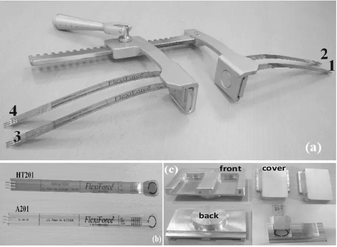
2 Materials
A commonly adopted straight sternal retractor, Finochietto type (Fig.1a), was equipped with both ultra-thin force sensors (Fig.1b and c) and electronic circuitry. The resulting electronic-engineered“sensory retractor”was tested by means of a home-made dummy.
back
front cover (c)
2.1 Ultra-Thin Force Sensors
We considered two different types of commercial piezo-resistive flexible ultra-thin (0.203 mm, 0.008 in.) off-the-shelf force sensors, the FlexiForce® A201 (these according to [17]) and the FlexiForce® HT201 (both types by Tekscan, Boston, USA), having a circular sensing area of 9.53 mm (0.375 in.) in diameter, on one edge, connected through a silver strip to the electric contacts, on the other edge (Fig.1b). The A201 type, with a polyester substrate, can measure forces up to 440 N, within a temperature oper-ating range of−9 °C to +60 °C (15 °F to 140 °F). The HT201 type, with a polyimide substrate, can measure forces up to 445 N, within−9 °C to +204 °C (15 °F to 400 °F).
2.2 Sensor Testing
To assess sensor characterization under known forces, we used a universal tensile test machine (LRX, by Lloyd Instruments, Berwyn, PA, US), showed in Fig.2a. It is a single column digital machine able to provide a constant compression/extension force, up to 2500 N depending on the used load cell (Fig.2b and c). Several parameters can be set, in particular the fall and rise speed, used to measure the repeatability and reproducibility of measurements, and the load cell sensibility. Machine operations are controlled by the NEXIGEN software, which can simultaneously analyze the test results sampled @1 kHz and acquired through the RS232 interface.
One sensor sample at a time was positioned under the load cell.
The used load cell was an electronic component (transducer), made of an elastic hard metal (e.g. stainless steel) to which is connected a Wheatstone bridge, with four strain gauges varying their resistance under traction, which generates a voltage signal depending on the cell deformation.
In such a way, the electric voltage value is referred to the force applied. The voltage signal is amplified, calibrated and compensated with temperature, then processed by an algorithm to correct the device nonlinearity. Consequently, the value of the applied force was determined, taking into account deformation and material characteristics. Two load cells of 50 N and 500 N, respectively (Fig.2a and b) were used for sensor characterization.
2.3 Data Acquisition
The electrical resistance values (outputs of the sensors) were converted into voltages by means of voltage dividers. Those voltage signals fed an electronic circuitry, based on an Arduino-compatible microcontroller board, which operated 10 bit digital conver-sions and sent data to a personal computer via USB port. The following data process was handled by ad-hoc home-made LabView (by National Instruments, TX, USA) and Matlab (by MathWorks, Massachusetts, USA) routines.
Front-End Electronics. The front-end electronic circuit was developed on the basis of a previous one, which was made to interfaceflex and electromyography sensors [18]. In particular, the electrical resistance values (outputs of the sensors) were converted into voltages by means of voltage dividers (Fig.3a), differently with respect to the inverting operational amplifier recommended by the manufacturer (Fig.3b). This was to reduce the circuit size rather than provide signal amplification. Moreover, the inverting amplifier needs a double supply, whereas this circuit shares the same +5 V bias supply of the following microcontroller board, taken from the USB cable con-necting to the personal computer (PC). The front-end circuit is a simple voltage divider, where the sensor is represented by the series resistor. A shunt capacitance of 100 nF was used tofilter noise coming from the bias supply (Fig.3a). When the force applied to each sensor increases, the sensor resistance decreases, and the corresponding output voltage, accordingly to Eq. (1), proportionally increases.
VOUT¼VBIAS RP
RSENSþRP
ð1Þ
The values of each shunt resistors RP was determined taking into account some needs. In particular, RP has to protect each sensor against excessive currents, and has to guarantee the largest-as-possible output voltage swing so to allow adequate resolution for the following digital conversion. With a force value ranging from 1 to 400 N, the sensor resistance span of 1 MXroughly. Since the maximum allowable current for the sensor is 2.5 mA, when the sensor is in short circuit, the RP value must be higher than
RP[ 5 V 2:5 mA
¼2 kX ð2Þ
The digital conversion is made by a 10 bit analog to digital converter (ADC), to which corresponds a voltage resolution given by Eq. (3)
VLSB ¼ 5 V
2101’4:9 mV ð3Þ
The minimum voltage from the front-end circuit, in case of zero applied force, calculated by Eq. (1) withRSENS = 1 MX, should be greater thanVLSB
VOUTmin¼5 V RP 1 MXþRP
[4
:9 mV ð4Þ
which impliesRP[1 kX. Finally, the selected value wasRP¼47 kX.
Digital Processing. The voltage signals fed an electronic board circuitry, based on Arduino Uno, which operated 10 bit digital conversions and sent data to a personal computer via USB port at a sampling rate of 175 Hz. Afterwards, the Arduino Uno board was replaced with Luigino328 (an Arduino-compatible microcontroller board based on an ATMega328 MCU). This was because Luigino328 allows switching the bias supply to an external one, without overcharge the USB port of a Personal Com-puter (PC), which could be damaged for supply current greater than 500 mA, whereas in Arduino the supply from the USB port has the highest priority. Luigino328, in fact, has also a small microcontroller (PIC16F) for the following tasks: (1) to handle the voltage selector, (2) to disconnect the serial port when programming the board, without remove shields using the serial port and (3) to exclude the SmartReset function, avoiding to reset the board every time a serial port is connected, allowing the running program to go on independently. This device allowed interfacing the LabView inte-grated development environment (IDE) without the sudden resets which occur in Arduino. Finally, the Luigino328 is equipped with the LM1117 voltage regulator, which is more reliable than the MC33269D of Arduino, especially for high supply currents.
Serial Communication. To start a serial communication, it is possible to select the transmission speed (baud rate) in bit per second (bps), within 300–115200 bps. The standard rate is 9600, whereas we used 19200 bps. The default data length is 8 bit, no parity and one stop bit. We adopted the RS-232 as standard communication protocol, reduced to 9 pin (usually COM), virtually implemented on a USB port, being COM ports unavailable on up-to-date PC.
The ADC converts the analog sample in a 10 bit digital string. For serial com-munication, however, the 10 bit string is divided into two bytes (8 bit). Three token bytes (all 1’s) were inserted before each data sample. Actually, two bytes would be enough for the token string, because the data bits cannot be all 1’s, coming from a 10 bit ADC, six bits are definitely 0’s. We added one more token byte to make the system more reliable with strong EM interferences. Considering the start (0) and stop (1) bit before and after each byte, the string length for each acquisition is 50 bit, as represented in Table1. When the acquisition system reads simultaneously the four sensor applied to the sternal retractor, the three token bytes are transmitted only once, then two bytes for each sensor, for a total number of 110 bits, according to the scheme in Table2.
2.4 Sternal Retractor
An aluminum straight Finochietto retractor (by Tekno-Medical Optik-Chirurgie GmbH Tuttlingen, Germany) was equipped with an array of four force sensors. Two sensors were placed on the blade of the mobile arm and two on the blade of the fixed arm (Fig.1a), the size of the blade being 44.4 mm (1.75 in.) in length and 30.9 mm (1.22 in.) in width. The sum of the single detected forces on each blade yielded the total force for both the fixed and the mobile arm. The ultra-thin force sensors were placed in ad-hoc smooth aluminum housings (Fig.1c).
Table 1. Serial packet transmission for data acquisition from a single sensor device.
0 Byte 1 0 byte 1 0 byte 1 0 byte 1 0 byte 1
3 token bytes (30 bits) 2 data bytes (20 bits)
50 bits
Table 2. Serial packet transmission for simultaneous data acquisition from four sensor devices.
3 token bytes sensor 1 30 bits 20 bits 20 bits 20 bits 20 bits
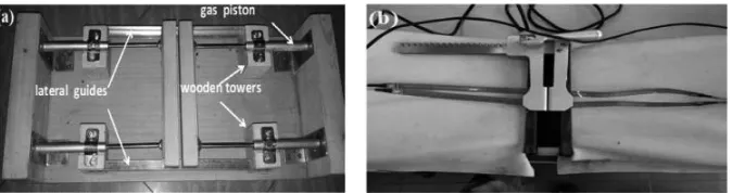
2.5 In-vitro Test
In-vitro tests of the instrumented Finochietto retractor were performed by means of a made-on-purpose dummy built up with four gas pistons (manufactured by Team Pro, Italy), two for each side, laterally anchored to a wooden shell (Fig.4a). Different set of gas pistons were evaluated, i.e., 150 N, 100 N and 80 N. On the basis of several opening/closing cycles performed by three different surgeons, the dummy equipped with the 80 N pistons offered the most realistic feeling with respect to the clinical practice. However, pistons can be easily replaced. The instrumented retractor, equipped with the four force sensors, was positioned into the dummy (Fig.4b).
The Authors are aware that the mechanics of the proposed dummy is very simple with respect to the complex biomechanics of the rib cage. Anyway, the idea was to realize a dummy able to support the test of the device and not meant to be taken as a biomechanical model of the rib cage.
3 Methods
3.1 Sensor Characterization
In this section the selected methods for static and dynamic characterization of the force sensors will be deeply described. Both of them were accomplished with the LRX testing machine with two load cells for compression up to 5 N and 500 N, respectively. Due to the sensor thinness, the sensor sample was placed on a stainless steel platform, and a 10 mm diameter steel disk was placed on the active area of the sensor, to achieve the required pressure on device during the jack fall down. References on sensor location help to replace the sample measurement in the same conditions.
Static Characterization. In the static characterization the descent rate of the jack was set to 5 mm/min and the static measurements were acquired @5 N, 10 N, 20 N, 30 N and 40 N with the 50 N load cell, and from 40 N to 400 N, step 40 N, with the 500 N load cell. Two minutes break were set before each step to acquire measurements, through the LabView-Arduino interface.
Considering a baud rate of 19200 bps for 2 min or 120 s, 50 bit for each sensor sample, the acquired resistance samples are (19200/50)∙120 = 46080. Waiting 52 s, when the sensor response was considered enough stable, 3000 resistance samples were considered for a duration of almost 7 s, of which the average and the standard deviation were calculated. In the same time interval, 30 samples of the force magnitude were considered among the only 384 samples stored by the test machine acquisition system in 120 s, because the sampling frequency is much smaller than that of Luigino/ LabView interface.
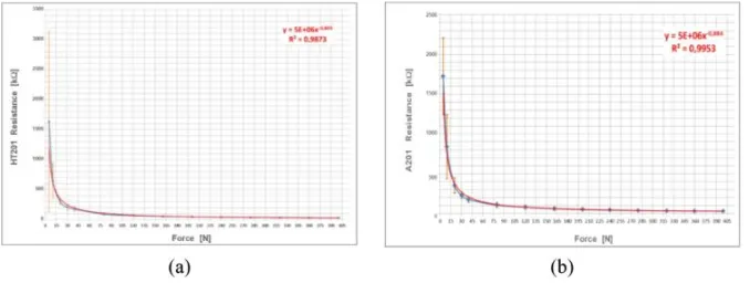
Figure5a and b shows the characterization of the HT201 and A201 devices, respectively. The sensor resistance is represented as the average and standard deviation among eight sensor samples as a function of the average compression force at each step. Ultrathin flexible force sensors HT201 and A201 both showed exponential resistance decay with the impressed force. Plots demonstrate the same behavior for A201 and HT201 sensors, but in the force range 30–440 N standard deviations are very small (7–42 kX, 8–51 kXrespectively), whereas in the range 5–30 N standard devi-ations become high (96–482 kX, 47–1000 kXrespectively).
In order to investigate if HT201 sensors can effectively support autoclaving con-ditions, these sensors were also characterized following the same procedure afterfive cycles of autoclave treatment (VaporMatic 770, Asal Srl, Milan, Italy). HT201 sensors did not show a significantly different behavior after five cycles of autoclave condi-tioning, which is a reasonable result since these sensors have been specifically designed for high temperature applications (up to 400 °F, approximately 200 °C). In any case, in the occurrence of degradation in performances, those sensors can be easily and con-veniently replaced.
and back for each speed rate, each time increasing and decreasing at a constant rate the traction force from 0 N to 400 N and down again to 0 N. The repetition period depends on the descent rate of the jack.
Results for the average sensor resistance against increasing force with different speed, superimposed for comparison with two equal and symmetrical static charac-teristics in Fig.6a and b show A201 sensors behave with lower repeatability than HT201 counterparts, changing the variation rate of the applied force.
Dynamic characterization was also repeated afterfive cycles of autoclave treatment for HT201 sensors, and characteristics do not show a significantly different behavior after treatment, as in the static case (so results are not presented for sake of brevity).
3.2 In-vitro Test
In the limited literature concerning the measurement of sternal forces, both on animals and corpses, a standard protocol to regulate the data acquisition and processing is missing, therefore it is impossible to compare the performance of systems developed by different research teams, because measurements results were obtained in different conditions, such as the speed, the aperture number and points, the human or animal subject under test. For this purpose, a statistical approach was applied [20] to sensory sternal retractor assessments, to compare different systems by evaluating mean, range and standard deviation tables for each tested subject. Since typical sternal apertures in cardiac surgery range from 5 to 10 cm, the standard conditions set out for the test applied to sternal force measurements are:
(1) 5 cm wide aperture (rest position) (2) 10 cm wide aperture (operating position)
Moreover, to assure the same conditions in different measurement sessions, special references were inserted on the dummy to make the retractor positioned always in the same way.
Test procedure consisted in four opening/closing cycles of the dummy by means of the instrumented retractor up to two different fixed widths, i.e. 5 cm (1.97 in.) and 10 cm (3.94 in.). On the basis of the feeling/practice of the surgeons, each opening/closing cycle was performed at a roughly constant rate of 2 s/cm, that is 10 s for 5 cm (1.97 in.) and 20 s for 10 cm (3.94 in.). The two final positions (5 cm, 1.97 in. and 10 cm, 3.94 in.) were held for 60 s so to evidence response decay, if any. The response of all the sensors in terms of force (F) versus time (t) was real-time acquired. Then, mean force (Fmean), maximum force (Fmax) and plateau force (Fplateau) were evaluated, the last as the mean value of the force recorded during 60 s in thefinal rest position. The distribution of the forces exerted along the two halves of the dummy was also determined.
4 Results and Discussion
4.1 Recent Findings
The investigated range of force (i.e., 5–400 N) includes the values reported by Bolotin et al. [21] and by Aigner et al. [17]. In more details, Bolotin et al. reported thefirst known successfully attempt to employ an instrumented retractor to monitor forces during car-diothoracic surgery. They equipped stainless steel curved profile retractor blades with strain gauges to measure applied forces during retraction, and reported results for lateral thoracotomy and median sternotomy on cadavers and sheep. The average force applied during force-controlled retraction was (77.88 N±38.85 N) and the maximum force displayed during force-controlled retraction (323.99 N±127.79 N).
Aigner et al. equipped a straight (SSR) (MTEZ 424 735; Heintel GmbH, Vienna, Austria) and a curved retractor (CSR) (Dubost Thoracic Retractor DC30000-00; Delacroix-Chevalier, Paris, France), with FlexiForce sensors, A201 type (Tekscan Inc). The blade of the mobile arm of the SSR (length 6.5 cm and width 4.5 cm) was equipped with two arrays of 4 sensors, and the mobile arm of the CSR (length 9.7 cm, width 4.8 cm, curvature radius 21 cm) was equipped with two arrays of 5 sensors. The sum of the single sensor forces yielded the total force. Force distribution, total force and displacement were recorded to a spread width of 10 cm in 18 corpses (11 males and 7 females). For every corpse, 4 measurement iterations were performed for both retractors; each retraction was performed in 14.3 s±6.2 s to reach 10 cm widespread. The Authors concluded that the shape of sternal retractors considerably influences the force distribution on the sternal incision. On the other side, it is reported that the total mean retraction force was not significant different between SSR and CSR (222.8 N±52.9 N versus 226.4 N±71.9 N). Nevertheless, the recorded mean total force was remarkably dependent on the gender. For the first retraction, it was 256.2 N±43.3 N for males and only 174.9 N±52.9 N for females.
Aigner et al. assessed that the force distribution did not change significantly for the other 3 retractions, for the different investigated spread widths (i.e. 5, 7.5, and 10 cm) and was not gender dependent. The maximum force for full retraction was 493.6 N, whereas the smallest maximum force was 159.0 N.
4.2 Finochietto In-vitro Results
Our results obtained for HT201 sensors are resumed in Table3 and the typical force (N) versus time (s) patterns are presented in Fig.7. In all cases, a high stability of the response to afixed exerted force was evidenced. In fact, the value of Fplateaushowed a mean standard deviation as low as 0.33 N±0.16 N. Some valuable information can be obtained from the acquired data.
It is interesting to observe during the retraction, the Finochietto experienced along the mobile arm a total Fmax(sensor#4 + sensor#3) that exceeded 200 N, ranging from 219.1 N±9.7 N for 5 cm spread and 266.6 N±25.4 N for 10 cm spread.
The force distribution along the retractor blade is also particularly interesting. In fact, in all cases, the highest maximum force (Fmax) was detected by sensor #4 posi-tioned on the mobile arm in proximal (cranial) position (Fig.1a), the value ranged between 156.4 N±12.5 N for 5 cm spread and 199.7 N±21.2 N for 10 cm spread. The lowest Fmaxvalues were 62.7 N±5.4 N for 5 cm and 66.9 N±4.3 N for 10 cm, registered in correspondence of sensor #3 of the mobile arm in distal (caudal) position. Interestingly, median sternotomy in corpses performed by means of a straight sternal retractor gave a comparable force distribution [17]. This result suggests that the made-on-purpose dummy enable to perform reliable test and thus it might also be employed by surgeons in order to assess their own learning curve for each specific instrumented retractor. Furthermore, sensor #4 detected also the highest value of (Fmax− Fmean), i.e. 114.6 N±12.9 N and 116.9 N ±16.9 N, respectively, for 10 cm and 5 cm opening. For all the other sensors, this value does not exceed 75.7 N±21.1 N, independently from the position on the retractor.
On the basis of these results, the presented implementation system can be con-sidered a valuable tool to evaluate intensity and distribution of retraction forces in human patients for conventional sternotomy procedures. On the basis of our knowl-edge, these data are not yet available in the Literature. As already previously suggested by Bolotin [16], thefinal goal is to develop clinical studies aimed at coherently cor-relating the biomechanical information obtained for a specific surgical procedure with the incidence of post-sternotomy chronic pain. In this respect, for example, the actual outcomes of cranial versus caudal positioning of the sternal retractor could be assessed. As far as we know, in the past decade such kinds of studies have not yet been performed probably due to the lack of an implemented user-friendly retractor suitable for conventional clinical sterilization process.
5 Conclusions
Median sternotomy is a surgical incision through the sternum, then after to allow access to the mediastinum a retractor is positioned. Wide opening of the hemi-sternum, by means of the retractor, guarantee better view and facilitate the surgical procedure. However, its increase the stress on the sternum halves and ribs, leading to partial or complete fractures and/or micro-fractures resulting in post-operative and chronic pain in a non-negligible number of patients.
By measuring the forces during different opening procedures, we demonstrated how it can be possible to understand and find the compromise between adequate surgicalfield and the risk for sternum and ribs fractures, aiming at improving patients postoperative coarse.
Within such a frame, this paper reports a new electronic-engineered sensory sternal retractor aimed at measuring the forces impressed by the plates when opening a dummy ribcage, so in an in-vitro median sternotomy condition.
We demonstrated that the impressed forces present“spikes”, i.e. sudden changes, with peak force values meaningfully higher of the plateau values which, from a mechanical point of view, can be the reason of the cracks/micro-cracks of the ribcage, and so of the persistent postoperative pain suffered by a number of patients after the surgical procedure.
References
1. Julian, O.C., Lopez-Belio, M., Dye, W.S., Javid, H., Grove, W.J.: The median sternal incision in intracardiac surgery with extracorporeal circulation: a general evolution of its use in heart surgery. Surgery42, 753–761 (1957)
2. Defalque, R.J., Bromley, J.J.: Poststernotomy neuralgia: a new pain syndrome. Anesth. Analg.69(1), 81–82 (1989)
3. Bruce, J., Drury, N., Poobalan, A.S., Jeffrey, R.R., Smith, W.C., Chambers, W.A.: The prevalence of chronic chest and leg pain following cardiac surgery: a historical cohort study. Pain112(3), 413 (2004)
Table 3. Values of the mean, maximum and plateau forces (expressed in N) measured by HT201 sensors positioned according to Fig.1a (i.e. S1, S2, S3, S4). The related standard deviation values are reported in parentheses [15].
Spread Force [N] S1 S2 S3 S4 S1+S2fixed
blade
S3+S4 mobile blade
4. Wildgaard, K., Kehlet, H.: Persistent Postsurgical Pain Syndromes, Chronic post-thoracotomy pain—what is new in pathogenic mechanisms and strategies for prevention? Tech. Reg. Anesth. Pain Manag.15(3), 83–89 (2011)
5. Riillo, F., Bagnato, C., Allievi, A.G., Takagi, A., Fabrizi, L., Saggio, G., Arichi, T., Burdet, E.: Ann. Biomed. Eng.44(8), 2431–2441 (2016)
6. van Gulik, L., Janssen, L.I., Ahlers, S.J.G.M., Bruins, P., Driessen, A.H.G., van Boven, W. J., van Dongen, E.P.A., Knibbe, C.A.J.: Risk factors for chronic thoracic pain after cardiac surgery via sternotomy. Eur. J. Cardiothorac. Surg.40, 1309–1313 (2011)
7. Hazelrigg, S.R., Cetindag, I.B., Fullerton, J.: Acute and chronic pain syndromes after thoracic surgery. Surg. Clin. N. Am.82, 849–865 (2002)
8. Mazzeffi, M., Khelemsky, Y.: Poststernotomy pain: a clinical review. J. Cardiothorac. Vasc. Anesth.25(6), 1163–1178 (2011)
9. Ochroch, E.A., Gottschalk, A., Troxel, A.B., Farrar, J.T.: Women suffer more short and long-term pain than men after major thoracotomy. Clin. J. Pain22, 491–498 (2006) 10. Zeitani, J., Penta de Peppo, A., Moscarelli, M., Gurrieri, L., Scafuri, A., Nardi, P., Nanni, F.,
Di Marzio, E., Chiariello, L.: Influence of sternal size and inadvertent paramedian sternotomy on stability of the closure site: a clinical and mechanical study. J. Thorac. Cardiovac. Surg.132, 38–42 (2006)
11. Healey, S., O’Neill, B., Bilal, H., Waterworth, P.: Does retraction of the sternum during median sternotomy result in brachial plexus injuries? Interact. CardioVasc. Thorac. Surg.17, 151–158 (2013)
12. Suzuki, S., Kikuchi, K., Takagi, K., Masuda, H., Yoshizu, H., Tanaka, S., Ogata, T.: Brachial plexus injury and fracture of thefirst rib as complications of median sternotomy. J. Japan. Assoc. Thorac. Surg.38(9), 1459–1462 (1990)
13. Zeitani, J., Penta de Peppo, A., Bianco, A., Nanni, F., Scafuri, A., Bertoldo, F., Salvati, A., Nardella, S., Chiariello, L.: Performance of a novel sternal synthesis device after median and faulty sternotomy: mechanical test and early clinical experience. Ann. Thorac. Surg.85(1), 287–293 (2008)
14. Baisden, C.E., Greenwald, L.V., Symbas, P.N.: Occult rib fractures and brachial plexus injury following median sternotomy for open-heart operations. Ann. Thorac. Surg.38(3), 192–194 (1984)
15. Saggio, G., Tancredi, G., Sbernini, L., Gaudio, C., Bianco, A., Zeitani, J.: In-vitro force assessments of an autoclavable instrumented sternal retractor. In: Proceedings of the 10th International Joint Conference on Biomedical Engineering Systems and Technologies (BIOSTEC 2017) - Volume 1: BIODEVICES, pp. 25–31 (2017)
16. Bolotin, G., Buckner, G.D., Campbell, N.B., Kocherginsky, M., Raman, J., Jeevanandam, V., Maessen, J.G.: Tissue-disruptive forces during median sternotomy. Heart Surg. Forum 10(6), 487–492 (2007)
17. Aigner, P., Eskandary, F., Schlöglhofer, T., Gottardi, R., Aumayr, K., Laufer, G., Schima, H.: Sternal force distribution during median sternotomy retraction. J. Thorac. Cardiovasc. Surg.146(6), 1381–1386 (2013)
18. Saggio, G., Orengo, G., Leggieri, A.: Sensory glove and surface EMG with suitable conditioning electronics for extended monitoring and functional hand assessment. In: Proceedings of the 9th International Joint Conference on Biomedical Engineering Systems and Technologies (BIOSTEC), co-located 9th International Conference on Bio-inspired Systems and Signal Processing (BIOSIGNALS), Rome, Italy (2016)
20. Orengo, G., Lagati, A., Saggio, G.: Modeling wearable bend sensor behavior for human motion capture. IEEE Sens. J.14(7), 2307–2316 (2014)
21. Bolotin, G., Buckner, G.D., Jardine, N.J., Kiefer, A.J., Campbell, N.B., Kocherginsky, M., Raman, J., Jeevanandam, V.: A novel instrumented retractor to monitor tissue-disruptive forces during lateral thoracotomy. J. Thorac. Cardiovasc. Surg.133(4), 949–954 (2007) 22. Ward, A.F., Grossi, E.A., Galloway, A.C.: Minimally invasive mitral surgery through right
mini-thoracotomy under direct vision. J Thorac Dis.5(6), 673–679 (2013)
23. Reser, D., Holubec, T., Caliskan, E., Guidotti, A., Maisano, F.: Left anterior small thoracotomy for minimally invasive coronary artery bypass grafting, Multimed. Man. Cardiothorac. Surg. 1–5 (2015).https://www.ncbi.nlm.nih.gov/labs/articles/26420246/ 24. Atluri, P., Stetson, R.L., Hung, G., Gaffey, A.C., Szeto, W.Y., Acker, M.A., Hargrove, W.C.:
Automated 3D-Printed Smartphone
Fraunhofer Portugal AICOS, Rua Alfredo Allen 455/461, 4200-135 Porto, Portugal
Instituto Nacional de Sa´ude Dr. Ricardo Jorge, Rua Alexandre Herculano 321, 4000-055 Porto, Portugal
3
INESCTEC and University of Porto, Rua Dr. Roberto Frias, 4200-465 Porto, Portugal
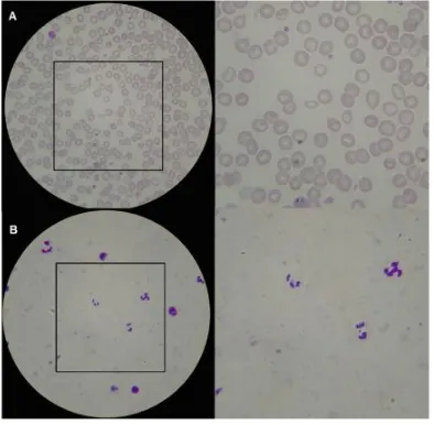
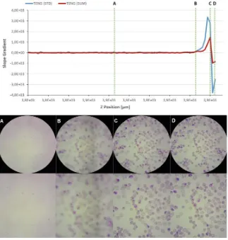
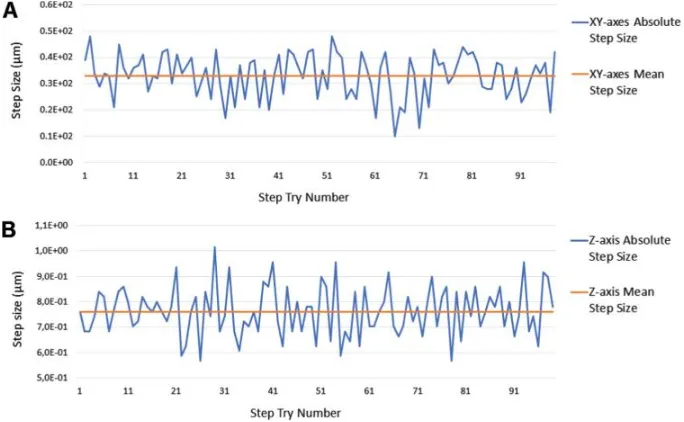
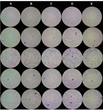
Abstract. Microscopic examination is the reference diagnostic method for several neglected tropical diseases. However, its quality and availabil-ity in rural endemic areas is often limited by the lack of trained personnel and adequate equipment. These drawbacks are closely related with the increasing interest in the development of computer-aided diagnosis sys-tems, particularly distributed solutions that provide access to complex diagnosis in rural areas. In this work we present our most recent advances towards the development of a fully automated 3D-printed smartphone microscope with a motorized stage, termed µSmartScope. The devel-oped prototype allows autonomous acquisition of a pre-defined number of images at 1000x magnification, by using a motorized automated stage fully powered and controlled by a smartphone, without the need of man-ual focus. In order to validate the prototype as a reliable alternative to conventional microscopy, we evaluated theµSmartScope performance in terms of: resolution; field of view; illumination; motorized stage perfor-mance (mechanical movement precision/resolution and power consump-tion); and automated focus. These results showed similar performances when compared with conventional microscopy, plus the advantage of being low-cost and easy to use, even for non-experts in microscopy. To extract these results, smears infected with blood parasites responsible for the most relevant neglected tropical diseases were used. The acquired images showed that it was possible to detect those agents through images acquired via theµSmartScope, which clearly illustrate the huge poten-tial of this device, specially in developing countries with limited access to healthcare services.
Keywords: Microscopy
·
Mobile devicesMotorized microscope stage
·
Developing countries·
Mobile healthc
1
Introduction
Neglected tropical diseases (NTDs) are a group of parasitic infectious diseases that affect over 1.5 billion of the world’s poorest population, including 875 mil-lion children [1]. The gold standard for detection of several NTDs is microscopic examination, particularly via the visualization of different types of human bio-logical products, like blood smears (e.g Malaria, Lymphatic filariasis, African Trypanosomiasis), stool smears (e.g. intestinal helminths), and urine smears (e.g. Schistosomiasis) [2]. Unfortunately, reliable identification of these parasitic infec-tions requires not only proper microscopic equipment, but also high-standard expertise for subsequent microscopic analysis. These requirements represents the most common practical difficulties experienced in rural health facilities, being closely related with the increasing interest of mobile health (mHealth) and computer-aided diagnosis solutions for those particular scenarios.
The mobile phone is currently Africa’s most important digital technology. In the year 2000 few Africans had a mobile phone, but today about three-quarters do [3]. So it becomes natural that mHealth is starting to play an important role when it comes to health in Africa, particularly through the usage of solutions that allow skipping over centralized laboratories [4]. For instance, the usage of advanced computer vision approaches coupled to the increasing processing capabilities of mobile devices is already showing promising results in the area of malaria diagnosis [5,6]. Moreover, considering the paramount importance of microscopic examination for NTDs detection, the development of new portable microscopic devices is an area that can greatly improve the chances of the suc-cessful deployment of innovative solutions for NTDs diagnosis in underserved areas [7]. To achieve that purpose, the constant advances and increasing pos-sibilities coming from additive manufacturing should certainly be taken into account, since 3D-printing currently allows faster and cheaper prototyping.
![Fig. 7. (a) and (b) Response of the four sensors (housed as shown in Fig.[N] versus time [s] during the 5 cm opening procedure](https://thumb-ap.123doks.com/thumbv2/123dok/3934637.1878057/34.439.44.382.221.493/fig-response-sensors-housed-shown-versus-opening-procedure.webp)
![Fig. 7. Prototype PCB to control the µStage [8].](https://thumb-ap.123doks.com/thumbv2/123dok/3934637.1878057/48.439.148.277.464.577/fig-prototype-pcb-to-control-the-ustage.webp)