Bioactivity of Ethanolic Extract of Propolis (EEP) in Balb/C Mice’s
CD4
+CD25
+and B220
+Lymphocyte Cells
Aisyah Zahroh Aden, Muhaimin Rifa’i
*Department of Biology, Faculty of Mathematics and Natural Sciences, University of Brawijaya, Malang, Indonesia
Abstract
This experiment was aimed to determine the bioactivity of ethanolic extract of propolis (EEP) against the changes in the quantity of CD4+CD25+ and B220+ lymphocytes and determine the optimal dose of EEP to increase the number of CD4+CD25+ and B220+ cells in Balb/c mice. Balb/c mice were divided into four treatment groups: control treatment, treatments of EEP at a dose of 50 mg.kg-1, 100 mg.kg-1, and 200 mg.kg-1 body weight of mice. All mice were dissected after two weeks post treatment. Profiles of lymphocytes from the spleen expressing CD4+CD25+ and B220+ cells were analyzed by flowcytometry using CellQuest software. Data was analyzed by Kruskall Wallis and Mann Whitney statistical test with P<0.05 using SPSS 16.0 for Windows. The results showed that the treatment of dose of 50 mg.kg-1 of EEP can increase relative number of CD4+CD25+ cells significantly, but those cells decrease significantly when we apply the dose of 100 and 200 mg.kg-1. The relative number of B220+ cells increase in the dose of 50 and 200 mg.kg-1 and decrease in the dose of 100 mg.kg-1 compared to the control. This experiment suggest that EEP has bioactivity to modulate the quantity of CD4+CD25+ and B220+ in dependent manner.
Keywords:B220+, CD4+CD25+, lymphocyte, propolis
INTRODUCTION
Propolis is a natural product from a mixture of plant derived products, which modified and used by bees for various needs. Bees use propolis in nature as antibiotics and protective materials from the threat of drought to their nests [1]. Propolis contains resin, fatty acids, waxes, proteins, polysaccharides, hydrocarbons, and various other organic compounds [2,3]. Propolis as a traditional medicine used by people as an anti-inflammatory, antibacterial, antiparasitic, antifungal, antitumor, antioxidant, and have immunomodulatory effects [1].
Immunomodulator is one mechanisms that needed in the body's immune system. Immuno-modulatory activity is a form of biological or pharmacological effects on various factors in the immune response. The purpose of immunomo-dulatory itself is to modulate the homeostasis of immune system. Immunomodulatory activity also used to treat and prevent various diseases originating from any occurred imbalance. The activities of immunomodulators are immuno-suppressants and immunostimulant [4]. Immu-nomodulatory effects of propolis was known better compared to other immunomodulatory
Correspondence author:
Muhaimin Rifa’i
Email : [email protected]
Address : Dept. of Biology, Faculty of Mathematic and Natural Sciences, University of Brawijaya, Jl. Veteran, Malang, 65145
substance that has only flavonoid components as main substances [5]. A number of clinical studies have proved that propolis has the ability to activate the body's immune system in mice and humans. This is supported by experiment data that showed an increase in the secretion of IL1, IL2, and IL4, increased antibody responses, cell proliferation of T lymphocytes, increased ratio of CD+/CD+, and activation of macrophages [1].
The effect of propolis on CD4 T cell differentiation process has not known yet [6]. CD4 T cell activation is also known to be influential in the process of B cell proliferation [7]. This study was conducted to determine the bioactivity of EEP on production of CD4+CD25+T cells and B220+ B cells in the spleen organ of Balb/c mice.
MATERIALS AND METHODS
This experiment was conducted on Septem-ber 2013 until June 2014 at Laboratory of Animal Physiology and Animal Room, Department of Biology; Biomedical Laboratory and Laboratory of Pharmacology, Faculty of Medicine, University of Brawijaya, Malang. There are four treatment groups that used in this experiment i.e. negative control, EEP dose 50, 100, and 200 mg.kg-1 body weight of mice.
Materials and Equipments
(EEP), Balb/c mice, mineral water, pellets BR-1, sterile distilled water, 70% alcohol, sterile PBS, and the monoclonal antibody (rat anti-mouse CD-4, CD-25, and B220). The equipments which were used in are cage of mice, masks, scales, spatulas, erlenmeyer glass, oral administration tool, sectio sets, surgical board, petri dish, micropipette, mikrotube, propylene tubes, yellow and blue tip, centrifugation, flowcytometry cuvette, and FACS CaliburTM flowcytometry.
Animal
Mice (Mus musculus) strains Balb/c 8 weeks old in healthy condition with ± 25 g body weight. Mice were purchased from mice farmers in Jember with pathogens free certification. Acclimatization mice were performed for seven days before giving the treatment.
Propolis Extraction
Propolis obtained from local bee keepers in Lawang, Malang, Indonesia. Propolis extraction process performed by maceration method. Clumps of propolis were collected from the hiveside. The propolis weighed as much as 200 grams and then maserated with 1 L of absolute ethanol. The solution of propolis maceration was filtered and evaporated for ± 1.5-2 hours. The resulting ethanolic extract of propolis was filtered and evaporated in a heated dish in the oven. The EEP is placed in a bottle and stored in a
refrigerator with temperature of 4ᵒC.
Oral Administration of EEP in Balb/c Mice
Each of mice in the treatment groups were not given oral administration of EEP treatment.
Animal Dislocation and Spleen Organ Isolation
Mice were killed by neck dislocation. Mice that had been killed was dissected in their dorsal part to isolate the spleen organ. Spleen which has been isolated then was washed twice and soaked in sterile PBS.
Flowcytometry Analysis
Spleen organ was crushed in a sterile petri dish containing sterile PBS. Spleen homogenates were filtered using wire and placed in propylene was then centrifuged at 2500 rpm speed, with
the temperature of 4ᵒC for 5 minutes. Pellets
were separated from the supernatant was then added with 1 ml of sterile PBS and homogenized. The second homogenates were then taken for 50 mL and placed in mikrotube containing 500 mL of sterile PBS. Homogenate in mikrotube was centrifuged again at 2500 rpm speed, with the temperature of 4ᵒC for 5 minutes. Pellets then added and incubated with solution of 50 mL monoclonal antibodies (rat anti-mouse CD4, rat anti-mouse CD25, and rat anti-mouse B220) respectively, which is specific to cells expressing CD4+CD25+ and B220+. The number of lymphocytes cells expressing CD4+CD25+ and B220+ was then calculated by inserting the cuvette containing pellet and antibodies was resuspended with 300 mL PBS into flowcytometry tool (FACS CaliburTM flowcyto-metry) that has been setting before.
Data Analysis
Data were analyzed with CellQuest software and were further tested with Kruskall Wallis test (p<0.05). The data show significant test results continue tested by Mann Whitney test. Data analysis was done using SPSS 16.0 for Windows.
RESULT AND DISCUSSION
Bioactivity of EEP on CD4+CD25+T Cells
Changes in the relative number percentage of CD4+CD25+ T cells to the lymphocyte cells population due to the effect of EEP between treatment groups generally show a decline pattern based on the results of flowcytometry
compared to the group of D1 by 0.98%.
Figure 1. Profile of relative number percentage of CD4+CD25+ T cells to the lymphocyte cells population flowcytometry analysis results between groups (K= control, D1 = EEP at dose of 50 mg.kg-1, D2 = EEP at dose of 100 mg.kg-1, and D3 = EEP at dose 200 mg.kg-1).
Figure 2. The relative number of CD4+CD25+ T cells to the
lymphocyte cells population between treatment groups of mice after the oral administration of EEP for two weeks.
Increased doses of EEP which is gived in mice based on this experiment may be will boost the number of other immunocompetent cells than the number of CD4+CD25+ T cells population. The total activation of other immunocompetent cell that occurs in the cell population of lymphocytes can be greater than the number of CD4+CD25+ T cells were activated itself. T reg cells beside derived from CD4 T cell populations are also known can be derived from the population of CD8+ cells by CD8+CD122+ marking. According to the experiment by Rifa'i et al. [8] CD8+CD122+ cells have the ability to control the activation of CD8+ and CD4+ T cells through the study of in-vivo and in-vitro were performed. CD8+CD122+
population thus able to be used to control the presence of abnormal T cells.
The content of compound in propolis there is known as an immunosuppressant. One such of that compound is Artepilin C (Art-C). Immuno-suppressive function of Art-C are acting in suppress the percentage of CD4+CD25+ T cells and IL-10 cytokine production in cultured lymphocytes in-vitro. The function of T reg cells CD4+CD25+FOXP3+ mediated through the production of IL-10 and TGFβ [9]. Cytokine TGF-β and IL-10 are known have a role on the process of differentiation of Treg cells [10]. Cytokine IL-10 may play a role in autocrine against Treg cells mainly serves to maintain FoxP3 expression [11]. Significant decrease in the relative number of CD4+CD25+ T cells in this experiment thus can also occur due to the content of Art-C compounds in propolis that has function as immunosuppressive as where the results of experiment conducted by Cheunga et al. [10].
Giving Brazilian EEP at a dose of 200 mg.kg-1 for 14 days in mice are known to inhibit the production of IL-1, IL-6, IFN-γ, IL-2 and IL-10 by spleen cells [1,12]. CAPE in propolis compounds known to inhibit IL-2 gene transcription and expression of IL-2R (CD25), and T cells ploriferation in humans. The process can occur by working CAPE compounds that inhibit the transcription factor NFAT and NF-Kb [13]. IL-2 is a cytokine that is able to stimulate the activation and differentiation of T reg cells [14]. T reg cells that stay in peripheral areas will be maintained through the presence of IL-2, although IL-2 does not play an important role against the development of T reg cells in the thymus [15]. Therefore, if the amount of IL-2 was reduced following by administration of EEP then it can be closely related to a decrease in the number of Treg cells in the lymphocyte cells population. Effect of EEP in mice against suppression to the relative number of CD4+CD25+ T cells in the lymphocyte cells population thus can be due to a content of compounds that are CAPE as imunosupresor.
expression of CD25+ [17]. Effect of EEP in this experiment but only up to the marking of CD4+CD25+ T cells and was not done until the identification of the presence of FoxP3. Therefore, the results of in experiment have not been able to explain in more specific about how the effect of EEP to change in the real relative number Treg cells CD4+CD25+FOXP3+ in lymphocyte cells population.
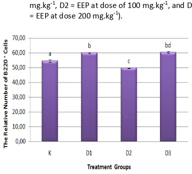
Bioactivity of EEP on B220+ Cells
Treatment of EEP in mice provide a diverse effect on the profile of the relative number percentage of B220+ cells in each treatment group (Figure 3). Effect of EEP was significantly (P<0.05) differences in the relative number of B220+ cells seen in mice in the treatment D1, D2, and D3 group when compared with the relative number of B220+ cells in the control treatment group (Figure 4).
The increase in the relative number of B220+ cells was significantly affected by EEP with D1 dose (50 mg.kg-1) and D3 dose (200 mg.kg-1). The relative number of B220+ cells on D1 increased compared to the control group, aproximately 59.90%. It is also not different from the relative number of B220+ cells on D3 group with a percentage of 60.02%. The increase in the relative number of B220+ cells due to the effect of EEP but did not different significantly between D3 group with D1 group. Effect of EEP in reducing the relative number of B220+ cells was significantly seen in D2 group with the relative number of B220+ cells was only aproximately 49.61%.
The results of this experiment showed that ther are instability of the patterns of change in the relative number of B220+ cells due to the influence of EEP. The increase in the relative number of B220+ cells in the lymphocyte population in the D1 and D3 treatment group showed that the content of compounds in propolis at that dose range that is assumed to be able to increase the relative number of B220+ cells in the lymphocyte cell population. Effect of EEP in decrease the relative number of B220+ cells significantly that occur in the D2 group indicate the presence of a suppressor mechanism that occurs in there.
Assumptions that can be awakened to explain this phenomenon is in the dose of 100 mg.kg-1 body weight of mice is a possible dose that can maximize suppressor molecules activity to reduce the relative number of B220+ cells in the lymphocyte cells population.
Figure 3. Profile of relative number percentage of B220+ cells flowcytometry analysis results between groups (K= control, D1 = EEP at dose of 50 mg.kg-1, D2 = EEP at dose of 100 mg.kg-1, and D3 = EEP at dose 200 mg.kg-1).
Figure 4. The relative number of B220+ cells between
treatment groups of mice after the oral administration of EEP for two weeks.
Experiment conducted by Draganova et al.
[18] showed that the ethanol extract of Bulgarian propolis at low concentrations 1 and 2.5 mg.L-1 was able to increase the proliferation of B cells in PBMC (Peripheral Blood Mononuclear Cells) cell culture. The use of high concentration 10 mg.L-1 on the other hand can induce apoptosis in B lymphocyte cells up to 56.08%. Ethanolic extract of propolis with low concentrations preferable for the entire population of cells to support proliferation activities and it will also provide protection for B lymphocytes cell. Therefore it can be assumed that the EEP with a low dose (D1) is the best dose to increase the relative number of B220+cells.
produce various kinds of cytokines, there are IL-1, IL-6, IL-12, and TNF-α. Cytokine product was produced by macrophages at the same time will stimulate T cell activation. Increased T cell activation will give possibility to improve the production of IFN-γ. Activation of CD4+ T cells will have an impact on the process of differentiation of CD4+ T cells into Th1 cells. Th1 cells it has contributed to the production of IL-2 and IFN-γ [12,19,20]. IFN-γ products are known to have activity in suppressing the B cell differentiation process [5]. The number of IFN-γ produced by
Giving ethanolic extract of propolis (EEP) with dose of 50 mg/kg, 100 mg/kg, and 200 mg/kg body weight of mice thus significantly influence changes in the quantity of CD4+CD25+ T cells and MS. as the reviwer; the entire team of Laboratory of Animal Physiology, Department of Biology, University of Brawijaya, Malang. This experiment was made possible through funding by Program
of Penelitian Unggulan Madya.
REFERENCES
[1] Fatahinia, M., A.R. Khosravi, H. Shokri. 2012. Propolis efficacy on TNF-α, IFN-γ and IL-2 Animal Husbandry, University of Brawijaya. Malang.
[3] Lopes, A.A., S.F. Thiago, T.N. Renata, L. Mannuella, M.P.P. Karla, M.S. Ari, M.B. Ricardo, J.R.S. Antonio, S.V. Samuel, C.P. Luís. 2014. Antioxidant action of Propolis on
mouse lungs exposed to short-term cigarette smoke. Bioorg. Med. Chem.
[4] Nazir, N. 2014. Imunomodulatory activity of Isoflavones isolated from Iris Kashmiriana: Effecton T-Lymphocyte Proliferation and Cytokine Production in Balb/c Mice. J.
Biomed. Prev. Nutr. 3. 151–157.
[5] Mustafiah, S.E., F. Dina, Y. Iwang. 2011. Peritoneal Macrophage Phagocytic Power Index after administration of Propolis on Mice (Mus musculus). Faculty of Medicine, Islam University of Sultan Agung. Semarang. [6] Okamoto, Y., H. Takazumi, E. Tatsuya, F. Takashi, M. Toshiyuki. 2014. Brazilian Propolis Ameliorates Trinitrobenzene Sulfo-nic Acid-Induced Colitis in Mice by Inhibiting Th1 differentiation. J. Int.
Immunophar-macol. 16. 178–183. Cell Homeostasis. J. Exp. Med. 9. 1123-1134. [9] Lee, Y.H., M. Rifa'i. 2011. CD4+CD25+ FOXP3+
Regulatory T Cells in Allogeneic Hemato-poietic Cell Transplantation. J. Trop. Life Sci.
2. 69-75.
[10] Cheunga, K.W., M.Y.S. Daniel, K.C. Wing, D. Rui-Xia, T. Wenwei, C.F.C. Godfrey. 2011. Brazilian green Propolis and it’s constituent, Artepillin C inhibits Allogeneic Activated Human CD4 T Cells expansion and activation. J. Ethnopharmacol. 138. 463– 471.
[11] Murai, M., T. Olga, K. Gisen, M. Rajat, L.K. Christopher, C. Hilde, K. Mitchell. 2009. Interleukin 10 acts on Regulatory T Cells to maintain expression of the Transcription Factor FoxP3 and suppressive function in Mice with Colitis. Nat. Immunol. 11. 1178– 1184.
[12] Rifa’i, M. CD4+CD25+. 2013. Regulatory T Cells prevent detrimental autoimmune reactions International Journal. The Open
Autoimmun. J. 5. Bentham OpenPublisher.
[14] Nelson, H. Brad. 2004. IL-2, Regulatory T Cells and Tolerance. J. Immunol. 172. 3983-3988.
[15] Maloy, K.J., P. Fiona. 2005. Fueling Regulation: IL-2 Keeps CD4+ Treg Cells Fit.
Nat. Immunol. 6. 1071 – 1072.
[16] Campbell, D.J., S.F. Ziegler. 2007. FOXP3 modified Thephenotypic and functional properties of Regulatory T Cells. Nat. Rev.
Immunol. 7. 305–310.
[17] Askenasy, N., K. Ayelet, Y. Shai. 2008. Mechanisms of T Regulatory Cell Function.
Autoimmun. Rev. 7. 370–375.
[18] Draganova, M.F., M. Nikolova, A. Mihova, L. Peychev, V. Sarafian. 2010. A pilot study on the Immunomodulatory effect of Bulgarian Propolis. Biotechnol. Biotechnol. 24. 119-124.
[19] Rifa’i, M., Z. Shi, S.Y. Zhang, Y.H. Lee, H. Shiku, K. Isobe, H. Suzuki. 2008. CD8+CD12+ Regulatory T Cells Recognize Activated T Cells via Conventional MHC Class I–αβTCR Interaction and become IL-10-Producing Active Regulatory Cells. Int. Immunol. 20. 937-947.
[20] Sforcin, J.M., B. Vassya. 2011. Propolis: is there a potential for the development of new drugs? Rev. J. Ethnopharmacol. 133. 253–260.