A
D V A
N
G
ENETIC
A
NALYSES FROM
A
NCIENT
DNA
Svante P¨a¨abo, Hendrik Poinar,
1David Serre,
Viviane Jaenicke-Despr´es, Juliane Hebler,
Nadin Rohland, Melanie Kuch,
1Johannes Krause,
Linda Vigilant, and Michael Hofreiter
Max Planck Institute for Evolutionary Anthropology, D-04013 Leipzig,
Germany; email: [email protected]; [email protected]; [email protected]; [email protected]; [email protected]; [email protected];
[email protected]; [email protected]; [email protected]; [email protected]
Key Words DNA damage, domestication, Neandertal, population history
■ Abstract About 20 years ago, DNA sequences were separately described from the quagga (a type of zebra) and an ancient Egyptian individual. What made these DNA sequences exceptional was that they were derived from 140- and 2400-year-old specimens. However, ancient DNA research, defined broadly as the retrieval of DNA sequences from museum specimens, archaeological finds, fossil remains, and other unusual sources of DNA, only really became feasible with the advent of techniques for the enzymatic amplification of specific DNA sequences. Today, reports of analyses of specimens hundreds, thousands, and even millions of years old are almost common-place. But can all these results be believed? In this paper, we critically assess the state of ancient DNA research. In particular, we discuss the precautions and criteria necessary to ascertain to the greatest extent possible that results represent authentic ancient DNA sequences. We also highlight some significant results and areas of promising future research.
CONTENTS
MOLECULAR DAMAGE AND CONTAMINATION . . . .646
Molecular Damage. . . .646
Contamination with Exogenous DNA . . . .654
GENETICS THROUGH TIME . . . .660
Older and Older DNA . . . .660
Antediluvian DNA . . . .661
WHAT IS ACHIEVABLE? . . . .661
Species Phylogenies. . . .661
1Current address: Department of Anthropology, Pathology and Molecular Medicine,
McMaster University, Hamilton, Ontario L8S, 4L9, Canada.
0066-4197/04/1215-0645$14.00 645
Population History and Phylogeography . . . .662
Hominids . . . .664
Diet and Behavior . . . .665
Sediments. . . .666
Medical Molecular Archaeology . . . .667
Origins of Domestication. . . .667
THE FUTURE . . . .670
MOLECULAR DAMAGE AND CONTAMINATION
The molecular cloning of DNA from a quagga (55) and an Egyptian mummy (103) were the first successes in the retrieval of ancient DNA sequences. However, they were in a sense precocious, since the amounts of DNA present in the old tissues were so small that the isolation of bacterial clones carrying the same DNA sequence was essentially impossible. The results could therefore not be repeated in order to verify their authenticity. Thus, the litmus test of experimental science— reproducibility—was hard or impossible to achieve.
This changed with the development of the polymerase chain reaction (PCR) (123, 124). The PCR made it possible to produce essentially unlimited numbers of copies from very few or even single original DNA copies. Therefore, the same DNA sequence could be amplified multiple times from the same specimen and ancient DNA studied in a scientifically rigorous way. In fact, the very first applications of the PCR to extracts of ancient DNA (104, 106) already hinted at the two technical complications that remain the main challenges to the study of ancient DNA. The first complication was evident from the fact that when PCR was used to reexamine the same quagga from which DNA had been cloned, two positions were shown to be incorrect in the original sequences (106). The second complication was evident from work (104) showing that contemporary DNA contaminates almost all ancient remains and many laboratory environments. Below, we discuss how molecular damage and DNA contamination give rise to erroneous DNA sequences and describe strategies to combat these problems.
Molecular Damage
DNA DEGRADATION AND PRESERVATION Within living cells, the integrity of DNA
TABLE 1 Overview over different types of damage in ancient DNA
Type of
damage Process Effects on DNA Possible solutions
Strand Degradation by Reduction of overall PCR of short length breaks microorganisms DNA amounts and overlapping
Nucleases in the Size reduction fragments postmortem cell
Other chemical processes
Oxidative Damage to bases Base fragmentation PCR of short length
lesions and overlapping
Damage to deoxiribose Sugar fragmentation fragments residues
Nucleotide modification Multiple independent PCRs Cloning and sequencing
of several clones DNA Reactions between DNA e.g., Maillard products PTB (N-phenylacyl
crosslinks as well as DNA and thiazolium bromide) other biomolecules
Hydrolitic Loss of amino groups Change of coding Multiple independent PCRs lesions 1. adenine ⇒ potential Cloning and sequencing
hypoxanthine of several clones 2. cytosine ⇒uracil
3. 5-methyl-cytosin ⇒thymin 4. guanine ⇒xanthin
to those that affect the DNA in the living cell. However, after death they are not counterbalanced by cellular repair processes and thus damage accumulates progressively until the DNA loses its integrity and decomposes, with an irreversible loss of nucleotide sequence information (Table 1). What the PCR has made possible is the occasional salvage of information from some rare samples in which the disintegration of DNA is not yet complete.
DNA DAMAGE IN ANCIENT SAMPLES The most obvious type of damage to DNA
similar to or slightly slower than base loss (31, 131). The extent of degradation by these processes depends upon the idiosyncrasies of preservation and can vary even among museum specimens of the same age. Sometimes, fragments as long as a few hundred base pairs (18, 19, 41) and sometimes even more than 1 kb (81) can be amplified. However, compared with contemporary DNA preparations from fresh tissues, ancient DNA is invariably of shorter length (Figure 1).
The length of the DNA sequences that can be amplified by the PCR is limited not only by strand breaks but also by lesions that present blocks to the elongation of DNA strands by theTaqpolymerase. Many such lesions are induced by free radicals such as peroxide radicals (.O2), hydrogen peroxide (H2O2), and hydroxy radicals (.OH), which are created by, among other causes, background radiation. Major sites of oxidative attack are the double bonds of both pyrimidines and purines, leading to ring fragmentation. In addition, the chemical bonds of the
deoxyribose residues are susceptible to oxidation resulting in fragmentation of the sugar ring (31, 85). DNA extracted from fossil remains is susceptible to cleavage with an enzyme, endonuclease III, which is specific for oxidized pyrimidines (104). It has also been shown that paleontological specimens from a diverse range of environments and ages contain oxidized base residues (65). Specifically, no DNA sequences could be amplified via PCR (65) from samples with higher amounts of two oxidized pyrimidines 5-hydroxy-5-methylhydantoin (5-OH-5-MeHyd) and 5-hydroxyhydantoin (5-OH-Hyd), which block theTaqDNA polymerase.
Another type of damage are cross-links, which also block the DNA polymerase and can even be observed directly by electron microscopy in ancient DNA prepa-rations (104). By pyrolysis gas chromatography–mass spectroscopy, volatile com-ponents formed from Maillard products have been identified in ancient fecal re-mains (coprolites) (114). Maillard products are formed by condensation reactions between sugars and primary amino-groups in proteins and nucleic acids. Treat-ment with a reagent, N-phenacylthiazolium bromide (153), which breaks Maillard products, allows DNA sequences to be amplified from some ancient remains that otherwise are not amenable to amplification, for example 20,000-year-old ground sloth coprolites (114) and>40,000-year-old Neandertal bones (78).
In addition to fragmentation and DNA modifications that hinder the extension of DNA polymerases, other known and unknown types of damage are common in ancient DNA. Some of these DNA modifications are problematic because al-though they allow the amplification of the template molecules, they cause incorrect bases to be incorporated during the PCR. The most common form of such mod-ification is the hydrolytic loss of amino groups from the bases adenine, cytosine, 5-methylcytosine, and guanine, resulting in hypoxanthine, uracil, thymine, and xanthine, respectively (31). The deamination products of cytosine (uracil), of 5-methylcytosine (thymine), and of adenine (hypoxantine) are of particular relevance for the amplification of ancient DNA since they cause incorrect bases (A instead of G, and C instead of T) to be inserted when new DNA strands are synthesized by a DNA polymerase.
NUCLEOTIDE MISINCORPORATIONS IN AMPLIFICATIONS OF ANCIENT DNA The
AR230-GE38-20.te
x
AR230-GE38-20.sgm
LaT
eX2e(2002/01/18)
P1:
IKH
AR
REVIEWS
IN
AD
V
ANCE10.1146/annure
v.genet.37.110801.143214
¨ABO
ET
AL.
and cause the incorporation of A residues rather than G residues by theTaqDNA polymerase (50).
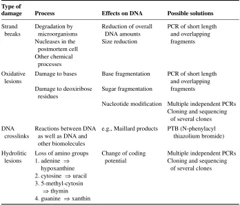
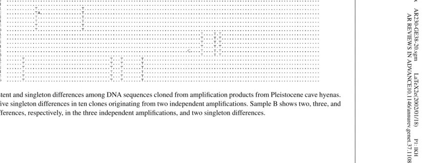
Such miscoding lesions in ancient DNA complicate the correct determination of ancient DNA sequences. To address this situation, it is necessary to distinguish between misincorporations induced by damage in the ancient DNA template and TaqDNA polymerase errors that occur in any PCR regardless of original DNA template quality. One way to do this is to perform multiple amplifications from DNA extracts containing just a few template molecules and clone the PCR products. Comparison of DNA sequences of multiple clones from such amplifications will reveal nucleotide differences that occur in all clones from one amplification but not in other amplifications from the same template preparation (Figure 2) (58). The vast majority of such “consistent” substitutions are due to errors occurring in the first cycles of PCR, which is when the original DNA extracted from an ancient specimen serves as a template. By contrast, additional substitutions seen in single clones that also carry consistent substitutions will be due to misincorporations that occurred later during the PCR when molecules synthesized during previous PCR cycles serve as a template (Figures 2 and 3). Thus, if the frequencies of misincorporations between these two classes of substitutions are compared, the difference between substitutions induced by damage in the original template can be discerned from the inherent error rate of the PCR under the conditions that occur in the exact same PCR reaction. Differences seen between clones where no consistent substitutions occur are less informative, since they represent a mixture of misincorporations that occur when an ancient DNA molecule served as a template and misincorporations that are due to errors during later PCR cycles when newly synthesized molecules are the main source of templates.
Figure 3 Schematic illustration of how consistent and singleton differences arise during an amplification starting from a single DNA molecule. In this example, deam-ination of a C residue has yielded a U residue in the ancient template. This results in the misincorporation of an A residue during the first cycle of the PCR. This error will be subsequently present in all molecules derived from this event. Misincorporations during later cycles of the PCR when newly synthesized molecules serve as templates occur in single or few of the resultant clones.
to such an extent that when C to T and G to A substitutions are disregarded, the error rate when ancient DNA templates are replicated does not differ from that when modern templates are replicated (58).
different ages are replicated by the approach outlined in Figure 3. Contamination with human DNA is common, and known and unknown modifications and other unexpected effects may occur in ancient DNA. For example, Pusch & Bachmann claimed that most extracts from ancient remains induce mutations even in mod-ern mitochondrial DNA added to the extracts and subsequently amplified by PCR (118). These authors therefore posit the existence of some uncharacterized factor that makes theTaqDNA polymerase error prone. In addition, they claim that such errors tend to fall at positions known to vary among human mitochondrial control region sequences. This scenario of mutagenic DNA extracts is presented without a plausible mechanistic framework and is highly questionable. First, we are un-able to reproduce their results using several extracts of ancient bones (129). Also, contaminating modern human DNAs often amplified from ancient remains fail to show a high frequency of misincorporations (80). Nevertheless, this claim under-scores the difficulty in excluding any particular misincoporation as “chemically impossible.” The advantage of the approach in which consistent and nonconsistent changes are analyzed in the same clones (Figures 2 and 3) is that misincorpora-tions that occur when ancient DNA template molecules are replicated can be largely distinguished from those that occur when intact newly synthesized DNA molecules are replicated in one and the same PCR reaction. Thus, this approach takes into account any hitherto unknown DNA modification as well as factors that influence the DNA polymerase’s fidelity.
RELIABILITY OF ANCIENT DNA SEQUENCES To what extent do nucleotide
misin-corporations cause incorrect DNA sequences to be determined from ancient re-mains? Clearly, the risk of this is great if amplifications start from single molecules and DNA sequences are determined from a single amplification. Under such con-ditions, any consistent misincorporation would result in an incorrect base being determined. For example, when mitochondrial DNA sequences are amplified from late Pleistocene cave bear remains (58), as many as a third of amplification prod-ucts carry consistent misincorporations. Consistent misincorporations should be minimized when amplifications start from many molecules. One ad hoc criterion to exclude effects of misincorporations may therefore be that if amplifications start from more than 1000 molecules (46), DNA sequences may be reliably determined from a single amplification.
(58). Since cytosine deamination is significantly more frequent than other forms of miscoding lesions (58), misincorporations should not pose a problem, provided that each position is determined from two or three independent amplifications, as outlined above.
However, if modifications fall preferentially at certain positions in the ancient DNA sequences, errors may pose a serious problem. In the cave bear DNA se-quences that have been studied extensively with respect to modifications of C residues, no evidence for hotspots for modifications was found (58). However, the power to detect such hotspots was small. Other attempts to identify hotspots us-ing amplifications of human mitochondrial control region sequences from ancient remains (34) suffer from the problem that the ubiquitous contamination with con-temporary human DNA (see below) may confound the results. Nevertheless, from a chemical point of view, DNA sequence context is expected to affect the frequency of most types of DNA damage and this is therefore a source of concern. One way to assess if errors induced by damage or some other mechanism are frequent in ancient DNA sequences is to ask if any apparent acceleration is observed in the rate of evolution of DNA sequences from ancient organisms relative to closely related extant organisms. Such an acceleration would result if the ancient DNA sequences shared substitutions induced by postmortem modifications at particular sites. For three species, cave bears, ground sloths, and Neandertals, DNA sequences have been determined using the criteria described above. When they are compared with extant brown bears, extant sloths, and extant humans (58), no such acceleration in the ancient species is seen. Thus, few if any fixed substitutions due to misincorpo-rations occur in the DNA sequences determined from these extinct creatures. Ob-viously, this does not mean that no errors at all are present in these DNA sequences (128). Therefore, whenever conclusions rely on the presence of any particular base at a certain position, care should be taken to reproduce the amplifications several times, preferably from extracts that contain many template copies.
Contamination with Exogenous DNA
PERVASIVENESS OF CONTAMINATING DNA Many ancient samples contain no
possible from laboratories where work with contemporary DNA is performed. All extraction work should be conducted with protective clothing and the work space cleaned regularly with oxidants such as bleach and irradiated with UV lights. Sec-ond, it was suggested that explicit criteria that support the authenticity of DNA sequences should be followed (104). Contamination remains the single most seri-ous concern in the study of ancient DNA (46, 47, 62, 74, 159), a reality reflected in the continuous evolution of techniques to avoid contamination as well as the addition to and modification of criteria of authenticity.
CRITERIA OF AUTHENTICITY The first published criteria of authenticity (104) were
limited to three points: (a) testing of control extracts should be performed in parallel with extracts from old specimens to detect contamination introduced from reagents and solutions during the extraction procedure; (b) more than one extract should be prepared from each specimen and both should yield identical DNA sequences; (c) there should be an inverse correlation between amplification efficiency and size of the amplification product, reflecting the degradation and damage in the ancient DNA template.
These criteria, although still useful, have been continuously extended (20, 45, 62, 86) as novel aspects of contamination and misincorporations have become obvious. A substantial list of criteria now exists (Table 2). Briefly, the rationales behind these are described as follows.
1. Amplification products should be routinely cloned and multiple clones se-quenced. This allows any heterogeneity in the amplification product to be unambiguously detected. It also allows the spectrum of errors to be estimated. 2. Blank extraction controls should be performed alongside extractions from ancient materials. Similarly, negative PCR controls should always be per-formed when ancient DNA templates are amplified. In fact, since contami-nants present in laboratory reagents may be of so low a quantity that they are detected only sporadically in negative controls, several amplifications with-out any added template should be performed in each experiment. We find it useful to routinely do three such controls. A further concern is that some extracts of ancient remains contain substances, such as sugars and microbial DNA, that may serves as a “carrier” during the PCR, allowing a contami-nant of low concentration to be amplified (105). Thus, a contamicontami-nant will become amplified when such an extract is added to the amplification but not in blank PCR controls, although it may be present there. To detect this effect, it is useful to add extracts from ancient species for which the primers used will not work to negative PCR controls to see if some amplification product appears.
TABLE 2 Criteria of authenticity for ancient DNA
1. Cloning of amplification products and sequencing of multiple clones
This serves to detect heterogeneity in the amplification products, due to contamination, DNA damage, or jumping PCR.
2. Extraction controls and PCR controls
At least one extraction blank that does not contain any tissue but is otherwise treated identically should be done. During each PCR blank, PCR controls should be performed to differentiate between contamination that occurs during the extraction and during the preparation of the PCR.
3. Repeated amplifications from the same or several extracts
This serves two purposes. First, it allows detection of sporadic contaminants (see main text). Second, it allows detection of consistent changes due to miscoding DNA lesions in extracts with extremely low numbers of template molecules.
4. Quantitation of the number of amplifiable DNA molecules
This shows whether consistent changes are likely to occur or not. If consistent changes can be excluded (roughly for extracts containing>1000 template molecules), a single amplification is sufficient. Quantitation has to be performed for each primer pair used as the number of amplifiable molecules varies dramatically with the length of the amplified fragment, the sensitivity of the specific primer pair used, and the base composition of the amplified fragment.
5. Inverse correlation between amplification efficiency and length of amplification
As ancient DNA is fragmented, the amplification efficiency should be inversely correlated with the length of amplification (Figure 1).
6. Biochemical assays of macromolecular preservation
Poor biochemical preservation indicates that a sample is highly unlikely to contain DNA. Good biochemical preservation can support the authenticity of an ancient DNA sequence. 7. Exclusion of nuclear insertions of mtDNA
It is highly unlikely that several different primer pairs all select for a particular nuclear insertion. Therefore, substitutions in the overlapping part of different amplification products are a warning that nuclear insertions of mtDNA may have been amplified. A lack of diversity in population studies can also be taken as an indication that nuclear insertions may have confounded the results is warranted.
8. Reproduction in a second laboratory
This serves a similar purpose as criteria 2 and 3, i.e., to detect contamination of chemicals or samples during handling in the laboratory. In our view this is not warranted in each and every study, but rather when novel or unexpected results are obtained. Note that contaminants that are already on a sample before arrival in the laboratory will be faithfully reproduced in a second laboratory.
4. Quantitation of the number of amplifiable DNA molecules (Table 2) present in an extract serves to determine if so few molecules initiate the PCR that consistent changes may occur (Figures 2 and 3). Note that PCR-based quanti-tation needs to be performed for each primer pair used since different primers may vary substantially in how efficiently they initiate amplifications. If a large number of molecules is present (>1000 may serve as a rule of thumb) (46), and only one type of DNA sequence is expected, there is no need to per-form several amplifications since consistent changes are extremely unlikely to occur. If fewer molecules are present, several amplifications are needed (criterion1). The most economical way to proceed is to first perform two am-plifications and sequence several clones from each. If a consistent difference between the two sets of sequences is observed (58), a third amplification is in general sufficient to determine which of the two sequence variants is re-producible, provided that what is studied are mitochondrial DNA sequences or other DNA sequences for which an individual is expected to carry only a single DNA type. If an autosomal sequence for which two alleles may exist is studied, the two amplifications should yield an approximately equal num-ber of the two alleles if the amplification starts from many molecules. If it starts from few molecules, multiple successive amplifications are necessary to distinguish homozygous individuals from heterozygous individuals (93, 94). However, if the genotype of the individuals is not of interest, two to three amplifications will suffice (38, 69).
5. An inverse correlation between amplification efficiency and length of the amplification is a very simple indicator of the extent of degradation and blocking lesions present in an ancient DNA template (Figure 1). There are large differences in the length of amplifications that can be achieved from different specimens. Thus, whereas most ancient remains will not allow the amplifications of more than 100 or 200 base pairs of mitochondrial DNA (104), a few thousand-year-old remains of New Zealand flightless birds allow as much as about 500 bp of mitochondrial DNA to be retrieved in a single amplification (18, 19), and amplifications up to 1.6 kb have been reported from permafrost remains (81). In general, if shorter fragments are not more readily amplified than longer ones when compared with modern DNA sequences, it is an indication that the source of the DNA is likely be a modern contamination. If longer DNA sequences are determined by shorter overlapping segments, variable positions in the overlap or the primer site should ensure that the two sequences are indeed linked.
experience has been gained. Thus, in our hands, the combination of to-tal amount of amino acids, the composition of amino acids, and their ex-tent of racemization is a useful proxy for DNA preservation in bones and teeth (80, 115, 130). Although the kinetics of racemization depend upon the position of the aspartic acid in the protein chain (15), specimens that contain very little amino acids, a composition of amino acids that indi-cates that their macromolecules have been replaced by microorganisms, or where amino acids are extensively racemized are unlikely to contain en-dogenous DNA. Alternative methods include the estimation of the ratio of peptide fragments to single amino acids via mass spectrometry (117), di-rect assessment of bone histology (6, 8, 16, 70), determination of DNA damage via gas chromatography/mass spectrometry (65), measurement of porosity and density in bone (95), and transmission electron microscopy (75). Large-scale studies of the correlation of each of these techniques with the preservation of unambiguously authentic ancient DNA would be very valuable.
7. DNA fragments derived from genomes of organelles such as the dria (9) are often present in the nuclear genome (148). Because mitochon-drial DNA is the molecule of interest in most ancient DNA projects, such nuclear integrations may occasionally be amplified by PCR and be mistaken for the organellar DNA sequences. This is particularly likely to happen if the primers used differ from the organellar DNA sequence in the individ-ual specimen but not from the version of the same sequence that exists as a nuclear insertion. Erroneous conclusions regarding intraspecific variation (143) as well as species phylogenies (152) will then result. To prevent this problem, different primer sets can be used to amplify the same overlapping and variable sequences since it is very unlikely that two primer sets would both preferentially amplify a particular nuclear insertion (80). However, in species where very large numbers of nuclear copies of mitochondrial DNA exist, multiple sequences may be obtained from all primer pairs, making the determination of mtDNA sequences impossible (143).
AN INTEGRATED APPROACH TO AUTHENTICATION In general, it is of paramount importance to consider all of the criteria in Table 2 as well as other potential sources of errors in every ancient DNA study. It is also of obvious importance to make all data, for example clone sequences, publicly available. However, a rigid adherence to each and every criterion in every case is not warranted because all sources of errors do not occur in all studies. Although extraction and PCR con-trols should always be performed, repeated amplifications from one and the same extract are wasted efforts if a quantitation reveals that amplifications from a spec-imen start from thousands of template molecules because consistent changes are not expected to occur. Biochemical analyses of preservation may also be super-fluous when specimens are obviously well preserved. However, when conclusions of great biological significance rely on the authenticity of a particular sequence of ancient DNA, many or all of the criteria in Table 2 should be fulfilled, including repetition in a second laboratory to exclude the unlikely event of a laboratory con-taminant not detected by blank extractions and extracts from irrelevant organisms. Thus, when the first Neandertal DNA sequence was determined (80), all the criteria were used to the extent that current understanding allowed, including repetition in a second laboratory. Repetition in a second laboratory was done also for the second Neandertal DNA sequence (101), but as subsequent Neandertal DNA sequences have been found to be similar to the first determined ones (78, 128, 130), repe-tition in another laboratory is, in our opinion, extravagant. However, an unusual or unexpected result of great consequence would clearly warrant repetition in a second laboratory. Such an example would be the detection of a Neandertal-like mitochondrial DNA sequence in an early anatomically modern human, a finding that would represent first direct proof of genetic interbreeding between these two groups of hominids.
Fulfillment of the criteria in Table 2 cannot be taken as proof that a DNA se-quence is genuinely ancient. For example, if a specimen is contaminated with a certain DNA sequence, then all the criteria, including repetition in second lab-oratory, can be fulfilled but the result would still be invalid. For example, an approximately 30,000-year-old tooth once belonging to a cave bear from China yielded reproducible human DNA sequences (62), as have several Pleistocene cave bear remains in Europe (130). In such cases, all of the criteria in Table 2 could, in principle, be fulfilled although the results are patently flawed. Thus, scientific judgment of the reliability of results is even more of a necessity in the study of ancient DNA than in many other areas of genetics.
HUMAN DNA SEQUENCES? As indicated above, human DNA is easily retrievable
where Cro Magnon DNA sequences (1, 14) or more modern human DNA sequences have been determined (99, 154, 172). The only possible exceptions are unusual instances in which relatively rare variants are expected that are not present in the investigators, including excavators, museum personnel, or laboratory researchers. This may in some cases apply to Native American remains, (see 116, 137–139; reviewed in 71) or to an isolated population such as the Andaman Islanders, east of India (27, 144). It may also be true for extremely well-preserved remains retaining large amounts of DNA, a very rare occurrence in temperate zones.
GENETICS THROUGH TIME
Older and Older DNA
After the cloning and subsequent amplification of DNA sequences from the quagga (55), the PCR was next applied to another extinct animal, the marsupial wolf, a carnivorous dog-like Australian animal (146). Short stretches of the mitochondrial cytochromebgene showed the Tasmanian wolf to be related to other Australian carnivorous marsupials but not to South American marsupials (76, 77, 146), as had also been suggested. This established the retrieval of DNA from museum specimens by PCR as a viable approach to the study of extinct animals.
DNA sequences were also soon extracted from species that became extinct so long ago that they are only found by archaeologists, speleologists, and others. The first results achieved were from the extinct moas (19), giant flightless birds from New Zealand that appeared to be related to the kiwis currently living in New Zealand as well as to the ostriches in Africa, the rheas in South America, and the emus and cassowaries in Australia. Mitochondrial DNA sequences from specimens of four species of moas, one of them dated to approximately 3550 years B.P., showed that the moas were related more closely to the Australian emus and cassowaries than to the kiwis. This indicated that New Zealand was colonized twice by flightless birds, once by the ancestors of moas and once by the ancestors of kiwis. Recently, this has been substantiated by the retrieval of complete mito-chondrial genomes in small (200–600-bp) overlapping fragments from four moas (18, 41). These technical achievements have also allowed a more exact dating of the divergences among this group of birds and suggested a late Cretaceous origin for these flightless birds as well as other avian orders.
Antediluvian DNA
To enthusiasts, it once seemed that there was no limit to what could be achieved when the PCR was applied to ancient remains. As a result, spectacular reports about DNA sequences dating back millions of years were published. First among these were chloroplast DNA sequences from Miocene plant compression fossils (35, 135), followed by DNA sequences from insects and plants in amber (13, 22, 113), a mitochondrial DNA from a Cretaceous dinosaur bone from Utah (170), DNA sequences from bacteria in the guts of amber-entombed insects (12), and bac-teria in salt crystals (29, 158). However, based on extrapolation from the rates of DNA damage, the idea that DNA can survive for millions of years was questioned (85, 107). There were also reports of the inability to repeat DNA sequences in the case of the Miocene plants (132) and the amber inclusions (5). The lack of preser-vation of other molecules, such as lignin in the Miocene plants (89) and chitin in amber-entombed insects (136), have also been used to argue against the preser-vation of DNA in these fossils. In one case, it was even shown that a putative mitochondrial DNA sequence from a dinosaur stemmed from a mitochondrial in-sertion in the nuclear human genome (2, 53, 54, 175). In our opinion, it is likely that all million-year-old DNA sequences are artifacts.
WHAT IS ACHIEVABLE?
Given that the chemical properties of DNA probably restrict the survival of any molecules to this side of a million years even in favorable environments where low temperatures and dry conditions slow the rate of chemical processes that degrade DNA (65, 133, 134, 164, 165), what has the study of ancient DNA achieved to date and what can be expected in the future? Below, we outline some broad areas where ancient DNA sequences have yielded novel insights and where further progress can be expected.
Species Phylogenies
An obvious avenue of research opened up by ancient DNA sequences is the ability to relate extinct species with extant species via molecular phylogenies. Australian marsupial wolves (76, 77, 146), New Zealand moas (18, 19, 41), American ground sloths (37, 64) and endemic Hawaiian geese (108) are examples of about 50 extinct animal species (Figure 4) for which this has been done. In fact, many natural history museums, realizing that their collections represent genetic repositories, have established guidelines for removal of samples for molecular analyses and even installed molecular laboratories to work on their collections (142).
Figure 4 Histogram showing the cumulative number of extinct species from which ancient DNA sequences have been retrieved.
the ability to resolve phylogenies of species that either diverged recently in time or so rapidly that different parts of the genome have different phylogenies. However, there are encouraging indications that this limitation can sometimes be overcome. For example, nuclear DNA sequences have been determined from several Pleis-tocene animals (38, 112) and from plants preserved in dry environments (36, 69). Recently, sex determination of moa samples using nuclear DNA sequences has revealed that several moa forms previously regarded as different species based on their morphology were, in fact, male and female birds of the same species (10, 68). Consequently, the number of moa species has been reduced from 11 to 9 (Figure 5).
Population History and Phylogeography
Figure 5 Phylogenetic tree for mitochondrial DNA of extinct moas from New Zealand (modified after 40). Numbers 1–3 represent three species that had been es-tablished based on morphological traits. However, sex determination based on ancient DNA showed that the putative species 1 were made up of exclusively of males, whereas the putative species 2 and 3 were made up of exclusively females. Together with the mtDNA analysis, this suggests that they represent a single species with a phylogeo-graphic division between the North and South Islands.
last 150 years, probably due to human influence (109). Other species for which population history has been followed over time are rabbits (51), pocket gophers (42), black-footed ferrets (167), sea otters (82), otters (110), grizzlies (92), red squirrels (44), and penguins (81, 122).
A landmark study used analysis of late Pleistocene brown bears to radically alter the view of bear population dynamics in Alaska (7, 83). Whereas mitochondrial brown bear lineages today are neatly distributed in different geographical areas of the world, this study showed that the same mitochondrial lineages coexisted in a single area about 35,000 years ago. This has potentially great implications for conservation genetics as it is often argued that mitochondrial lineages that are spatially separated today have been separated for much longer time periods and may represent “subspecies” adapted to different environments. As a consequence, it is often suggested that they should be managed separately and not allowed to mix in captivity or through enhancement of wild stocks. For bears, ancient bear DNA sequences have proved that contemporary samples do not reproduce long-term patterns. In the future, direct testing of the phylogeographic patterns of additional species will, it is hoped, clarify whether they are recent effects of random genetic drift in small populations or represent long-term separation of populations.
Hominids
The study of ancient DNA sequences has had relatively limited impact on our un-derstanding of recent human history, and this situation is unlikely to change in the near future (63). The reasons are the ubiquitous problems with contamination by modern human DNA and the fact that many modern human populations share iden-tical DNA sequences even in the rapidly evolving mitochondrial genome. Ancient DNA has, however, yielded insights into the relationship between anatomically modern humans, who spread from Africa to the rest of the world beginning around 100,000 years ago, and their forerunners in Europe, the Neandertals. Neandertals lived in Europe and western Asia from around 300,000 years ago until disappear-ing from the fossil record a little after 30,000 years ago. Usdisappear-ing fossil and cultural evidence, some paleontologists have argued for a substantial genetic contribution of Neandertals to the newly arrived modern human populations, making Neander-tals ancestral to modern Europeans (25, 52, 150, 168, 169), or even for continuity between Neandertals and modern Europeans. However, currently the majority of paleontologists (140, 141) interpret the same data to be consistent with a complete or almost-complete replacement of Neandertals when modern humans arrived in the area.
mtDNA sequences from the same individual (79), as well as by the determination of mtDNA sequences very similar to those of the type specimen from three addi-tional Neandertal individuals (78, 101, 128). Thus, it seems clear that Neandertals have not contributed mtDNA to current humans (80).
However, these results do not definitively resolve the question of a possible Neandertal contribution to the gene pool of modern humans since such a contri-bution might have been erased by genetic drift (80, 96) or the continuous influx of modern human DNA into the Neandertal gene pool (28). Furthermore, if some Neandertals carried mtDNA sequences similar to contemporaneous humans, such sequences may be erroneously regarded as modern contaminants when retrieved from fossils (149). We have recently started to address these issues by the analysis of 24 Neandertal and 40 early modern human remains (130). The biomolecular preservation of four Neandertals and of five early modern humans was similar, and good enough to suggest the preservation of DNA. Although the DNA sequences present in the early modern humans cannot be determined because of the aforemen-tioned contamination problem, for all specimens we tried to amplify a fragment of mtDNA that is known to carry two particular substitutions in previously studied Neandertals. All four Neandertals yielded “Neandertal-like” mtDNA sequences, whereas none of the five early modern humans contained such mtDNA sequences, even though they were as well-preserved as the Neandertals. This information, in combination with reasonable assumptions about population history, was used to construct a statistical model that excludes any genetic contribution by Neandertals to early modern humans larger than 25%. However, any direct evidence of such a contribution has yet to be found, so it is quite possible that no such contribution took place.
Diet and Behavior
plants were identified, showing that the ground sloth was feeding on trees as well as on herbs and grasses. Furthermore, the types of plants in the boluses indicated that the climate at 11,000 years B.P. was dryer than at 20,000 and 28,500 years B.P. However, the sloths seem to have fed at water sources more frequently at 11,000 B.P. than at earlier times. Thus, the feeding habits and the environment before, during, and after the last glaciation can be studied through molecular co-proscopy. This can be extended also to human coprolites for which the identities of not only plants but also ingested animals can potentially be determined (116).
Sediments
A further step toward a molecular genetic archaeology was the demonstration that sediments in the permafrost, as well as in caves, often contain amplifiable animal DNA that can be amplified by PCR (59, 164). In addition, plant cpDNA has been retrieved in permafrost sediments that go back 300,000 to 400,000 years in time (164), and bacteria DNA sequences have been found in sediments that go back over half a million years (166). This opens up the exciting possibility of detecting the presence of organisms even when no macroscopically identifiable remains are present.
However, the realization that such sediments can contain DNA sequences has also added an unexpected level of complexity to the analysis of both coprolites and of the sediments themselves. It is impossible to know to what extent movements of particles or molecules downwards and upwards between layers, perhaps associated with percolation of water, may have occurred. Thus, the dating of any sequence is uncertain, even if the sediment level in which it occurs is dated (59). This problem would be minimized in frozen and dry sediments, but even under such circumstances it is currently unclear to what extent movement of DNA can be excluded for the entire time since deposition. As a consequence, it is also not clear to what extent such DNA sequences in sediments can penetrate a coprolite. Thus, whereas bones and teeth have the advantage that they can yield one and only one mtDNA sequence of the relevant animal, coprolites and sedimental samples that yield several different mtDNA sequences represent a problem of interpretation. Only extensive, systematic studies can establish if coprolites and sediments are sources of reliably dated DNA sequences.
and the paleobotanical record is taken into account can a more precise putative identification be achieved (56). If a single mismatch was accepted, equally many ambiguous and incorrect classifications as correct ones were seen. If simply the closest match in GenBank is used (164), the rate of misclassification is expected to be very high.
Medical Molecular Archaeology
A potentially attractive application of ancient DNA retrieval is the study of path-ogens such as bacteria and viruses. A large number of papers report the retrieval of bacteria such as Mycobacterium tuberculosis(4, 23, 30, 40, 125, 173, 174) andYersinia pestis(24, 119), as well as influenza virus from the great epidemic of 1918 (120). This is a potentially very exciting field because the evolution of some pathogens can be expected to be fast enough to allow genetic change to be followed over decades or centuries. However, potential sources of contamination may often exist. For example, soil bacteria may carry DNA sequences similar to M.tuberculosis, and some of these studies have been subject to well-reasoned skepticism (32). Thus, a series of well-controlled and rigorous studies that address technical issues and establish reliability criteria is still needed.
Origins of Domestication
Domestication of animals and plants occurred in several regions of the world starting around 10,000 years ago. It involved the initial selection of certain traits in wild ancestral populations and the continuous selection of these and other traits as the domesticate was adapted to its new role. In addition, out-crossing with wild species and the spread of the domesticate over larger regions often occurred. Variable genetic loci that were not selected during domestication, such as mtDNA, can be used to examine whether many different wild populations have contributed to the gene pool of current domesticates or if domestication originated from only one region. Genes selected during domestication can be identified by their low variation compared with the wild ancestor. Once such genes are identified, one can, in principle, determine the time at which various traits were selected by analyzing the variation in ancient samples.
Contemporary cow mtDNA sequences have been compared with those of the extinct wild ancestor, the aurochs, from Europe (6, 151). The results show that the European aurochs carried mtDNAs different from current cows, which were pre-sumably domesticated in the Near East and did not interbreed with local wild cows when introduced by early Neolithic farmers to Europe (Figure 6). Unfortunately, auroch samples from the Middle East have not yet yielded any DNA sequences and so the wild ancestral cow population has not been identified.
Figure 6 Phylogenetic tree for mitochondrial DNA sequences of domestic cows and European aurochs. The analysis indicates that the European auroch has not contributed mtDNA to contemporary cows. The tree is rooted with a zebu (Bos indicus) (modified after 149).
Dogs are domesticated versions of wolves, and comparisons of the mtDNA diversity in the two species show that dogs retain much of the diversity of wolves (126, 157). There may be indications that the domestication event took place in Asia (126) but if so, later interbreeding has allowed additional wolf mtDNAs to become incorporated into the dog. This apparently did not happen when dogs arrived in the New World with Native Americans because pre-Columbian dogs in the Americas differ from American wolves with respect to their mtDNA (84). Pigs (161, 162), goats (72), and rabbits (51) are other domesticated animal species for which studies of both contemporary and ancient mtDNA have begun to shed light on the domestication process.
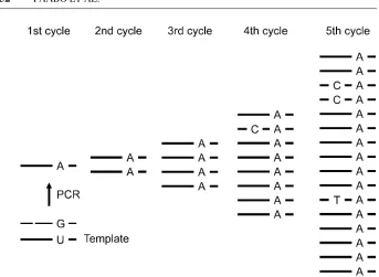
Figure 7 Phylogenetic tree for selected (left) and unselected (right) parts of the gene tb1in maize and its wild ancestor teosinte (modified after 158). The selected part of the gene carries drastically less diversity in maize than in teosinte and is derived from one or a few teosinte alleles.
selection at one of the genes may not yet have been complete. Because corn cobs are abundant at many archaeological sites in the Americas and additional genes involved in properties unique to maize will be identified, one can hope that a detailed understanding of where and when particular genetic variants of maize developed will become possible in the future. Similar analyses in other important domesticates would also be interesting.
THE FUTURE
The study of ancient DNA has the allure of time travel and attracts much attention and many practitioners. However, the generation of results that are reliable, repro-ducible, and interesting requires more than the mere application of methods that are commonplace in most molecular laboratories. The first prerequisite of any ancient DNA project should be a clear understanding of the biological question at hand and how analysis of ancient DNA is an essential aspect of addressing the question. Attention should be paid to the expected outcome. For example, an investigator proposing to study mtDNA variants in individuals from a 1000-year-old graveyard should realize that very few, if any, mutations could be expected to have appeared in that time and so little information of value may be gained, whereas contamination would be problematic. Other projects such as ancient DNA analyses of public per-sonalities such as Christopher Columbus, Jesse James, or former U.S. presidents may be novel and of interest to the public. However, they are devoid of any larger scientific contribution and sometimes ethically questionable (3). Moreover, the power of the PCR, the key molecular technique in ancient DNA research, is such that even with laborious, pain-staking precautions, erroneous results are common. Therefore, the most important prerequisite for successful ancient DNA research is a highly skeptical attitude to one’s own work. The criteria detailed in this paper are a mere framework for validation of results, and their efficacy depends wholly upon their integrated use in a project characterized by clear scientific reasoning. With this in mind, the analysis of ancient DNA offers the unique possibility to allow long-deceased individuals and extinct species to contribute to our understanding of molecular genetic evolution.
TheAnnual Review of Geneticsis online at http://genet.annualreviews.org
LITERATURE CITED
1. Adcock GJ, Dennis ES, Easteal S, Hut-tley GA, Jermiin LS, et al. 2001. Mi-tochondrial DNA sequences in ancient Australians: implications for modern hu-man origins.Proc. Natl. Acad. Sci. USA
98:537–42
2. Allard MW, Young D, Huyen Y. 1995.
Detecting dinosaur DNA. Science 268: 1192
3. Andrews LB, Buenger N, Bridge J, Rosenow L, Stoney D, et al. 2004. Con-structing ethical guidelines for biohistory.
Science304:215–16
Holcomb TA. 1995. Pre-Columbian tu-berculosis in Northern Chile—molecular and skeletal evidence.Am. J. Phys. An-thropol.98:37–45
5. Austin JJ, Ross AJ, Smith AB, Fortey RA, Thomas RH. 1997. Problems of reproducibility—does geologically an-cient DNA survive in amber-preserved in-sects?Proc. R. Soc. London Ser. B264: 467–74
6. Bailey JF, Richards MB, Macaulay VA, Colson IB, James IT, et al. 1996. Ancient DNA suggests a recent expansion of Eu-ropean cattle from a diverse wild progen-itor species.Proc. R. Soc. London Ser. B
263:1467–73
7. Barnes I, Matheus P, Shapiro B, Jensen D, Cooper A. 2002. Dynamics of Pleisto-cene population extinctions in Berin-gian brown bears. Science 295:2267– 70
8. Barnes I, Young JPW, Dobney KM. 2000. DNA-based identification of goose species from two archaeological sites in Lincolnshire.J. Archaeolog. Sci.27:91– 100
9. Bensasson D, Zhang DX, Hartl DL, He-witt GM. 2001. Mitochondrial pseudo-genes: evolution’s misplaced witnesses.
Trends Ecol. Evol.16:314–21
10. Bunce M, Worthy TH, Ford T, Hoppitt W, Willerslev E, et al. 2003. Extreme re-versed sexual size dimorphism in the ex-tinct New Zealand moa Dinornis.Nature
425:172–75
11. Burger J, Rosendahl W, Loreille O, Hem-mer H, Eriksson T, et al. 2004. Molecular phylogeny of the extinct cave lion Pan-thera leo spelaea.Mol. Phylogenet. Evol.
30:841–49
12. Cano RJ, Borucki MK. 1995. Revival and identification of bacterial-spores in 25-million-year-old to 40-million-year-old Dominican amber.Science268:1060– 64
13. Cano RJ, Poinar HN, Pieniazek NJ, Acra A, Poinar GO. 1993. Amplification and sequencing of DNA from a
120–135-million-year-old weevil.Nature363:536– 38
14. Caramelli D, Lalueza-Fox C, Vernesi C, Lari M, Casoli A, et al. 2003. Evidence for a genetic discontinuity between Nean-dertals and 24,000-year-old anatomically modern Europeans.Proc. Natl. Acad. Sci. USA100:6593–97
15. Collins MJ, Waite ER, van Duin ACT. 1999. Predicting protein decomposition: the case of aspartic-acid racemization ki-netics.Philos. Trans. R. Soc. London Ser. B354:51–64
16. Colson IB, Richards MB, Bailey JF, Sykes BC, Hedges REM. 1997. DNA analysis of seven human skeletons excavated from the terp of Wijnaldum.J. Archaeolog. Sci.
24:911–17
17. Constable JJ, Packer C, Collins DA, Pusey AE. 1995. Nuclear DNA from primate dung.Nature373:393
18. Cooper A, Lalueza-Fox C, Anderson S, Rambaut A, Austin J, Ward R. 2001. Com-plete mitochondrial genome sequences of two extinct moas clarify ratite evolution.
Nature409:704–7
19. Cooper A, Mourer-Chauvire C, Cham-bers GK, von Haeseler A, Wilson AC, P¨a¨abo S. 1992. Independent origins of New Zealand moas and kiwis.Proc. Natl. Acad. Sci. USA89:8741–44
20. Cooper A, Poinar HN. 2000. Ancient DNA: Do it right or not at all.Science
289:1139
21. Debruyne R, Barriel V, Tassy P. 2003. Mitochondrial cytochrome b of the Lyakhov mammoth (Proboscidea, Mam-malia): new data and phylogenetic anal-yses of Elephantidae. Mol. Phylogenet. Evol.26:421–34
22. Desalle R, Gatesy J, Wheeler W, Grimaldi D. 1992. DNA sequences from a fos-sil termite in Oligomiocene amber and their phylogenetic implications.Science
257:1933–36
complex DNA in calcified pleura from remains 1400 years old. Lett. Appl. Microbiol.27:265–69
24. Drancourt M, Aboudharam G, Signoli M, Dutour O, Raoult D. 1998. Detection of 400-year-oldYersinia pestisDNA in hu-man dental pulp: an approach to the di-agnosis of ancient septicemia.Proc. Natl. Acad. Sci. USA95:12637–40
25. Duarte C, Mauricio J, Pettitt PB, Souto P, Trinkaus E, et al. 1999. The early Up-per Paleolithic human skeleton from the Abrigo do Lagar Velho (Portugal) and modern human emergence in Iberia.Proc. Natl. Acad. Sci. USA96:7604–9 26. Eglinton G, Logan GA. 1991. Molecular
preservation.Philos. Trans. R. Soc. Lon-don Ser. B333:315–27; discussion 27–28 27. Endicott P, Gilbert MTP, Stringer C, Lalueza-Fox C, Willerslev E, et al. 2003. The genetic origins of the Andaman Is-landers.Am. J. Hum. Genet.72:178–84 28. Enflo P, Hawks J, Wolpoff M. 2001. A
simple reason why Neanderthal ancestry can be consistent with current DNA in-formation.Am. J. Phys. Anthropol.114: 62
29. Fish SA, Shepherd TJ, McGenity TJ, Grant WD. 2002. Recovery of 16S ribo-somal RNA gene fragments from ancient halite.Nature417:432–36
30. Fletcher HA, Donoghue HD, Holton J, Pap I, Spigelman M. 2003. Widespread occurrence ofMycobacterium tuberculo-sisDNA from 18th–19th century Hungar-ians. Am. J. Phys. Anthropol. 120:144– 52
31. Friedberg EC, Walker GC, Siede W. 1995.
DNA Repair and Mutagenesis. Washing-ton, DC: ASM Press. 698 pp.
32. Gilbert MTP, Cuccui J, White W, Lyn-nerup N, Titball RW, et al. 2004. Absence of Yersinia pestis-specific DNA in hu-man teeth from five European excavations of putative plague victims. Microbiology-Sgm150:341–54
33. Gilbert MTP, Hansen AJ, Willerslev E, Rudbeck L, Barnes I, et al. 2003.
Char-acterization of genetic miscoding lesions caused by postmortem damage. Am. J. Hum. Genet.72:48–61
34. Gilbert MTP, Willerslev E, Hansen AJ, Barnes I, Rudbeck L, et al. 2003. Distri-bution patterns of postmortem damage in human mitochondrial DNA.Am. J. Hum. Genet.72:32–47
35. Golenberg EM, Giannasi DE, Clegg MT, Smiley CJ, Durbin M, et al. 1990. Chloro-plast DNA sequence from a miocene Mag-nolia species.Nature344:656–58 36. Goloubinoff P, P¨a¨abo S, Wilson AC. 1993.
Evolution of maize inferred from se-quence diversity of an adh2 gene segment from archaeological specimens. Proc. Natl. Acad. Sci. USA90:1997–2001 37. Greenwood AD, Castresana J,
Feldmaier-Fuchs G, P¨a¨abo S. 2001. A molecular phylogeny of two extinct sloths.Mol. Phy-logenet. Evol.18:94–103
38. Greenwood AD, Capelli C, Possnert G, P¨a¨abo S. 1999. Nuclear DNA sequences from Late Pleistocene megafauna.Mol. Biol. Evol.16:1466–73
39. Greenwood AD, Lee F, Capelli C, DeSalle R, Tikhonov A, et al. 2001. Evolution of endogenous retrovirus-like elements of the woolly mammoth (Mammuthus prim-igenius) and its relatives.Mol. Biol. Evol.
18:840–47
40. Haas CJ, Zink A, Molnar E, Szeimies U, Reischl U, et al. 2000. Molecular evidence for different stages of tuberculosis in an-cient bone samples from Hungary.Am. J. Phys. Anthropol.113:293–304
41. Haddrath O, Baker AJ. 2001. Complete mitochondrial DNA genome sequences of extinct birds: ratite phylogenetics and the vicariance biogeography hypothesis.
Proc. R Soc. London Ser. B268:939–45 42. Hadly EA, Kohn MH, Leonard JA, Wayne
RK. 1998. A genetic record of popula-tion isolapopula-tion in pocket gophers during Holocene climatic change. Proc. Natl. Acad. Sci. USA95:6893–96
AM. 1994. DNA from ancient mammoth bones.Nature370:333–34
44. Hale ML, Lurz PW, Shirley MD, Rush-ton S, Fuller RM, Wolff K. 2001. Impact of landscape management on the genetic structure of red squirrel populations. Sci-ence293:2246–48
45. Handt O, H¨oss M, Krings M, P¨a¨abo S. 1994. Ancient DNA: methodological challenges.Experientia50:524–29 46. Handt O, Krings M, Ward RH, P¨a¨abo
S. 1996. The retrieval of ancient human DNA sequences. Am. J. Hum. Genet.
59:368–76
47. Handt O, Richards M, Trommsdorf M, Kilger C, Simanainen J, et al. 1994. Molecular genetic analyses of the Ty-rolean Ice Man.Science264:1775–78 48. H¨anni C, Begue A, Laudet V, Stehelin D,
Brousseau T, et al. 1995. Molecular typing of Neolithic human bones.J. Archaeolog. Sci.22:649–58
49. H¨anni C, Laudet V, Stehelin D, Taberlet P. 1994. Tracking the origins of the cave bear (Ursus spelaeus) by mitochondrial DNA sequencing.Proc. Natl. Acad. Sci. USA91:12336–40
50. Hansen A, Willerslev E, Wiuf C, Mourier T, Arctander P. 2001. Statistical evi-dence for miscoding lesions in ancient DNA templates.Mol. Biol. Evol.18:262– 65
51. Hardy C, Callou C, Vigne JD, Casane D, Dennebouy N, et al. 1995. Rabbit mitochondrial DNA diversity from pre-historic to modern times. J. Mol. Evol.
40:227–37
52. Hawks JD, Wolpoff MH. 2001. The accre-tion model of Neandertal evoluaccre-tion. Evo-lution55:1474–85
53. Hedges SB, Schweitzer MH. 1995. Detecting dinosaur DNA. Science 268: 1191–92
54. Henikoff S. 1995. Detecting dinosaur DNA.Science268:1192
55. Higuchi R, Bowman B, Freiberger M, Ryder OA, Wilson AC. 1984. DNA se-quences from the quagga, an extinct
mem-ber of the horse family.Nature312:282– 84
56. Hofreiter M, Betancourt JL, Sbriller AP, Markgraf V, McDonald HG. 2003. Phylogeny, diet, and habitat of an ex-tinct ground sloth from Cuchillo Cura, Neuquen Province, southwest Argentina.
Quat. Res.59:364–78
57. Hofreiter M, Capelli C, Krings M, Waits L, Conard N, et al. 2002. Ancient DNA analyses reveal high mitochondrial DNA sequence diversity and parallel morpho-logical evolution of Late Pleistocene cave bears.Mol. Biol. Evol.19:1244–50 58. Hofreiter M, Jaenicke V, Serre D,
Hae-seler Av, P¨a¨abo S. 2001. DNA sequences from multiple amplifications reveal ar-tifacts induced by cytosine deamination in ancient DNA.Nucleic Acids Res.29: 4793–99
59. Hofreiter M, Mead JI, Martin P, Poinar HN. 2003. Molecular caving.Curr. Biol.
13:R693–95
60. Hofreiter M, Poinar HN, Spaulding WG, Bauer K, Martin PS, et al. 2000. A molec-ular analysis of ground sloth diet through the last glaciation.Mol. Ecol.9:1975–84 61. Hofreiter M, Rabeder G, Jaenicke-Despres V, Withalm G, Nagel D, et al. 2004. Evidence for reproductive isolation between cave bear populations.Curr. Biol.
14:40–43
62. Hofreiter M, Serre D, Poinar HN, Kuch M, P¨a¨abo S. 2001. Ancient DNA.Nat. Rev. Genet.2:353–59
63. Hofreiter M, Vigilant L. 2003. Ancient human DNA: phylogenetic applications. In Nature Encyclopedia of the Human Genome, ed. DN Cooper, pp. 116–19. London: Nature Publ. Group
64. H¨oss M, Dilling A, Currant A, P¨a¨abo S. 1996. Molecular phylogeny of the extinct ground sloth Mylodon darwinii. Proc. Natl. Acad. Sci. USA93:181–85 65. H¨oss M, Jaruga P, Zastawny TH,
66. H¨oss M, Kohn M, P¨a¨abo S, Knauer F, Schr¨oder W. 1992. Excrement analysis by PCR.Nature359:199
67. H¨oss M, P¨a¨abo S, Vereshchagin NK. 1994. Mammoth DNA sequences.Nature
370:333
68. Huynen L, Millar CD, Scofield RP, Lam-bert DM. 2003. Nuclear DNA sequences detect species limits in ancient moa. Na-ture425:175–78
69. Jaenicke-Despres V, Buckler ES, Smith BD, Gilbert MTP, Cooper A, et al. 2003. Early allelic selection in maize as revealed by ancient DNA.Science302:1206–8 70. Jans MME, Nielsen-Marsh CM, Smith
CI, Collins MJ, Kars H. 2004. Charac-terisation of microbial attack on archae-ological bone.J. Archaeolog. Sci.31:87– 95
71. Jones M. 2003. Ancient DNA in pre-Columbian archaeology: a review.J. Ar-chaeolog. Sci.30:629–35
72. Kahila Bar-Gal G, Khalaily H, Mader O, Ducos P, Kolska Horwitz L. 2002. Ancient DNA evidence for the transition from wild to domestic status in Neolithic goats: a case study from the site of Abu Gosh, Israel.Ancient Biomolecules4:9–17 73. Kohn MH, Wayne RK. 1997. Facts from
feces revisited. Trends Ecol. Evol. 12: 223–27
74. Kolmann CJ, Tuross N. 2000. Ancient DNA analysis of human populations.Am. J. Phys. Anthropol.111:5–23
75. Koon HEC, Nicholson RA, Collins MJ. 2003. A practical approach to the identi-fication of low temperature heated bone using TEM.J. Archaeolog. Sci.13:1393– 99
76. Krajewski C, Buckley L, Westerman M. 1997. DNA phylogeny of the marsupial wolf resolved.Proc. R. Soc. London Ser. B264:911–17
77. Krajewski C, Driskell AC, Baverstock PR, Braun MJ. 1992. Phylogenetic relation-ships of the thylacine (Mammalia: Thy-lacinidae) among dasyuroid marsupials: evidence from cytochrome b DNA
se-quences. Proc. R. Soc. London Ser. B
250:19–27
78. Krings M, Capelli C, Tschentscher F, Geisert H, Meyer S, et al. 2000. A view of Neandertal genetic diversity.Nat. Genet.
26:144–46
79. Krings M, Geisert H, Schmitz RW, Krainitzki H, P¨a¨abo S. 1999. DNA se-quence of the mitochondrial hypervari-able region II from the Neandertal type specimen.Proc. Natl. Acad. Sci. USA96: 5581–85
80. Krings M, Stone A, Schmitz RW, Krainitzki H, Stoneking M, P¨a¨abo S. 1997. Neandertal DNA sequences and the origin of modern humans.Cell90:19–30 81. Lambert DM, Ritchie PA, Millar CD, Hol-land B, Drummond AJ, Baroni C. 2002. Rates of evolution in ancient DNA from Adelie penguins.Science295:2270–73 82. Larson S, Jameson R, Etnier M, Fleming
M, Bentzen P. 2002. Loss of genetic di-versity in sea otters (Enhydra lutris) asso-ciated with the fur trade of the 18th and 19th centuries.Mol Ecol.11:1899–903 83. Leonard JA, Wayne RK, Cooper A.
2000. Population genetics of Ice Age brown bears.Proc. Natl. Acad. Sci. USA
97:1651–54
84. Leonard JA, Wayne RK, Wheeler J, Valadez R, Guillen S, Vila C. 2002. An-cient DNA evidence for Old World origin of New World dogs.Science298:1613–16 85. Lindahl T. 1993. Instability and decay of the primary structure of DNA.Nature
362:709–15
86. Lindahl T. 1993. Recovery of antediluvian DNA.Nature365:700
87. Lindahl T, Karlstro O. 1973. Heat-induced depyrimidination of deoxyri-bonucleic acid in neutral solution. Bio-chemistry12:5151–54
88. Lindahl T, Nyberg B. 1972. Rate of depurination of native deoxyribonucleic acid.Biochemistry11:3610
Northern Idaho, USA. Geochim. Cos-mochim. Acta59:751–63
90. Loreille O, Orlando L, Patou-Mathis M, Philippe M, Taberlet P, H¨anni C. 2001. Ancient DNA analysis reveals divergence of the cave bear, Ursus spelaeus, and brown bear,Ursus arctos, lineages.Curr. Biol.11:200–3
91. Matsuoka Y, Vigouroux Y, Goodman MM, Sanchez GJ, Buckler E, Doebley J. 2002. A single domestication for maize shown by multilocus microsatellite geno-typing. Proc. Natl. Acad. Sci. USA99: 6080–84
92. Miller CR, Waits LP. 2003. The history of effective population size and genetic diversity in the Yellowstone grizzly ( Ur-sus arctos): implications for conservation.
Proc. Natl. Acad. Sci. USA100:4334–39 93. Morin PA, Chambers KE, Boesch C, Vig-ilant L. 2001. Quantitative polymerase chain reaction analysis of DNA from non-invasive samples for accurate microsatel-lite genotyping of wild chimpanzees (Pan troglodytes verus).Mol. Ecol.10:1835–44 94. Navidi W, Arnheim N, Waterman MS. 1992. A multiple-tubes approach for accu-rate genotyping of very small DNA sam-ples by using PCR—statistical considera-tions.Am. J. Hum. Genet.50:347–59 95. Nielsen-Marsh CM, Hedges REM, Mann
T, Collins MJ. 2000. A preliminary in-vestigation of the application of differen-tial scanning calorimetry to the study of collagen degradation in archaeological bone.Thermochim. Acta365:129–39 96. Nordborg M. 1998. On the probability of
Neanderthal ancestry.Am. J. Hum. Genet.
63:1237–40
97. Noro M, Masuda R, Dubrovo IA, Yoshida MC, Kato M. 1998. Molecular phylo-genetic inference of the woolly mam-mothMammuthus primigenius, based on complete sequences of mitochondrial cy-tochrome b and 12S ribosomal RNA genes.J. Mol. Evol.46:314–26
98. Oota H, Saitou N, Matsushita T, Ueda S. 1995. A genetic-study of 2,000-year-old
human remains from Japan using mito-chondrial DNA sequences.Am. J. Phys. Anthropol.98:133–45
99. Oota H, Saitou N, Matsushita T, Ueda S. 1999. Molecular genetic analysis of re-mains of a 2,000-year-old human popu-lation in China—and its relevance for the origin of the modern Japanese population.
Am. J. Hum. Genet.64:250–58
100. Orlando L, Bonjean D, Bocherens H, Thenot A, Argant A, et al. 2002. Ancient DNA and the population genetics of cave bears (Ursus spelaeus) through space and time.Mol. Biol. Evol.19:1920–33 101. Ovchinnikov IV, G¨otherstr¨om A,
Ro-manova GP, Kharitonov VM, Liden K, Goodwin W. 2000. Molecular analysis of Neanderthal DNA from the northern Cau-casus.Nature404:490–93
102. Ozawa T, Hayashi S, Mikhelson VM. 1997. Phylogenetic position of mammoth and Steller’s sea cow within Tethytheria demonstrated by mitochondrial DNA se-quences.J. Mol. Evol.44:406–13 103. P¨a¨abo S. 1985. Molecular cloning of
ancient Egyptian mummy DNA.Nature
314:644–45
104. P¨a¨abo S. 1989. Ancient DNA: extrac-tion, characterizaextrac-tion, molecular cloning, and enzymatic amplification.Proc. Natl. Acad. Sci. USA86:1939–43
105. P¨a¨abo S. 1990. Amplifying ancient DNA. In PCR-Protocols and Applications—A Laboratory Manual, ed. MA Innis, DH Gelfand, JJ Sninsky, TJ White, pp. 159– 66. San Diego: Academic
106. P¨a¨abo S, Wilson AC. 1988. Polymerase chain reaction reveals cloning artefacts.
Nature334:387–88
107. P¨a¨abo S, Wilson AC. 1991. Miocene DNA sequences—a dream come true? Curr. Biol.1:45–46
109. Pergams ORW, Barnes WM, Nyberg D. 2003. Rapid change in mouse mitochon-drial DNA.Nature423:397
110. Pertoldi C, Hansen MM, Loeschcke V, Madsen AB, Jacobsen L, Baagoe H. 2001. Genetic consequences of population de-cline in the European otter (Lutra lutra): an assessment of microsatellite DNA vari-ation in Danish otters from 1883 to 1993.
Proc. R. Soc. London Ser. B268:1775– 81
111. Piperno DR, Flannery KV. 2001. The ear-liest archaeological maize (Zea maysL.) from highland Mexico: new accelerator mass spectrometry dates and their impli-cations. Proc. Natl. Acad. Sci. USA98: 2101–3
112. Poinar H, Kuch M, McDonald G, Martin P, P¨a¨abo S. 2003. Nuclear gene sequences from a Late Pleistocene sloth coprolite.
Curr. Biol.12:1150–52
113. Poinar HN, Cano RJ, Poinar GO. 1993. DNA from an extinct plant. Nature
363:677
114. Poinar HN, Hofreiter M, Spaulding WG, Martin PS, Stankiewicz BA, et al. 1998. Molecular coproscopy: dung and diet of the extinct ground sloth Nothrotheriops shastensis.Science281:402–6
115. Poinar HN, H¨oss M, Bada JL, P¨a¨abo S. 1996. Amino acid racemization and the preservation of ancient DNA.Science
272:864–66
116. Poinar HN, Kuch M, Sobolik KD, Barnes I, Stankiewicz AB, et al. 2001. A molec-ular analysis of dietary diversity for three archaic Native Americans. Proc. Natl. Acad. Sci. USA98:4317–22
117. Poinar HN, Stankiewicz BA. 1999. Pro-tein preservation and DNA retrieval from ancient tissues.Proc. Natl. Acad. Sci. USA
96:8426–31
118. Pusch CM, Bachmann L. 2004. Spiking of contemporary human template DNA with ancient DNA extracts induces mutations under PCR and generates nonauthentic mitochondrial sequences.Mol. Biol. Evol.
21:957–64
119. Raoult D, Aboudharam G, Crubezy E, Larrouy G, Ludes B, Drancourt M. 2000. Molecular identification by “suicide PCR” ofYersinia pestis as the agent of Medieval Black Death.Proc. Natl. Acad. Sci. USA97:12800–3
120. Reid AH, Fanning TG, Hultin JV, Tauben-berger JK. 1999. Origin and evolution of the 1918 “Spanish” influenza virus hemagglutinin gene. Proc. Natl. Acad. Sci. USA96:1651–56
121. Richards MB, Sykes BC, Hedges REM. 1995. Authenticating DNA extracted from ancient skeletal remains.J. Archae-olog. Sci.22:291–99
122. Ritchie PA, Millar CD, Gibb GC, Baroni C, Lambert DM. 2004. Ancient DNA en-ables timing of the Pleistocene origin and Holocene expansion of two Adelie pen-guin lineages in Antarctica. Mol. Biol. Evol.21:240–48
123. Saiki RK, Gelfand DH, Stoffel S, Scharf SJ, Higuchi R, et al. 1988. Primer-directed enzymatic amplification of DNA with a thermostable DNA polymerase.Science
239:487–91
124. Saiki RK, Scharf S, Faloona F, Mullis KB, Horn GT, et al. 1985. Enzymatic amplifi-cation of beta-globin genomic sequences and restriction site analysis for diagnosis of sickle cell anemia.Science230:1350– 54
125. Salo WL, Aufderheide AC, Buikstra J, Holcomb TA. 1994. Identification of My-cobacterium tuberculosisDNA in a Pre-Columbian Peruvian mummy.Proc. Natl. Acad. Sci. USA91:2091–94
126. Savolainen P, Zhang YP, Luo J, Lunde-berg J, Leitner T. 2002. Genetic evidence for an East Asian origin of domestic dogs.
Science298:1610–11
127. Schaaper RM, Kunkel TA, Loeb LA. 1983. Infidelity of DNA-synthesis asso-ciated with bypass of apurinic sites.Proc. Natl. Acad. Sci. USA80:487–91 128. Schmitz RW, Serre D, Bonani G, Feine