• Tidak ada hasil yang ditemukan

DNA EXTRACTION METHOD FOR MOLECULAR DETECTION METODE EKSTRAKSI DNA UNTUK DETEKSI MOLEKULER

N/A
N/A
Protected

Academic year: 2018

Membagikan "DNA EXTRACTION METHOD FOR MOLECULAR DETECTION METODE EKSTRAKSI DNA UNTUK DETEKSI MOLEKULER"

Copied!
7
0
0

Teks penuh

(1)

DNA EXTRACTION METHOD FOR MOLECULAR DETECTION

METODE EKSTRAKSI DNA UNTUK DETEKSI MOLEKULER

R Hutami1a, N Idzni1, R Ranasasmita2, and M Suprayatmi1

1 Food Technology and Nutrition Department, Faculty of Halal Food Science, Djuanda University,

Indonesia.

2 The Assessment Institute for Foods, Drugs and Cosmetics, the Indonesian Council of Ulama, Indonesia a Correspondence: Rosy Hutami, E-mail: rosy.hutami@unida.ac.id

(Diterima: 09-08-2017; Ditelaah: 10-08-2017; Disetujui: 26-10-2017)

ABSTRACT

In molecular detection technique such as Loop-Amplification Mediated Polymorphism (LAMP)

and Polymerase Chain Reaction (PCR), right upstream preparation of deoxyribonucleic acid

(DNA) is very important. Liquid phase extraction and solid phase extraction are some of DNA

extraction methods those are available. The purpose of this research was to characterizedthe

method and product of DNA extraction based on DNA purity, DNA visualization, DNA

concentration, and processing time of DNA extraction methods. Extraction methods evaluated

included phenol-chloroform method (Method A)as liquid phase extraction and Surefood

Extraction kit method (Method B) as solid phase extraction. Result showed that Method A could

be performed on samples with very low DNA concentrations ranging from 7.00 to 9.45 ng/µl

with a good purity (1.80 to 2.10). Although, it showed no DNA isolates bands on gel agarose 1%

and need ± 30 hours processing time. Method B had a good performa in extracting sample with

high concentration DNA (49.67 to 357.28 ng/µl) with a good purity (1.93 to 2.07). This method

showed bands for each DNA samples on gel agarose 1% and need about ± 1 hour processing

time. Both methods can be used for sample preparation in molecular analysis including halal

authentication purposes.

Keywords: phenol, chloroform, Surefood kit, LAMP.

ABSTRAK

Dalam teknik deteksi molekuler seperti Loop-Amplification Mediated Polymorphism (LAMP) dan

Polymerase Chain Reaction (PCR), pembuatan hulu asam deoksiribonukleat (DNA) sangat

penting. Ekstraksi fase cair dan ekstraksi fasa padat merupakan beberapa metode ekstraksi DNA

yang tersedia. Tujuan dari penelitian ini adalah untuk mengkarakterisasi metode dan produk

ekstraksi DNA berdasarkan kemurnian DNA, visualisasi DNA, konsentrasi DNA, dan waktu

pemrosesan metode ekstraksi DNA. Metode ekstraksi yang dievaluasi meliputi metode

fenol-kloroform (Metode A) sebagai ekstraksi fasa cair dan metode Ekstraksi Surefood kit (Metode B)

sebagai ekstraksi fasa padat. Hasil penelitian menunjukkan bahwa Metode A dapat dilakukan

pada sampel dengan konsentrasi DNA sangat rendah berkisar antara 7,00 sampai 9,45 ng / μl

dengan kemurnian yang baik (1,80-2,10). Meski tidak menunjukkan DNA isolat band pada gel

agarosa 1% dan membutuhkan waktu pemrosesan ± 30 jam. Metode B memiliki performa yang

baik dalam mengekstraksi sampel dengan DNA dengan konsentrasi tinggi (49,67 sampai 357,28

ng / μl) dengan kemurnian yang baik (1,93 sampai 2,07). Metode ini m

enunjukkan band untuk

setiap sampel DNA pada gel agarose 1% dan membutuhkan waktu ±1 jam. Kedua metode

tersebut dapat digunakan untuk preparasi sampel dalam analisis molekuler termasuk tujuan

otentikasi halal.

(2)

Hutami

R, N Idzni, R Ranasasmita, and M Suprayatmi. 2017. DNA Extraction Method for

Molecular Detection. Jurnal Pertanian 8(2): 106-112.

INTRODUCTION

Liquid and solid phase DNA extraction

method have a different characteristics in the

processand obtained products. Those DNA

extraction methods determined the success of

molecular detection technique including

Loop-Amplification Mediated Polymorphism

(LAMP) and Polymerase Chain Reaction

(PCR). By characterizing DNA extraction

method and its products, we can choose the

most suitable one for our research purposes.

Factors to be considered when selecting

nucleic acid extraction methods for molecular

detection include sample background, lysis,

appropriate preparation chemical, required

detection limits, and requirements for

particular application.

In the As previous study, DNA/RNA

preparation for molecular detection have

been reviewedwhich reported the various

method of nucleic acid extraction

1

. Evaluation

of DNA and RNA extraction methods was also

reported. It showed comparison of some

extraction methods

2

.Steps of the methods

included extracting, separating, purificating,

and concentrating DNA.

The aim of this research was to report the

characters of method and products of

phenol-chloroform DNA extraction as liquid phase

extraction method and Surefood Kit DNA

extraction as solid phase extraction method.

MATERIALS AND METHODS

Materials

Porcine tissue, chicken tissue, goat tissue, beef

tissue, fish tissue, reagen for

phenol-chloroform extraction method, R Biopharm

AG (kit SureFood

®

PREP Basic), agarose, TBE,

loading

dye,

microtube,

micropippetes,

centrifuge, electrophoresis instrument.

Method A

A total of 250 μL animal tissue samples were

prepared and fed into the microfuge tube and

then 300 μL STE, 40 μL SDS 10%, and 20 μL

proteinase-K were incubated at 55oC and

homogenized for 2 h. A further 400 μL of

phenol, 400 μL of CIAA (chloroform

isoamyl

alcohol), and 40 μLNaCl 5M were added.

Then,it was incubated at room temperature

and homogenized for 1 hour. Next it was

centrifuged for 5 minutes and the supernatant

was taken. Subsequently the sample was

incubated at -20 ° C for 1 night. Then it was

centrifuged for 5 minutes and the alcohol part

was removed. Then it was centrifuged for 5

minutes, the alcohol was removed and the

tube was dried for 1-2 hours.

Method B

Extraction were done by 50 mg fresh meat

using kit manufactured by R Biopharm AG (kit

SureFood® PREP Basic) with modification.

Intially, sampel were lysed with lysis buffer

containing Proteinase-K. DNA was then bind

using binding buffer and purified through 2

types of spin filter (clear and yellow) with the

aid of washing buffer (pre-wash and wash

buffer). DNA was then diluted by elution

buffer. The SureFood DNA extraction kit itself

basically adopt silica mini column since it is a

common approach for DNA isolation.

Gel for Electrophoresis

Gel was made from 0.3 grams of agaroseand

30 ml of buffered (0.5 x TBE) solution being

heated. The agarose solution was left slightly

cold while stirring with a stirrer magnet, then

added 1.8 μldye of etidium bromide A total of

5

μ

l DNA samples were dissolved in 1

μ

lloading dyes Electrophoresis wascarried out

for 40 minutes at a constant voltage of

100volts until the blue bromtimol reaches the

bottom of the gel.

RESULT AND DISCUSSION

Result

(3)

concentration.DNA

isolates

quality

assessment was performed on the basis of

DNA purity and the results of DNA

visualization on agarose gel qualitatively.

Method A

In method A, the samples used were porcine

DNA residues on washed food equipments

after washing by different cleaning agents

based on sharia. The samples were coded as A

to E. The results obtained refer to Table 1.

Table 1 Measurement of DNA concentration and purity of sample in Method A

Sample

code

DNA concentration (ng/µl)

Purity (λ260/280)

Repetition

1

Repetition

2

Mean ± SD

Repetition

1

Repetition

2

Mean ± SD

A

8.80

8.70 8.75 ± 0.07

2.08

2.12

2.10 ± 0.03

B

7.80

8.00 7.90 ± 0.14

1.90

1.70

1.80 ± 0.14

C

7.10

6.90 7.00 ± 0.14

1.96

1.97

1.97 ± 0.01

D

9.20

9.70 9.45 ± 0.35

1.71

1.92

1.82 ± 0.15

E

9.50

9.30 9.40 ± 0.14

1.90

1.92

1.91 ± 0.01

The DNA concentration of fifth samples

ranged from 7.00 to 9.45 ng/µl. The purity of

samples

ranged

from

1.80

to

2.10.

Visualization of extracted genome DNA based

on gel electrophoresis was shown as Figure 1.

The DNA bands from the porcine positive

control extraction appearedsuccessfully on

1% agarose gel by electrophoresis, while the

DNA bands from the fifth samples did not

appear.

Method B

In method B, the samples used were raw meat

animal tissue of several species. The samples

which were coded as A, B, C, D, and Ewere

porcine, beef, chicken, goat, and fish tissue-

respectively. The results obtained refer to

Table 2.

Figure 1. Visualization of extracted genome

DNA on 1% agarose gel. M : Marker

100 pb, P : positive control of

porcine DNA, 1-2: residue of

porcine DNA on sample A, 3-4 :

residue of porcine DNA on sample

B, 5-6 : residue of porcine DNA on

sample C, 7-8 : residue of porcine

DNA on sample D, 9-10 residue of

porcine DNA on sample E.

Table 2 Measurement of DNA concentration and purity of sample in Method B

Sample

DNA concentration (ng/µl)

Purity (λ260/280)

Repetition

1

Repetition

2

Mean ± SD

Repetition

1

Repetition

2

Mean ± SD

A

165.64

214.61

190.13 ± 34.63

1.97

1.95

1.96 ± 0.01

B

53.18

46.15

49.67 ± 4.97

1.87

2.03

1.95 ± 0.11

C

455.76

258.80

357.28 ± 139.27

2.07

1.99

2.03 ± 0.06

D

163.20

148.62

155.91 ± 10.31

2.09

2.04

2.07 ± 0.04

E

210.56

153.34

181.95 ± 40.46

1.82

2.03

1.93 ± 0.15

The DNA concentration of samples ranged

from

49.67

to

357.28

ng/µl. The purity of

(4)

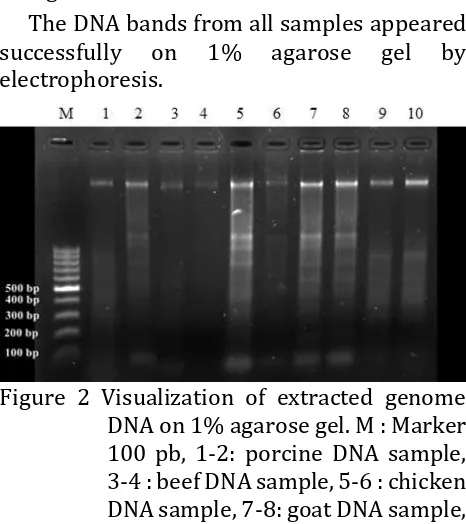
DNAbased on gel electrophoresis was shown

as Figure 2.

The DNA bands from all samples appeared

successfully

on

1%

agarose

gel

by

electrophoresis.

Figure 2 Visualization of extracted genome

DNA on 1% agarose gel. M : Marker

100 pb, 1-2: porcine DNA sample,

3-4 : beef DNA sample, 5-6 : chicken

DNA sample, 7-8: goat DNA sample,

9-fish DNA sample.

2.07.DNA isolates can be said to be pure and

have met the requirements for molecular

analysis when the ratio

λ

260/280ranges from

1.8 to 2.0

3,4

. DNA has maximum absorption in

λ

260 and protein has maximum absorption in

λ

280. So that, the products of both DNA

extraction methods can be used for molecular

analysis.

DNA Visualization

In method A, the DNA bands from the porcine

positive control extraction were successfully

visible on 1% agarose gel, but the DNA bands

from the fifth samples did not present. The

isolate DNA from fifth samplesthat did not

present on the gel can be caused by very small

amounts of DNA (<50 ng/

μl).

But, the

appearance of band of porcine positive

control and total DNA purity within the

normal range indicates that the

phenol-chloroform method can be used for molecular

analysis especially in sample with low DNA

concentration.

In method B, bands appeared in all of

samples well, although they had a various

thickness. The various band thickness can be

cause of different concentration of DNA.

Athick or dense and solid DNA band showed a

high concentration and good integrity of

DNA

5

. It was also showed DNA smear in

sample gel agarose visualization.DNA smear

showed that DNA was broken/sheared during

extraction process

5

.

DNA Concentration

DNA isolation resulted in varying yield from

7.00 to 9.45 ng/µl in method A and 49.67 to

357.28 ng/µl in method B of DNA

concentration. This yield is determined by

DNA

isolation

time

and

lysis

buffer

composition. Timing is essential since if it

takes too long, there is possibility of DNA

precipitation

6

.DNA concentration of the

extraction products were not uniform.

Therefore, the concentration of DNA that has

been obtained is uniformed by dilution, thus

DNA needs to be diluted to a certain level

concentration.

The

suggested

DNA

concentration is 10 ng to

1 μg DNA per μl for

PCR reaction

7

.

Method Principles

Method A

(5)

enzyme

8,9

. Sodium Tris EDTA (STE) roles in

inactivating DNase enzymes that can denature

isolated DNA, EDTA inactivates nuclease

enzymes by binding magnesium and calcium

ions required as cofactors of DNAse enzyme

10

.

The second step is separating DNA from

protein using proteinase-K. Proteinase-K is

one of the primary protease used for DNA

separation. It is used to reduce protein

background and aid in lysis by digesting

membrane or capsid proteins. This protease

will break proteins into smaller molecules by

cleaving peptide bonds

11

.

Nucleic acid is a hydrophilic molecule and

it is soluble in water. In addition, the protein

also contains a hydrophobic residue that

causes the protein to dissolve in organic

solvents. Based on this nature, there are

several deproteinization methods based on

the selection of organic solvents. In this

research, the method used was phenol

-chloroform containing 4% isoamyl alcohol

(CIAA). The use of chloroform isoamyl alcohol

(CIAA) is based on differences in the nature of

organic solvents. Phenols denatures proteins,

which stay in the organic phase, whereas the

DNA is in the aqueous phase. The addition of

chloroform and isoamyl alcohol helps to

separate the phases and prevent foaming

12

.

Chloroform can not mix with water and its

ability to deproteinized based on the ability of

denatured polypeptide chains to enter or

mobilize into phases between chloroform -

water. High protein concentrations in the

intermediate phase can cause the protein to

undergo precipitation. While lipids and other

organic compounds will be separated in the

chloroform layer

13

. The third step was

precipitation

using

alcohol

and

high

concentration of salt

14

. Precipitation also

serves to remove chloroform residues derived

from the extraction stage. The fourth step was

concentrating the DNA by centrifugation and

manual manipulation by drying the DNA.

According to Thatcher

1

, this liquid extraction

method produces very clean product, though

some molecules or particles can coprecipitate

with the DNA. This method need about 30

hours processing time.

Method B

Method B used refers to the extraction

methods released by R Biopharm AG

(SureFood® PREP Basic kit) by making some

modifications. This method of extraction or

isolation of DNA used a ready-to-use

extraction buffer. In Surefood Kit, filtration

tool used is spin filter there were two types of

spin filter that was clear spin filter and yellow

spin filter. The cell was broken by lysis buffer,

then the protein cell component was

degraded by using Proteinase-K as protease

enzyme. The next step was binding of nucleic

acids on spin filter by using buffer bindings,

filtering and washing the DNA by washing

buffer (pre-wash buffer and wash buffer), at

the final stage the DNA was dissolved into

elution buffer. The SureFood commercial

extraction kit has the same principle as other

commercial

extractions

that

use

the

minicoloumn principle or DNA filtration. DNA

extraction using the minicoloumn principle is

the most common method of extraction

because the results obtained are very good

with lesser processing when compared with

the phenol-chloroform method and the cost is

considerably cheaper when compared to the

magnetic bead method

15

.

Method B is chategorized as solid phase

extraction method. Solid phase extraction

method have become the most common

method of nucleic acid isolation for several

reason. They include minimal hazardous

chemicals,

fewer

and

easier

manual

manipulations, automation capability, and

increased throughput. Solid phase methods

used for nucleic acid separation include 3

principle technique : size exclusion by gel

filtration, ion exchange chromatography by

charge-based reversible adsoption, and

affinity chromatography

1

.

(6)

preparation hasfour basic steps; lysis, binding,

washing,and eluting. Unexpectedly, similar

chemicals can be used for lysis and

surface-binding. Chaotropic salts canbe used for cell

lysisand binding to a silica surface

17

. A liquid

sample passes through by centrifugation (spin

filter), pressure (syringe filter), or vacuum.

Kits with silica spin filters are fast to perform

and do not require hazardous chemicals.

CONCLUSION AND IMPLICATION

The phenol-chloroform method (Method A)

can be used for extracting sample with very

low DNA concentration (7.00 to 9.45 ng/µl).

The extraction product of phenol-chloroform

was very clean. The purity of DNA isolates was

ideal to be used in molecular analysis (1.80 to

2.10). Although in the DNA visualization, that

was no bands appeared in the gel according to

the low concentration of DNA isolates. But it

need long time process (about 30 hours)and

some hazardous chemical were still used. The

Surefood Kit method (Method B) were able to

extract sample with high concentration DNA

samples (49.67 to 357.28 ng/µl) with good

DNA purity (1.93 to 2.07), good visualization

in gel agarose, and less processing time (1

hour)

compared

to

phenol-chloroform

method.

ACKNOWLEDGMENTS

We would like to thanks for the support of

Direktorat Riset dan Pengabdian Masyarakat

Direktorat Jenderal Penguatan Risetdan

Pengembangan

Ministry

of

Research

Technolgy

and

Higher

Education

(Kemenristek-Dikti) for gave the grant of this

reseach in accordance with the Research

Contract No. 1598/K4/KM/2017. Research

grant scheme of

Program Kerjasama Antar

Perguruan Tinggi year 2017.

REFERENCE

Thatcer S A 2015 Clinical Chem.61 90

Shiaw C S E, Shiran M S, Path M, Cheah Y K, Tan

G C, Path M, Sabariah A R, Path M 2010

2010 Med. J. Malaysia65 137

Sambrook J, Fritsch F, Miniatis T 1989

Molecular Cloning Laboratory Manual. 3rd

Edition. (New York : Cold Spring Harbor

Laboratory Press)

Muladno 2010 Teknologi Rekayasa Genetika.

(Bogor : IPB Press)

Irmawati 2003

Perubahan Keragaman Genetik

Ikan Kerapu Tikus Generasi Pertama Pada

Stok

Hatchery

[Thesis]

(Bogor:

InstitutPertanian Bogor)

Komalasari K 2009

Pengaruh perbandingan

volume darah dan lisis buffer serta

kecepatan sentrifugasi terhadap kualitas

produk DNA pada sapi Frensian Holstein

(FH) [Undergraduate Thesis] (Bogor:

Institut Pertanian Bogor)

Sambrook J and Russell D W 2001 Molecular

Cloning: A Laboratory Manual, 3rd Edition

(New York : Cold Spring Harbor Laboratory

Press)

Birnboim H C, Doly J A 1979 Rapid alkaline

extraction

procedure

for

screening

recombinant plasmid DNA.

Nucleic Acids

Res7:1513

23

Switzer 1999

Experimental

Biochemistry

(Oxford : Blackwell Scientific Pub)

Corkill G, Rapley R 2008 The Manipulation of

Nucleic Acids: Basic Tools &Techiques in

Molecular Biomethods Handbook Second

Edition.

Ed: Walker J M, Rapley R (NJ :

purification chip to selectively isolate DN

A

or RNA from low copy/single bacterial cells

in minute sample volume followed by

direct on-chip quantitative PCR assay. Anal

Chem85:1484

91

Clark D P 2010

Molecular Biology: Updated

Edition (AP Cell Press)

Eickbush T H and Moudrianakis E N 1978 The

compaction of DNA helices into either

continuous supercoils or folded-

fiberrods

and toroids. Cell 13:295

306

(7)

diphteriae

untuk

pemeriksaan

PCR.

PenelitiKesehat. 42(2):85-92

Vogelstein B and Gillespie D 1979 Preparative

and analytical purification of DNA from

agarose. ProcNatlAcadSci76:615

9

Boom R, Sol C J, Salimans M M, Jansen C L,

WertheimvanDillen P M, van der Noordaa J

1990 Rapid and simple method for

Gambar

Table 2 Measurement of DNA concentration and purity of sample in Method B
Figure 2 Visualization of extracted genome  DNA on 1% agarose gel. M : Marker 100 pb, 1-2: porcine DNA sample, 3-4 : beef DNA sample, 5-6 : chicken DNA sample, 7-8: goat DNA sample, 9-fish DNA sample

Referensi

Dokumen terkait

Aset keuangan yang dimiliki hingga jatuh tempo adalah aset keuangan nonderivatif dengan pembayaran tetap atau telah ditentukan dan jatuh temponya telah ditetapkan

PROGRAM MAGISTER TEKNIK SIPIL UNIVERSITAS ATMA JAYA YOGYAKARTA..

Walaupun regulasi telah menentukan pendekatan statistik sebagai acuan dalam menghitung tahanan slip, kondisi terburuk bisa saja terjadi yaitu tahanan slip yang sebenarnya

Memang secara khusus tidak ada teks yang menyebutkan bahwa injil ini ditulis untuk golongan tertentu, tapi yang pasti injil ini ditulis oleh Rasul Yohanes kepada semua orang

dapat menyelesaikan skripsi yang berjudul “ Pengaruh Risiko Usaha Terhadap Kecukupan Modal Inti Pada Bank Umum Swasta Nasional Go Public ”. Penulis menyadari bahwa

Intisari: Telah dilakukan analisis kualitatif dan kuantitatif asam lemak tak jenuh Omega-3 dan Omega-6 pada minyak ikan patin ( Pangasius pangasius ) dengan menggunakan

selaku Ketua Departemen Matematika Fakultas Matematika dan Ilmu Pengetahuan Alam Universitas Sumatera Utara dan juga selaku dosen pembimbing yang senantiasa membimbing

Gedung H, Kampus Sekaran-Gunungpati, Semarang 50229 Telepon: (024)