Keberadaan Protein A pada Permukaan Sel Bakteri
Staphylococcus Aureus
Menggunakan Teknik “Serum
Soft Agar”
The Presence of Protein A on The Surface of
Staphylococcus Aureus Bacterial Cells Using Serum Soft
Agar Techniques
Titiek Djannatun1, I Wayan Teguh Wibawan2
1Department of Microbiology, Faculty of Medicine, YARSI University 2Laboratory of Microbiology, Department of Infectious Disease and
Veterinary Public Health, Faculty Veterinary Medicine, Bogor Agricultural University
KATA KUNCI KEYWORDS
ABSTRAK
Soft agar; serum soft agar; protein A; staphylococcus Soft agar; serum soft agar; protein A; staphylococcus
Protein A merupakan komponen protein spesifik di permukaan Staphylococcus aureus (S. aureus). Bakteri yang memiliki protein A banyak dimanfaatkan dalam biomedis. Penelitian ini bertujuan untuk menemukan metode sederhana mendeteksi keberadaan protein A pada S aureus.
Penelitian menggunakan 15 isolat S. aureus dari kasus lapangan, S. aureus cowan I sebagai kontrol positif, Staphylococcus epidermidis dan Micrococcus sp sebagai kontrol negatif. Bacteria diisolasi pada 10 ml medium Todd Hewith Broth, medium soft agar (SA), dan pada 10 ml serum soft-agar/SSA (SA yang ditambahkan serum kelinci atau ayam), agitasi menggunakan vortex dan diinkubasi pada 37oC selama 18-24 jam.
Bentuk koloni yang tumbuh diamati dan dikategorikan sebagai koloni kompak dan difus. Koloni yang diduga mengandung protein A dilakukan konfirmasi tes dengan Dot Blot.
ABSTRACT Protein A is a specific proteins on the surface of some Staphylococcus aureus (S. aureus) strains. Staphylococcus bacteria which have protein A has widely used in the biomedical. Aims of this study to find a simple method of detecting the presence of S. aureus protein A.
15 S. aureus isolates from field cases, S. aureus cowan I as positive control, Staphylococcus epidermidis dan Micrococcus sp as negatif control were used. Bacteria was inoculated into 10 ml Todd Hewith Broth medium, Soft Agar (SA) and into 10 ml of serum soft-agar (SSA), agitated using a vortex and incubated at 37oC for 18-24 h. Form of
colonies that grew was observed and categorized as compact and diffuse colonies. The colonies that suspected contain protein A conducted confirmation tests by Dot Blot.
7 isolates showed the change of colony formation from diffuse to compact in SSA (SA added rabbit or chicken serum), and 8 isolates remain diffuse.
Dot Blot test positive.Protein A has the ability to bind the Fc fraction of IgG but not the IgY from birds. The binding of S. aureus and IgG could be demonstrated by the change of colony growth from diffuse before the addition of serum containing IgG in SA to compact after the presence of serum. Protein A on the bacterial cell surface bind the Fc-IgG caused steric hinderance of bacteria and expressed as compact colony formation in SA. SSA using mammalian sera can be used to discriminate the bacterial strains with or without protein A.
Staphylococcus bacteria which have protein A has a very important benefit in the biomedical world, because of its use in the preparation of a diagnostic kit, the purification of biological material such as immunoglobulins, antigens, enzymes, hormones and other important substances. Therefore, it is necessary to look for the Staphylococcus aureus (S. aureus) or any other staphylococcus bacteria type, which has and has rich a protein A. Currently, Currently, S. aureus Cowan I is known as a rich protein AS. aureusstrain.
The protein A containing on the bacteria surface can be reduced by the getting older isolate’s age and because the isolates have been passage repeatedly. The use of serum soft agar technique in determination of the streptococcus B group serotype and in determination of the presence of cα and
cß proteins of Streptococcus agalactiae
have also been reported (Wibawan and Laemmler, 1990a; Wibawan and Laemmler, 1990b) and it was used to differentiate encapsulated and not encapsulated staphylococcus bacteria which isolated from human clinical cases and also cow mastitis patients (Opdebeeck et al., 1985, Opdebeeck et al., 1987, Opdebeeck et al., 1988). The serum soft agar technique has also been used to determine the serotypes of
Staphylococcus epidermidis, using specific serum against certain serotypes candidate. Homologous S. epidermidis
strains will show changes from diffuse to compact colonies form, while heterologous strains will remain diffuse colonized (Yoshidaet al., 1972).
Correspondence:
Titiek Djannatun, Department of Microbiology, Faculty of Medicine, YARSI University, Jakarta
Protein A as biologicaly acts has a role as a bacterial virulence factor (DeDentet al., 2007; Yaoet al., 2010) that is capable to bind the Fc fraction of almost all immunoglobulin G subclasses except IgG3 (human), IgG1 (mouse), IgG1, IgG2a , IgG2b (rat), as well as chicken immunoglobulin Fc (Djannatun, 2002).
The interaction of IgG with protein A using a variety methods have been reported. They are a double gel
immunodiffusion method,
immunofluoresensi, indirect hemagglutination, ELISA, slide hemagglutination test, microplate hemagglutination test (Huang and Chang, 2004; Cox et al., 1986; Takeuchi
et al., 1988, Takeuchi et al., 1995), single radial immunodiffusion, solid-phase radio immunoassay (Cheung et al., 1987), PCR (Wenwen et al., 2016). In this study showed the using of serum soft agar technique using rabbit serum to differentiate S. aureus that has and do not has protein A.
MATERIALS AND METHODS
Time and place
This research was conducted in March 2000 until November 2001 in Fakultas Kedokteran Hewan Institut Pertanian Bogor and Biofarma bandung.
Bacterial Isolated
In this study, 15 Staphylococcus aureus isolates from field cases were used. All isolates had been identified previously as S. aureus, and existed as a collection of Fakultas Kedokteran Hewan Institut Pertanian Bogor.
Staphylococcus aureus Cowan I, which has rich protein A, and Staphylococcus epidermidis, which has no protein A, used as reference strains.
Reidentification ofS. aureusIsolates All S. aureus isolates was reidentificated using standard procedures for these bacteria, that are the type of colony formation and type of haemolysis on blood agar medium, shape and composition of bacterial cells microscopically with Gram stain (Carter, 1986), and the ability to utilize glucose and mannitol and the expression of catalase and coagulase activities (Qianet al., 2007).
The soft agar technique
The S. aureus bacteria was inoculated into 10 mlTodd Hewith Broth
(THB) medium at 370C for 18-24 h, then Soft Agar (SA) was made consisting of 0.15% agar base in 10ml of Brain Heart Infusion medium (BHI). SA was boiled on the water until the liquid homogeneous, while boiling. After that it was cooled to about 37-40oC temperature.
The suspension of bacteria in media THB was taken with ½ cm oese needle, then was inoculated into 10ml NaCl physiological sterile saline. The solution and then was homogenized so that bacterial cells were spread evenly and bacterial suspension (1/2 cm oese needle) was inoculated into 10 ml of
soft-agar, agitated using a vortex and incubated at 37oC for 18-24 h. Form of colonies that grew observed and categorized as compact colonies and colonies diffuse Wibawan & Lämmler, 1990a; Wibawan & Lämmler, 1990b).
Bacteria Growth in Serum Soft Agar (SSA)
has been described, and then added 100 ul of rabbit serum or other avian serum. Bacterial suspension (1/2cm oese needle) was inoculated into 10 ml of serum soft-agar, agitated using a vortex and incubated at 37oC for 18-24 h. Form of colonies that grew was observed and categorized as compact colonies and colonies diffuse as well as in SA (Wibawan dan Laemmler, 1990b).
Dot Blot
The presence of protein-A on the cell surface of S. aureus bacteria was confirmed with the dot blot test. Isolates of S. aureus Cowan I, Sa53, 2 S. aureus selected isolates that showed a compact colonies in SSA and S. epidermidis were dropped onto a nitrocellulose membrane, dried with hair dryer. After that a nitrocellulose membrane submerged in skimmed milk which had been diluted 10 times for 45 minutes at room temperature, washed with 5 ml PBS (pH 7,5) 2x, and incubated with rabbit serum or chicken serum each for an hour followed by 2x washing with 5ml PBS. Incubated again in anti-rabbit conjugate or mice-anti-chicken (25 l conjugates + 5 ml PBS solution) for 60 min, followed by washing with PBS solution. The interaction of protein-A and IgG was visualized by adding 5ml of -chloronapthol (9ml -chloronapthol + 3 ml metanol + 25ml PBS) and 200 µl of H2O2. Positive reaction was characterized by the presence of black color on bacterial suspension droplets which were tested (Wibawan, 1993).
RESULTS
Characterization ofS. aureus
Staphylococcus aureus isolates which were grown on blood agar medium was studied through its colony characteristic and its growth characteristic on BHI (Brain Hearth Infusion) medium after incubated at 35 ± 10 C for 24-48 h. Besides that, the morphology and composition of bacterial cells microscopically was studied by Gram staining. For identification (determination of species) was studied by catalase test, coagulase and oxidase test as well as the ability of bacteria to fermentation mannitol. The
S. epidermidis and micrococcus bacteria used as a comparison, as bacteria, that have no protein A.
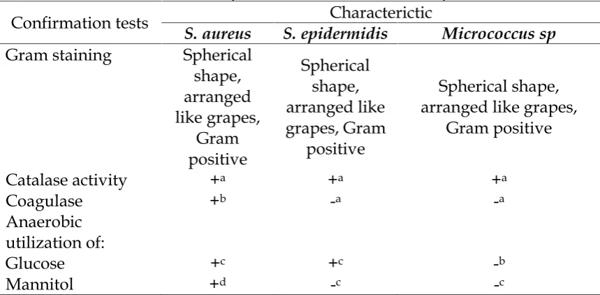
Table 1. Characteristic ofS. aureusbacteria in some tests compared with
S.epidermidisdanMicrococccus sp.
Confirmation tests Characterictic
S. aureus S. epidermidis Micrococcus sp
Gram staining Spherical shape, arranged like grapes,
Gram positive
Spherical shape, arranged like grapes, Gram
positive
Spherical shape, arranged like grapes,
Gram positive
Catalase activity +a +a +a
Coagulase +b -a -a
Anaerobic utilization of:
Glucose +c +c -b
Mannitol +d -c -c
Explanation: +a= produce air bubbles, +b= rabbit plasma agglutinate, -a= non rabbit plasma agglutinate, +c= glucose fermented, -b= non glucuse fermented,
+d= mannitol fermented, -c= non mannitol fermented.
Determination of the Rich Protein A S. aureus
To determine S. aureus isolate containing rich protein A, Serum soft agar technique was used. The changes of colony formation from diffuse to compact are indicators of the presence of protein A on the bacterial cell surface. Isolat 3, S.a-4, S.a-5, S.a-6, S.a-11, S.a-53, S.a-54 and S.a Cowan I showed the change
of colony formation from diffuse to compact colonies in SSA, while others
S. aureus isolates showed remain diffuse colonies in SSA. Serum soft agar (SSA) using rabbit serum were able to distinguish groups of S. aureus
isolates compact colonized and diffuse colonized in SSA (Table 2 and figure 1).
Diffuse colony form Compact colony form
Figure 1. The changes of colony diffuse (before addition of rabbit serum) to compact (after addition of rabbit serum) inS. aureuswhich has protein A with serum soft-agar
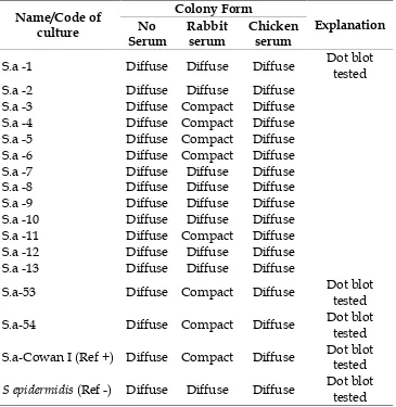
Table 2. Detection the presence of protein A on the surface of Staphylococcus aureus
bacterial cells using serum soft-agar.
Name/Code of culture
Colony Form
Explanation No
Serum
Rabbit serum
Chicken serum
S.a -1 Diffuse Diffuse Diffuse Dot blot tested
S.a -2 Diffuse Diffuse Diffuse
S.a -3 Diffuse Compact Diffuse
S.a -4 Diffuse Compact Diffuse
S.a -5 Diffuse Compact Diffuse
S.a -6 Diffuse Compact Diffuse
S.a -7 Diffuse Diffuse Diffuse
S.a -8 Diffuse Diffuse Diffuse
S.a -9 Diffuse Diffuse Diffuse
S.a -10 Diffuse Diffuse Diffuse S.a -11 Diffuse Compact Diffuse S.a -12 Diffuse Diffuse Diffuse S.a -13 Diffuse Diffuse Diffuse
S.a-53 Diffuse Compact Diffuse Dot blot tested
S.a-54 Diffuse Compact Diffuse Dot blot tested
S.a-Cowan I (Ref +) Diffuse Compact Diffuse Dot blot tested
S epidermidis(Ref -) Diffuse Diffuse Diffuse Dot blot tested Explanation: S.a =Staphylococcus aureus
Furthermore, based on the quality of the colonies form changes were selected S.a-53 and S.a-54 to be confirmed the presence of protein A on the bacteria cells surface used Dot Blot assay (Table 3). The Dot blot assay showed the interaction of S.a-53 and S.a-54 isolates with rabbit IgG. It were characterized by the presence of black color on paper nitrselulose that were dropped two bacterial suspension. The same reaction was shown also by S.a Cowan I, while S.a-1 and S. epidermidis
showed no reaction (Table 3). The S.a-53 and S.a-54 isolates showed the presence of protein A and S.a-1 showed no protein A. The results indicate that bacteria have a protein A have Dot Blot positive reaction. Otherwise bacteria that do not have protein A have Dot blot negative reaction. These results indicated also in bacteria reference,
Table 3. A positive reaction to the dot blot assay relating to compact colonies form on serum soft agar (SSA)
Isolate Dot Blot Reaction Colony Form (SSA)
S.a-53 + Compact
S.a-54 + Compact
S.a Cowan I + Compact
S.a-1 - Diffuse
S. epidermidis - Diffuse
Explanation: S.a =Staphylococcus aureus, + = has protein A, - = No protein A
DISCUSSION
S. aureusCharacterisation
Based on the results of biochemical tests obtained 12 strains of
Staphylococcus aureus (Cowan and Steel, 1973; Hof and Dörries, 2002; Brooks et al., 2010; Bien et al., 2011; Fehrmannaet al., 2013). The S. epidermidis and micrococcus bacteria used as a comparison and a differentiator other cocci-shaped bacteria (Table 1).
Detection of The presence of The Rich of Protein AS. aureus
From 15 S. aureus samples of fields isolates, 7 isolates showed the change of colony formation from diffuse to compact colonies after the presence of rabbit sera in serum soft agar (SSA), similar result was found by The reference using S. aureus Cowan I strain. No comparable results were found for 8 isolates and for S. epidermidis as a negative control strain, the colonies remained diffuse after the addition of rabbit sera in SSA. No change of colony formation was detected after the addition of chicken sera for all bacteria isolates used in this study (Table 2, figure 1).
The SAA technique can be used to discriminate the S. aureus bacterial strains with or without protein A on the bacteria surface cell. The results in Table 2 show that SSA using rabbit
serum were able to distinguish groups of S. aureus isolates compact colonized and diffuse colonized in SSA. The changes of colonies form in this interaction was not caused by coagulase reaction or clumping factor because all S. aureus isolates, which were used in this study, had positive reaction in coagulase test. The change of colony formation was related with the interaction between a protein on the surface of bacterial cells with IgG in the rabbit sera. It was confirmed by the addition of chicken serum on soft agar not to cause a change in the bacterial colony formation.
bacteria have a protein A have Dot Blot positive reaction. Otherwise bacteria that do not have protein A have Dot blot negative reaction. These results indicated also in bacteria reference,
Staphylococcus aureus Cowan I, which has rich protein A, and Staphylococcus epidermidis, which has no protein A.
SSA is a practical, simple and inexpensive method to detect the presence of protein A Staphylococcus aureus as compared to PCR and Elisa methode (Huang and Chang, 2004; Wenwenet al., 2016).
CONCLUSION
The SSA Technique using rabbit serum can be used to determine the presence of protein A on the surface of
Staphylococcus aureus bacterial cell through the changes of the colony form
from diffuse to compact.
Immunoglobulin Y (IgY) chicken was not able interact with protein A of S. aureus.
REFERENCES
Bien J, Sokolova O, and Bozko P 2011. Characterization of Virulence Factors of Staphylococcus aureus: Novel Function of Known Virulence Factors That Are Implicated in Activation of Airway Epithelial Proinflammatory
Response. Review Article. J. of
Pathogens. 13 pages (online). Article ID 601905. doi:10.4061/2011/601905. Brooks GF, Carroll KC, Butel JS, Morse, SA,
dan Mietzner TA 2010, Jawetz,
Melnick & Adelberg’s Medical
Microbiology. ed.25th. Pp: 223-228. Carter GR 1986. Essentials of Veterinary
Bacteriology and Mycology 3rd Ed. Lea and Febiger, Philadelphia. USA. Cheung AL, Bayer AS, Peters J, and Ward
JI 1987. Analysis by Gel
Electrophoresis Western Blot and
Peptide Mapping of Protein A
Heterogenity inS. aureus Strain. Infect. Immun. 55 (4):843-847.
Cowan ST and Steel KJ 1973. A Manual of the Identification of Medical Bacteria. Second Edition. Cambridge University Press.
Cox HL, Schmeer N, Newman SS 1986. Protein A in Staphylococcus intermedius
Isolates From Dogs and Cats. Am. J. Vet. Res. 47 (9): 1881-1986.
DeDent AC, McAdow M, and Schneewind O 2007. Distribution of Protein A on the Surface of Staphylococcus aureus. J. Bacteriol 189 (12): 4473-4484.
Djannatun T 2002. Metode Sederhana dan Praktis Pengujian Keberadaan Protein A Staphylococcus aureus Isolat Asal
Manusia dan Sapi Perah Serta
Aplikasinya dalam Pembuatan
Perangkat Diagnostik (Disertation). Bogor (ID). Institut Pertanian Bogor. Fehrmanna C, Jurkc K, Bertling A, Seidela
G, Fegelera W, Kehrelb BE, Petersa G, Beckera K, Heilmann C 2013. Role for
the fibrinogen-binding proteins
Coagulase and Efb in the
Staphylococcus aureus–Candida interaction. International journal of microbiology02 (011).
Hof, H und R. Dörries 2002. Medizinische Mikrobiologie. 2., Korrigierte Auflage. Georg Thieme Verlag Stuttgart. Pp: 275-285.
Larson A, RM Balow, TL Lindahl, and POForsberg 1993. Chicken Antibodies: Taking Advantages of Evolution. A Review. Poultry Science.72: 1807-1812. Opdebeeck JP, O'Boyle D, and Frost Aj
1985. The expression of capsule in
serum-soft agar by Staphylococcus
aureus isolated from human clinical sources.J Med Microbiol.20: 275-278 Opdebeeck JP, Watson DL, Frost AJ 1988.
Colony morphology ofStaphylococcus
aureusin serum-soft agar following in vivo and in vitro growth.Veterinary microbiology.16 (1): 87-91.
Staphylococcus aureusisolated from bovine mastitis .Veterinary
microbiology. 13 (3): 225-234. Qian Q, Eichelberger K, Kirby JE 2007.
Rapid Identification ofStaphylocoocus aureusin blood cultures by use of the direct tube coagulase test.J Clin Microbiol45: 2267- 2269.
Huang Su-Hua, Chang Tsung-Chain 2004. Detection ofStaphylococcus aureusby A Sensitive Immuno-PCR Assay.
Clinchem. 50.
Takeuchi S, Kobayashi Y, Morizumi T, and Mori Y 1988. Protein A in
Staphylococcus hyicus subsp. Hyicus
Isolates from Pigs, Chickens and Cows. Jpn.J. Vet. Sci. 50 (1): 153-157.
Takeuchi S, Matuda K, and Sasano K 1995. Protein A inStaphylococcus aureus
Isolates From Pigs. Am. J. Vet. Sci. 57 (3): 581-582.
Wenwen JX, Wang Y, Weijun Z, Zhenxing K, Zhifeng Fu W 2016. Ultra-sensitive
chemiluminescent detection of
Staphylococcus aureus based on
Competitive binding of
Staphylococcus protein A-Modified Magnetic Beads to Immunoglobulin G.
Micro. Chim. Ica ACTA. Issue 4. 183: 1507-1512.
Wibawan IWT, and Lämmler C 1990a. Properties of group B Streptococci with Protein Surface Antigens X and R.J. Clin. Microbiol. 28: 2834-2836.
Wibawan IWT, and Lämmler C 1990b.
Demonstration of alpha and beta components of group B streptococcal
protein antigen c in serum-soft agar. Zentbl.Vet.B. J.Vet. Med.37 (9): 680-683.
Wibawan IWT 1993. Typenantigene von Streptokokken der Grupe B und deren
Bedeutung als Virulenzfaktoren.
(Disertation). Justus Liebig
Universitat.Gieβen.
Yoshida, K., Smith R., and Naito Y.
1972. Serological Typing of
Staphylococcus epidermidis Strains by the Serum-Soft Agar Technique. Infect
Immun.5 (1): 8-11.