ULTRAVIOLET MUTAGENESIS OF LOCAL ISOLATE
Trichoderma
sp. T065 FOR IMPROVING CELLULASES ACTIVITY
Trisanti Anindyawati 1 a, Eddy Jusuf a,Haznan Abimanyu b
a Research Center for Biotechnology - LIPI, Jl. Raya Bogor Km. 46 Cibinong 16911 a Research Center for Chemistry- LIPI, Kawasan PUSPIPTEK, Serpong, Tangerang Selatan 15314
Naskah diterima: 7 April 2016, Revisi akhir: 22 Juni 2016, Disetujui terbit: 24 Juni 2016
MUTAGENESIS ISOLAT LOKAL
Trichoderma
sp. T065 MENGGUNAKAN
ULTRAVIOLET UNTUK MENINGKATKAN AKTIVITAS SELULASE
ABSTRAK
Mutagenesis isolat lokal kapang Trichoderma sp. T065 dilakukan dengan sinar UV pada laminar air flow dan UV crosslinker untuk meningkatkan aktivitas selulase. Tiga puluh empat kapang mutan diuji kapasitas pertumbuhannya pada mineral agar dengan beberapa jenis sumber karbon yaitu kertas
saring Whatman no.1, 1% carboxymethylcellulose (CMC), 2% serbuk selulosa, 1% avicel dan 4%
tandan kosong sawit (TKS) dengan ukuran granula 200 mesh. Tiga mutan (1.1, 1.2 dan UV-1.3) mempunyai zona pertumbuhan yang lebih besar pada substrat selulosa dengan sumber karbon 4% TKS daripada isolat asli Trichoderma sp. T065. Aktivitas selulase tertinggi adalah 0,65 FPU/mL dan 0,57 FPU/mL berturut-turut dari mutan UV-1.1 dan UV-1.3 yang lebih tinggi dari isolat aslinya yaitu 0,038 FPU/mL.
Kata kunci : Trichoderma sp. T065, mutasi, sinar UV, sumber karbon, aktivitas selulase
ABSTRACT
Mutagenesis of indigenous fungal isolates Trichoderma sp. T065 was achieved by UV light in a laminar air flow and UV crosslinker to increase cellulase activity. Thirty-four mutants were tested for their growth capacity in mineral agar with several carbon sources: Whatman filter paper no.1, 1% carboxymethylcellulose (CMC), 2% cellulose powder, 1% Avicel and 4% delignified oil palm empty fruit bunches (DOPEFB) with granule size of 200 mesh. Three mutants (UV-1.1, 1.2-UV and UV-1.3)
showed bigger growth zone on cellulose substrate of 4% DOPEFB than that of wild type Trichoderma
sp. T065. The highest cellulase activities were 0.65 FPU/mL and 0.57 FPU/mL from UV-1.1 and UV-1-3, respectively higher than wild type that is equal to 0.038 FPU/mL.
Keywords: Trichoderma sp. T065, mutations, UV light, carbon source, cellulase activity
INTRODUCTION
Cellulases are enzymes which hydrolyse the 1,4-glucosidic linkages of cellulose, a biocatalysts composed of endo-β-1,4-D glucanase (EG, EC 3.2.1.4), exo- β-1,4-D-glucanase or cellobiohydrolase (CBH, EC 3.2.1.91) and β-1,4-D-glucosidase (BGL, EC 3.2.1.21), which were used to convert cellulose fibers to soluble sugar. Such enzymes are often produced from fungal species, such as Trichoderma sp. and Aspergillus
sp. Some bacteria are also effective in producing
cellulolytic enzymes, such as Cellulomonas sp.,
Bacillus sp., and Clostridium thermocellum (Suto and Tomita, 2001).
UV-light, nitrosoguanidine and catabolite depression by resistance to the antimetabolite 2-deoxyglucose (2DG) (Peterson and Nevalainen, 2012). Besides that, mutant T. atroviride 102C1 produced high endoxylanase which can assist to degrade lignocellulosic material (de Olivera et al., 2014)
Plant biomass is the most abundant renewable bioresource on earth, particularly in Indonesia, and it is considered to play the same role in the coming year as oil (Lynd, 2002; Merino and Cherry 2007). Cellulose and hemicellulose are the major constituents of plant biomass, when subjected to enzymatic hyrolysis, these polysaccharides are transformed into glucose and other fermentable carbohydrates, which might further be converted to liquid fuel and may other useful chemicals. Ethanol production from lignocellulosic biomass through the biological route seems very attractive and sustainable due to several reasons among which the renewable and ubiquitous nature of biomass and its non-competitiveness with food crops are the major ones (Singhania et. al. 2010).
About 2.9×10³ million tons of lignocellulosic
residues are available from cereal crops and 3×10³
million tons from pulse and oil seed crops. Also 5.4×10² million tons is produced annually from
plantation crops worldwide (FAOSTAT, 2006). Participating to overcome possibility the scarcity of fuel in the future, this work is destined to obtain the strains highly producing cellulases from a local isolates, from which the enzyme will be produced. The potential strain will be subjected to many genetic improvement for better cellulase production, which is in this case, carried out by mutagenesis was using UV-light. Mutant strains of T. reesei are considered indisputable champions in cellulase production among biomass-degrading fungi. Most R&D projects on bioethanol production from lignocellulosics have been based on using
Trichoderma cellulases strain’s (Gusakov, 2011). A local isolate Trichoderma sp. T065 has tested the activity of the cellulases produced from many local strains. The activity of the cellulolytic of each strain obtained have been performed towards carboxymethylcellulose (Fahrurrozi et al., 2010), but comparing with commercial cellulases is still too low. To obtain new strains having high cellulolytic activity and productivity was subjected to many genetic improvement by the means of the selection after UV light treatment. Through mutation by UV light, some strains
capable to produce high activity and quantity of enzymes to convert cellulosic materials from agricultural waste to sugar and it can be used in different applications.
MATERIALS AND METHODS
Materials
Trichoderma sp. strain T065 was obtained from Biotechnology Culture Collection (BTCC), Research Center for Biotechnology, Indonesian Institute of Sciences (LIPI).
Methods
Preparation of Microorganism
The cultures were cultivated on potato dextrose agar plates at 28°C for 7-10 days. After sporulation, the spores were resuspended in sterile NaCl solution (9 g/L) and 30% (v/v) sterile glycerol was added. This mixture was stored in
1.8 mL tubes at −20oC.
Preparation of Inoculum and Production Medium
The inoculum and production medium were
prepared according to Mandels et al. (1976),
consisting of (%): 0.05 KH2PO4, 0.05 K2SO4, 0.1 (NH4)2SO4, 0.01 MgSO4 7H2O, 0.1 CaCl2 and 0.6 NaCl. The amount of 0.01% glucose as carbon source and 0.01% yeast extract as source of growth factor were used for inoculum. Several carbon sources being used for the experiments were Whatman filter paper no.1 qualitative 12.5 cm, 2% cellulose powder (Sigma EC no. 232-674-9), 1% carboxymethylcellulose sodium salt (CMC) (Sigma C 5678), 1% (w/v) Avicel PH101 (Fluka, Buchs, Switzerland) and 4% the delignified oil palm empty fruit bunches powder (DOPEFB). To prevent degradation and precipitation, the separate solutions containing carbon sources, 0.01% yeast extract, Rose Bengal 33 ppm and 0.1% (v/v) Tween 80, was sterilized separately and aseptically added to the other components.
Preparation of Mutants
suspended in distilled water, diluted properly
and then counted to 108 conidia /mL with the
cell counting plate under the microscope. A stock of each strain of mutants and wild type
in microtubes were maintained at 4oC for any
purpose of inoculation and treatment.
Ultraviolet treatment, using the methods
described by Li et al. (2010) two sources of
ultraviolet light, one that in Laminar air-flow (Germ free Laboratories Incorporated, Miami, Florida, Model BBF 4, Class II type A, with common UV lamp λ = 260 nm) and the other one, an UV-crosslinker apparatus (Hoefer) having power 20 W, wavelength 254 nm and the distance 20 cm. The energy was stated 1200 x 100 micro Joule/cm2, and treatment was given to the spore
for 1, 2 and 5 min, both with a distance of 15 cm from the UV light to the 10 mL spore suspension in the 9 cm diameter of Petri with 10 mL buffer phosphate pH 7.0 containing 10.000 conidia. The UV treatment for each time was replicated five times. The Petri dish was covered thouroughly with alumunium foil, the Petri cover was opened only as the time of treatment. The suspensions were then spread about for the selection in the mineral agar containing 1% CMC and incubated in room temperature for four days. The colonies appeared from every UV treatment were counted and compared with those 200 conidia of non-UV-treated T065 spread on the same agar. The UV mutants were obtained according to method
described by Awad et al. (2005) with slight
modifications. The plates without lids containing
spore suspension of wild Trichoderma sp.
T065 was exposed to the suspension of spores prepared in normal saline (0.9%) of 106 spore/
mL concentration. The sporulation was evaluated using Neubaur chamber.
Screening of Trichoderma sp. T065 Mutants
Producing High Cellulolytic Acitivity
After four days of incubation, the hydrolysis halos were made visible by Congo red staining. The fatality rate of spore was counted giving percentage of survival among spores. The qualitative cellulase acitivities were judged by the ratio of hydrolysis halo diameter to the mutant strain colony diameter (H/C). The mutant strains producing the bigger ratio (H/C > 1.5) were selected. The mutants obtained were inoculated on PDA slant agar where after sporulation were conserved in 4oC.
The mutants obtained were then introduced onto five cellulosic substrates mentioned above. The growth agar medium in Petri dish was made in two layers, the bottom layer was the 1.2% agar mineral of Mandel and the upper part was the cellulosic substrate in 0.8% agar containing Rose Bengal 33 ppm, yeast extract 0.01% and 0.01% Tween 80 for facilitating of dispersion. From each mutant tested, about 10 µL spore suspension
containing 104 conidia was inoculated on the
center of petri dish and incubated in the dark chamber at room temperature and observed after 3 days until 7 days in measuring the diameter of colony and halo zone development. The highest colonies and halo zones diameter were taken for further works.
Growth Observation of Mutants Selected
The mutants selected from the result of the work described above were then reintroduced onto five cellulosic substrates described above in the Petri dish of 15 cm of diameter. The same growth agar medium was also made in two layers. From each mutant tested, about 10 µL spore suspension containing 104 conidia was exactly
inoculated on the center of Petri dish perforated into mineral agar layer and incubated in the dark chamber at room temperature. The observation was performed every 24 hours in studying its growth characteristics, colony and halo zone development. The mycelial development and the characteristic to degrade cellulosic material was studied under microscope.
Observation of Fungal Morphological Changes in Different Carbon Sources
Microscope Leica SFC 310 FX (Leica Microsystem GmbH) completed with computer and monitor was used for studying morphology and cellulases character of the selected Trichoderma
Extraction and Cellulase Activities Assays
The sporulating of strains on the PDA slant agar were pourred with 5 mL sterilized aquadest, after being rigorously shaked by vortex were inoculated into 250 mL Erlenmeyer flask containing 20 gram of wheat bran and 20 mL of water then incubated in 30oC for 7 days. The
enzymes were prepared by adding 100 mL of 0.02 M phosphate buffer, mixed and incubated in 4oC for 2 hours. After being squeezed, the liquid
obtained was centrifuged 8.000 rpm and filtered to yield the crude enzyme. We used IUPAC to determine overall cellulase activity that is called FPU assay (Estubauer et al., 1991). Fifty millig Whatman filter paper is incubated with enzyme solution at pH 4.8 and temperature 50oC for 60
minutes. The liberated sugars are determined colorimetrically by dinitrosalicyclic acid reagent and expressed as glucose equivalent. International filter paper unit (FPU) is defined as amount of enzyme which form 1 mmol (equal 0.18 mg) glucose min-1 under standard condition.
RESULTS AND DISCUSSION
Isolation and Mutants Selected
UV treatment resulted the mutant selected are mostly from 1 minute of radiation. UV
treatment againts 104 spore in each treatment
showed negative correlation between the time of UV treatment and irradiated conidia survival on Mandel mineral agar containing carboxy methyl cellulose. The selected mutants obtained from the
treatment by UV light of laminar air flow were two colonies from 1 minute UV treatment (code number UV-1.1 and UV-1.2), six colonies from 2 minutes UV treatment each UV-2.1, UV-2.2, UV-2.3, UV-2.4, UV-2.5 and UV-2.6; none was obtained from 5 minutes of treatment.
Addition of 0.1% glucose to medium of the UV-treated conidia seems to increase the number of conidia to grow in forming a colony, as seen in the Table 1. Thirty four colonies were appeared from each treatment into fresh mineral agar containing CMC. After four days in room temperature incubation, the colonies giving the larger size than colonies control of Trichoderma
sp. T065. The mutants obtained from the treatment by UV light of UV-crosslinker apparatus were more numerous than one discribed above. The one minute of UV treatment gave 13 numbers of mutant, each named as mutant 1-1, 1-2, 1-3, 1-4, 1-5, 1-6, 1-7, 1-8, 1-9, 1-10, 1-11, 1-12 and 1-13; from two minutes UV treatment gave seven mutants, each: 2-1, 2-2, 2-3, 2-4, 2-5, 2-6 and 2-7; and from five minutes of UV treatment were obtained six mutants, each: 5-1, 5-2, 5-3, 5-4, 5-5 and 5-6. The 34 mutants from those works was obtained.
The result indicate that the enhancement of enzyme production by the mutant strain was not because of increase in growth but to the enhancement in the production and secretion of the enzyme and the mutant of ultraviolet treatment are stable after 9 generations. Gresik et al. (1991) declare that the result of light exposure, contribute to the observed rise in cAMP, which in turn promited conidiation.
Table1. The Results of The UV Treatment to Trichoderma sp. T065
UV source Time of UV treatment Addition 0.1% glucose % of conidia survive
Air-Laminar Flow
1 minute Yes 11.45
No 1.84
2 minutes Yes 0.71
No 0.49
5 minutes Yes 0.37
No 0.15
UV-Cross-linker Apparatus (Hoefer)
1 minute No 4.84
2 minutes No 0.12
In our work showed that the addition of glucose on Mandels mineral agar growing
culture of mutagenized Trichoderma sp. T065
indicated of decline killing rate of the conidia but giving two mutant UV-1.1 and UV-1.2 from 11.45 survival. Meanwhile the same condition but radiation being performed in UV cross-linker apparatus give mutant 1-3 from 4.84 survival.
Selection the Best Mutants Producing High Cellulolytic Activity
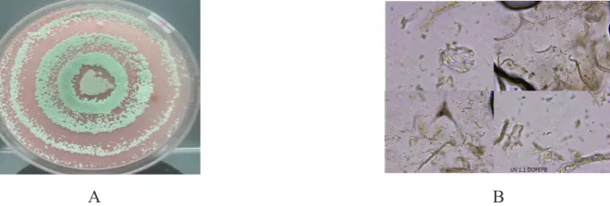
Each mutant tested showed different manner to grow depending on the which substrate being utilized. Mutant UV-1.1 on Whatman paper filter covered all paper surface after three days of incubation and became thick with green colour after five days (Fig. 1.A). On the agar containing CMC, all the 9 cm diameter Petri dish had been covered by the hyphae of colony after four days, where on cellulose powder the flask were entirely covered by hyphae after six days. Mineral agar containing DOPEFB powder showed the highest growth, where the Petri surface was covered in three days by thick colony of hyphae, meanwhile on mineral agar containing Avicel was similar as on cellulose powder. This mutant grew well on CMC and covered the medium surface after five days, where on α-cellulose powder was after six days. The mutant UV-1.2 on the Whatman showed almost the same profile as UV-1.1 but less dense (Fig.1.B), on CMC grew poorly, reached only in average 30 mm diameter of colony, where on cellulose powder in six days, on the DOPEFB powder the colony had covered the medium surface after seven days, and on Avicel at least seven days incubation reached only average 35 mm of colony diameter. The mutant 1-3 had covered Whatman paper almost paper surface
with thin layer of hyphae (Fig.1.C). Meanwhile, the other 31 mutants tested showed no significant differences with wild type T065. The control, wild
type Trichoderma sp. T065 on Whatman paper
showed only about 8 mm of colony diameter after four days of incubation and little augmentation to be 11 mm after seven days (Fig.1.D), where on CMC reached about 17 mm, on cellulose powder 25 mm, on the DOPEFB powder 30 mm and on Avicel was 37 mm, respectively.
According to Lejuene et al. (1995),
Trichoderma cultured on certain medium, its colony morphology is dependent on the kinds of the substrate given. On the media with limited nutrition, the colony seems to be transparent, and when the nutrition is added, the colony becomes too white. Conidiae could be formed in one week and the colour would be yellow, green or whitish.
Steyaert et al. (2010) suggest that there are
considerable and expected difference between
Trichoderma isolates in their conidiation and growth response to pH. The colony of all mutants tested showed the nearly similar growth profile, having the shape of piled rings up, where the perfect circular ring forms crowded around the center point of colony. Generally the colonies were well-done circular form with several rings layered surrounding in different colours, the center point was grey, the middle rings were green and the outer ring were white. The mutant UV-1.1 showed the fastest growth among the strains tested, followed by mutant UV-1.2 and mutant 1-3.
Growth Characterization of Mutants Selected
The screening medium was found to give fairly readible indication of elevated cellulolytic activities.
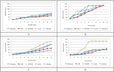
A B C D
The growth wild type Trichoderma sp. T065 on the five different substrates, Whatman filter paper, CMC, DOPEFB, Avicel and cellulose powder showed no significant different pattern. The best growth obtained on the cellulose powder, followed those on Avicel, then on Whatman filter paper and DOPEFB. The growth of colony on CMC in first day until eleven days was lowest. All mutants showed the rapid growth mainly on DOPEFB where the mutant 1-3 showed extra ordinary growth on that substrate while the colonies on other four substrates seem to be same speed of growth. The colony of mutant UV-1.1 grown quickly on DOPEFB where in four days has reached diameter 86 mm followed by the colony growing on cellulose powder in five days who gave the same diameter. But after seven days all of the Petri surface (135 mm of diameter) of medium containing DOPEFB has been covered by the colony of this mutant.
The colony growing on the CMC grew mostly slow among those growing on other substrates. The colony on Whatman paper had covered the Petri surface in seven days as well as the colony on DOPEFB. The colony on cellulose powder covered the Petri surface after 9 days of incubation. As well
as the mutant UV 1.1, the colony of mutant UV 1.2 on its growth seems to be fastest on DOPEFB than on the other substrate, which in this case the Petri surface of medium has been covered in 7 days of incubation. The colony on cellulose placed in third position after the coloy on the avicel who grown faster after eleven days of incubation. The colony on Whatman paper grown very slowly where after eleven days incubation reached only 41 mm of diameter, where CMC grown faster and reached diameter 64 mm. The growth of mutant 1-3 on five substrates was different with the other mutants and its wild type, where that mutant grown significantly fast on DOPEFB. Started from third day incubation within 17 mm of colony diameter, in five days has reached 69 mm and sixth day the colony diameter has already reached 127 mm and in seven days all Petri surface has been covered.
Mutant Colonies and Its Cellular Development in Five Different Carbon Sources
The wild type Trichoderma sp. T065 growth
on the Whatman filter paper, CMC, DOPEFB,
Whatman CMC DOPEFB Avicel Cellulose
0
Whatman 2 CMC 5 DOPEFB 6 Avicel 0 Cellulose 0
A B
Whatman CMC DOPEFB Avicel Cellulose
0
Whatman CMC DOPEFB Avicel Cellulose
C D
The colony was firstly observable after four days of incubation in the form of a whitist compact
perfect circular and fixed until elevendays of
incubation. Microscopically, on the Whatman paper appeared the somatic hyphae non-septate, straightly growing in the agar, branched in certain hyphae length area and stucked straightly on the fibre of paper and until eleven days of growth, conidiophore were not found. On CMC, the intensed mycelia were observed, but the granula of CMC were rarely found. The hyphae seems to wind arround the granule and the conidophore were also not found. Only on DOPEFB conidiae were formed. Non septate hyphae have many branch to wind arround the fibre of DOPEFB, in eleven days of growth the substrates have been almost entirely digested and the green oval conidiae found to be dispersed in the media. On Avicel, the hyphae started to penetrate the granules in six days of growth. The hyphae were slim like straightly thread without conidophore formation eventhough has passed eleven days of growth. On the α-cellulose, the non-septate hyphae found mostly in the fibre, but condiophore were not formed.
The mutant UV 1.1 on the Whatman filter paper, formed a grey colony at the center with green irregular circularon its surroundings. The colony form has been already observed after three days of incubation significantly within its diameter 45 mm with 55 mm lytic zone, and in seven days all Whatman disk surface of 135 mm has fully covered by the colony. On the CMC medium, showed the extraordinary growth, where since third day the circular campact colony appeared already within grey coloured of 19 mm of diameter with lytic zone of 24 mm. The colony continued to develop constantly in the grey
circular compact form surrounded by white small colonies guild. The surface of CMC medium was entirely covered by this mutant colony after eleven days of incubation. The mutant UV-1.1 also grown well on medium containing Avicel and cellulose powder as well as on DOPEFB where on both those substrates, it gave the similar profile of colony growth (Fig. 3A). The prominent colony was little and thin surrounded by a greenish dense ring in the outside. The colony has covered the 135 mm surface of cellulose medium in the nine days and the surface of Avicel in the eleven days. The mutant UV-1.1 growing on Whatman paper formed the conidiae in five days of growth. There were two type hyhae, non-septate hyphae and pseudohyphae forms. The conidiophore and phialides seems to be gloomy covered by the conidiae. On CMC, there were observed the hyphae non-spetate where conidiae sticked on hypha and lined up along, and also on the apical of hypha. On DOPEFB medium, the hypha of mutant UV-1.1 in the form of septate hyphae, and in eleven days the fibre of substrate has been entirely digested by this mutant strain, until the hyphae appeared with the debris. The oval form of conidia seems to disperse among the mycelia and the debris. On Avicel in eleven days of growth there were the debris of substrate granules within dispersed individually oval conidiae. The hyphae were straight non septate, stucked on the whole substrate. On the cellulose powder, the hyphae non septate to form gloomy conidiophore. Conidiae gathered 2 or 4 in a group on the apical of hypha. Some parts of hypha thread seems to form septate, whereon the apex of hypha there were a form of conidiae in composition, such an arrangement of conidiae, but on its arrangement the short hypha was formed and to form a troop of conidiae again.
A B
Figure 3. A. Colony Growth Profile of Mutant UV-1.1 on The Mandels Agar Containing 200 Mesh DOPEFB Powder B. Microscopic View of The Mutant Non-Septate Hyphae Degrading the Fibres by
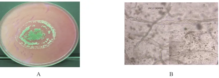
Meanwhile, the colony of mutant UV-1.2 on Whatman filter paper grown as well as its wild type T065, the colony appeared clearly colored slightly white. After eight days, the colony became a guild spreading of small colonies. Contrary, it grown slowly in the form of scattered irregular yellow greenish colony. The mutant UV-1.2 grew moderately speed on Avicel and cellulose powder where the colonies formed irregular shape in the prominent but the ring of colonies on the outside was rerely perfect circular. The mutant UV-1.2 growing on the Whatman paper in five days of growth showed the dense mass of hyphae outside of fibre. The conidiophore was hiden and non observable but some conidae were found. On CMC, the hyphae were slim and long, non septate. On the medium containing DOPEFB, after seven days of growth was observed the non-septate hyphae bandaged the fibre surrounded by some hidden piles, that would be conidiophore. The conidiae found to be dispersed in whole field sight of microscope. On Avicel after seven days of growth was observed long non-septate hyphae in the outside of substrate granules. It is probable that the hyphae penetrated into avicel granules and liberated the conidie from its surfaces. On α-cellulose fibre after seven days of growth was observed the long non-septate hyphae stucked straightly on the fibre where in certain location branched and penetrated into the fibre. The conidiophore and phialides were not clearly observed.
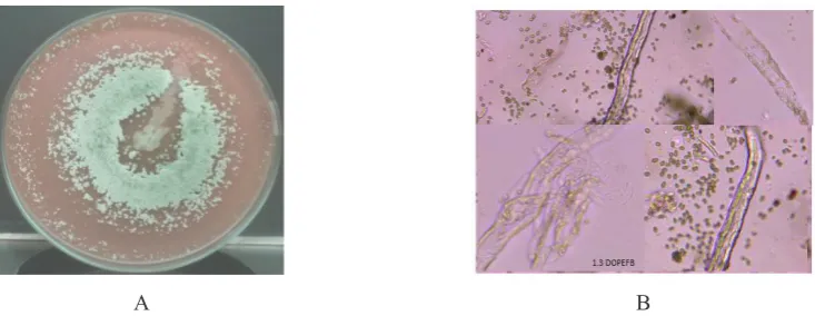
The mutant 1-3 on Whatman filter paper gave the appearance of visible colony afer five days with diameter 35 mm and halo zone of 45 mm. After eleven days of incubation, the form of colony became perfectly compact circular
within diameter 53 mm with 60 mm diameter of halo zone but covered by some yellowish white colonies around. It grown slowly on CMC medium in the form of perfect circular creamy colony with 62 mm of diameter. This mutants of showed the different hypha form with the other mutants and the wild type T065 and also varied which depend on substrate consumed. On the Whatman paper, we observed the presence of branched hyphae in the form non-septate and pseudohyphae bandaged the substrate fibre. In five days of growth the indifferently conidiophores and phialides were observed but none of conidiae. On the certain apical of hypha, an arrangement such as troop of conidiae were observed, but we suppose it would like a form of septate hyphae with clamp connections. We did not observe the dispersion of conidiae even though the growth has passed eight days, giving us the estimation that the conidiae stucked each other to form a troop at the tip of hyphae or were not liberated from phialides. On the CMC, the hyphae were varied between non-septate straight and branched septate hyphae. The hyphae bandaged the substrate granules and its banch penetrate into granule. The conidiophore, phialides and conidia were not observed. On DOPEFB, the hyphae found were varied from non septate to septate form with clamp connection in some portions. We observed the presence of conidiophore in five days of growth with small amount conidiae. The conidiae increased after eight days, dispersed around the substrate fibre that started to loose. On Avicel, the hyphae were straight, the septa have a distance each other, and in certain length branched in two like a fork which than bandaged the substrate granules.
A B
Figure 4. A. Colony Growth Profile of Mutant UV-1.2 on The Mandels Agar Containing 200 Mesh DOPEFB Powder B. Microscopic View of The Mutant UV-1.2 Where The Hyphae Seems to Wind
Until eight days of growth no conidia was found. On the cellulose, almost like as on Avicel, the non septate hyphae straight which then bandaged the substrate fibre and the branch penetrae into fibre. Until eight days of growth the conidiophore and the conidiae were not observed.
The ability of filamentous fungi to secrete large amounts of proteins has motivated their extensive use for the production of industrial enzymes. However, the morphology, mechanisms of cell growth, and product formation are not well known. Fungal fermentation is widely recognized as a complex process with several problems, some of which are related to the diversity of fungal morphologies under cultivation (Cui et al., 1998). However, there is a limited knowledgement on the effect of the culture conditions on the morphology of T. reesei
and the possible relations between morphology,
growth and enzyme production (Lejuene et
al., 1995). The fungal morphology determines
the medium rheology and cell growth as well. For most fungal fermentations, productivity is dependent on an optimal morphology. To achieve and control the optimal morphology, the relation between operational conditions and fungal
morphology must be known (Cui et al., 1998).
The synergism between T. reesei RUT-C30 and A. niger NL02 and the addition of surfactants were enhanced cellulolytic enzyme production (Fang
et al., 2013). It is believed that these surfactants can increase the enzyme transport across cell
membrane (Pardo, 1996; Domingues, et al.,
2000). Acording to Domingues et al. (2000),
high inoculum spore leads to high mycelium,
low concentration inoculum produce pellet. Meanwhile, low inoculum size, pellet growth is dominant and the majority of pellets were large. Increase inoculum, concentration pellet growth became prevalent. The use Tween 80 inhibited the pellet formation and the condition that induce pellet formation decrease protein concetration and filter paper activity. In the medium without Tween 80, at low inoculum size, the majority of the pellet were large and well individualized, in contrast, at higher inoculation densities small flocs were obtained with higher production of soluble protein and higher filter paper activity. The presence of Tween 80 in fermentation medium inhibited the pellet formation of the strain of Trichoderma.
Cellulolytic Activity After Mutation
The capacity of the mutant to degrade the paper seems to be augmented by comparing its wild type. The result work showed the mutant UV-1.2 has given the lowest activity amount the other mutants and not far from its wild type, where UV-1.2 gave 0.048 FPU/mL and wild type T065 was 0.038 FPU/mL. The mutant UV-1.1 give highest activity, 0.648 FPU/mL followed with mutant 1-3 giving activity 0.573 FPU/mL both were far higher than wild type and mutant UV-1.2. The capacity of the mutant to degrade the paper seems to be augmented by comparing its wild type. Mutagenesis on T.
reesei PTCC 5142 was induced by gamma and UV radiation to improve cellulase production (Shahbazi et al., 2014).
A B
Figure 5. A. Colony Growth Profile of Mutant 1-3 on The Mandels Agar Containing 200 Mesh DOPEFB Powder B. Microscopic View of The Mutant 1.3 Where The Hyphae Seems to Wind Around
On the other side, cellulase production in T. reesei was increased by a the carbon catabolite repressor 1 by transformation of T. reesei QM6aΔtmus53 using the truncated creI
of Rut-C30 amplified with primers RG 186
and RG187 and Escherichia coli hph marker
gene amplified with primers RG188 and hph3′
fw using the plasmid pRLMex30 as template
(de Sousa et.al., 2014). Kovacs et al. (2008) denoted from his work that cellulosic activity of enzyme produced from a fungal strain is different depending on the carbon source contained in the fermentation media. The filter paper activity from mutants obtained with variation of cellulosic substrates ranging from 0.73 to 1.09 FPU/mL. Yan et al. (2012) reported that cellulase production using the mutant
T. reesei YC-108 increased the FPU activity nearly five times to 15.82 IU/mL. Meanwhile, the mutan NU-6 showed approximately 1.8 fold increase activity of FPase (4.7 U/mL) after the strain of T. reesei Rut C-30 was treatment with nitrosoguanidine followed by UV irradiation
(Jun et al., 2009). Even though the mutants
obtained in this work grown on solid medium of wheat bran was still giving rather lower value on filter paper activity compared with previous work, but optimization with different media could give better activity. The result of this work has shown that all three mutants can grow and degrade DOPEFB which could be interpreted that enzymes activity againts palm fruit waste would be greater than filter paper activity.
CONCLUSION
Three mutants yielded after UV-light
mutation of a local isolate Trichoderma sp.
T065, UV-1.1, UV-1.2 as well as 1-3 were selected as the best among 34 mutants giving lytic zone greater than its wild type, investigated from its signifcant growth particularly on the DOPEFB. The mutant UV-1.1 and mutant 1-3 showed relatively high value of filter paper unit, 0.648 FPU/mL and 0.573 FPU/mL, respectively higher than its wild type that is equal to 0.038 FPU/mL.
ACKNOWLEDGMENT
This work was supported by National Priority Project of Fiscal Year 2013.
REFERENCES
Arun, P., Suhas, V. B., Naveen, S., Ravishanka, H. N. (2014). Study on the synergistic action of cellulase systems from Trichoderma and
Aspergillus mutants on carboxy methyl cellulose. The Scietech J. 1(1), 25-28
Awad, G., Florence, M., Yannick, C., Lebrihi A. 2005. Characterization and regulation of new secondary metabolites from Aspergillus ochraceus M18 obtained by UV mutagenesis.
Can. J. Microbiol. 51, 59-67
Cui,Y.Q., Ouwenland, J.N.W., Van Der Lans, R.G.J.M., Giuseppin, M.L.F., Luyben, K.C.A.M. 1998. Aspects of the use of complex media for submerged fermentation of Aspergillus awamori. Enzyme Microb. Technol. 23, 168 –77
De Olivera, M.M.Q., Grigorevski-Lima, A.L.G. Fanco-Cirigliano, M.N., do Nascimento, R.P., da Silva Bon E.P., Coelho, R.R.R. 2014.
Trichoderma atroviride 102C1 mutant: A high endoxylanase producer for assisting
lignocellulosic material degradation. J.
Microb. Biochem. Technol. 6(5), 236-241 De Sousa, T.M.M., Gorsche, R., Rassinger, A.,
Pocas- Fonceca, M.J., Mach R.L., Mach-Aigner, A.R. 2014. A Truncated form of the carbon catabolite repressor 1 increases cellulase production in Trichoderma reesei.
Biotechnol. for Biofuels 7:129, 1-12
Domingues, F.C, Queiroz, J.A., Cabral, J.M.S., Fonseca, L.P. 2000. The influence of culture conditions on mycelial structure and cellulase production by Trichoderma reesei Rut C-30
Enzyme Microb. Technol. 26, 394–401 Estubauer, H., Steiner, W., Labudova, I., Hermann,
A., Hayn, M. 1991. Production of cellulase in laboratory and pilot scale. Biores. Technol.
36, 51-65.
Fahrurrozi, Ratnakomala, S., Anindyawati, T., Lisdiyanti, P. Sukara, E. 2010. Rapid assesment of diverse Trichodermal isolatea of Indonesian origin for cellulase production.
Ann. Bogorienses 14 (1), 39-44
Fang, H., Zhao, C., Song, X-Y., Chen, M., Chang, Z., Chu, J. 2013. Enhanced cellulolytic enzyme production by synergism between
Trichoderma reesei RUT-C30 and Aspergillus niger NL02 and by the addition of surfactans.
Biotechnol. and Bioprocess Eng. 18, 390-398 FAOSTAT, 2006. Statistical data base. http://
faostat.fao.org
Gresik, M., Kolarovaand, M., Farkas, U. 1991. Hyperpolarization and intracellular
acidification in Trichoderma viridae as a
Gusakov, A.V. 2011. Alternatives to Trichoderma reesei in biofuel production. Trends in Biotechnol. 29 (9), 419-425.
Jun, H., Bing, Y., Keying, Z., Xuemei, D., Daiwen, C. 2009. Strain improvement of
Trichoderma reesei Rut C-30 for increased cellulase production. Indian J. Microbiol. 49, 188-195
Kovacs, K., Megyeri, L., Szakacs, G., Kubicek, C.P., Galbe, M., Zacchi, G.
2008. Trichoderma atroviridae mutant
with enhanced production of cellulae and
β-glucosidase. Enzyme andMicrob. Technol.
43, 48-55
Lejuene, R., Nielsen, J., Baron, G. 1995.
Morphology of Trichoderma reesei QM
9414 insubmerged cultures. Biotechnol.
Bioeng. 47, 609-615.
Li, X-H., Yang, H-J., Roy, B., Park, E.Y., Jiang, L-J., Wang, D., Miao, Y-G. 2010. Enhanced
cellulase production of the Trichoderma
viridae mutated by microwave and ultraviolet. Microbiol. Res. 16 (3), 190-198. Lynd, L.R. 2002. Microbial cellulose utilization:
fundamentals and biotechnology. Microbiol. Molec. Biol. Rev. 66, 506-577.
Mandels, M., Andreotti, R., Roche, C. 1976. Measurement of saccharifying cellulase.
Biotech. Bioeng. Symp. 6, 21-23.
Merino, S.T., Cherry, J. 2007. Progress and challenges in enzyme development for
biomass utilization. Adv. Biochem. Eng.
Biotechnol. 108, 95-120.
Pardo, A.G. 1996. Effect of surfactants on cellulase production by Nectria catalinensis.
Curr. Microbiol. 33(4), 275–8.
Peterson, R., Nevalainen, H. 2012. Trichoderma reesei RUT-C30 - thirty years of strain improvement. Microbiology 158, 58-68. Shahbazi, S., Ispareh, K., Karimi, M., Askari,
H., Ebrahimi, M.A. 2014. Gamma and UV radiation induced mutagenesisi in
Trichoderma reesei to enhance celulases enzyme activity. Int’l J. of Fam. and Alli Sci. 3(5), 543-554.
Singhania, R.R., Sukumaran, R.K., Patel, A.K., Larroche, C. Pandey, A. 2010. Advancement and comparative profiles in the production technologies using solid-state and submerged fermentation for microbial
cellulases. Enzyme Microb. Technol. 46,
541-549.
Steyaert, J.M., Weld, R.J., Stewart, A. 2010. Ambient pH intrinsically influences
Trichoderma conidiation and colony morphology. Fungal Biology 114, 193-208. Suto, M., Tomita, F. 2001. Induction and
catabolite repression mechanisms of cellulase in fungi. J. Biosci. Bioeng. 92 (4), 305-311.
Yan, Z-L., Cao, X-H., Liu, Q-D., Yang, Z-Y., Teng, Y-O., Zhao, J. 2012. A Shortcut to the optimization of cellulase production using
the mutant Trichoderma reesei YC-108.