responsibility of every practitioner to evaluate the appropriateness of a particular opinion in in the context of actual clinical situations and with due considerations to new developments. The author, editors, and the publisher cannot be held responsible for any typographical or other errors found in this book.
ISBN: 0-306-48675-X eISBN: 0-306-48676-8
C
2005 by Kluwer Academic/Plenum Publishers, New York 233 Spring Street, New York, New York 10013 CopyrightC 1998, 2000 by ICMSF
(Formerly published by Chapman & Hall, ISBN 0-7514-0430-6) http://www.wkap.nl 10 9 8 7 6 5 4 3 2 1 A C.I.P. record for this book is available from the Library of Congress. All rights reserved No part of this work may be reproduced, stored in a retrieval system, or transmitted in any form or by any means, electronic, mechanical, photocopying, microfilming, recording, or otherwise, without written permission from the Publisher, with the exception of any material supplied specifically for the purpose of being entered and executed on a computer system, for exclusive use by the purchaser of the work. Permissions for books published in Europe:[email protected]
Permissions for books published in the United States of America:[email protected]
2 Poultry products 107 VI Minced fish and surimi products 214 VII Cooked crustaceae (frozen or XI Fully dried or salted products 231
VIII Fermented and acidified fruits 348 II Dry soup and gravy mixes 372
A Definitions 372
B Initial microflora 372 C Primary processing 373 D Control (dry soup and gravy mixes) 374
III Soy sauces 374
IV Fish and shrimp sauces and pastes 382
A Definitions 382 VIII Breakfast cereals and snack foods 423
D Pathogens 536 C Primary processing of raw water 576
D Pathogens 580 D Control (process or product water) 585
III Raw milk for direct consumption 650 A Effects of handling of raw milk
on microorganisms 650
The second edition ofMicrobiology of Foods 6: Microbial Ecology of Food Commoditieswas written by the ICMSF, comprising 16 scientists from 11 countries, plus consultants and other contributors to chapters.
The intention of the second edition was to bring the first edition (published in 1996) up to date, taking into account developments in food processing and packaging, new products, and recognition of new pathogens and their control acquired since the first edition.
The overall structure of the chapters has been retained, viz each covers (i) the important properties of the food commodity that affect its microbial content and ecology, (ii) the initial microflora at slaughter or harvest, (iii) the effects of harvesting, transportation, processing, and storage on the microbial content, and (iv) an assessment of the hazards and risks of the food commodities and (v) the processes applied to control the microbial load.
In 1980s, control of food safety was largely by inspection and compliance with hygiene regulations, together with end-product testing.Microorganisms in Foods 2: Sampling for Microbiological Analysis: Principles and Specific Applications (2nd ed. 1986)put such testing on a sounder statistical basis through sampling plans, which remain useful when there is no information on the conditions under which a food has been produced or processed, e.g. at port-of-entry. At an early stage, the Commission recognized that no sampling plan can ensure the absence of a pathogen in food. Testing foods at ports of entry, or elsewhere in the food chain, cannot guarantee food safety.
This led the Commission to explore the potential value of HACCP for enhancing food safety, partic-ularly in developing countries.Microorganisms in Foods 4: Application of the Hazard Analysis Critical Control Point (HACCP) System to Ensure Microbiological Safety and Quality (1988)illustrated the procedures used to identify the microbiological hazards in a practice or a process, to identify the crit-ical control points at which those hazards could be controlled, and to establish systems by which the effectiveness of control could be monitored. Recommendations are given for the application of HACCP from production/harvest to consumption, together with examples of how HACCP can be applied at each step in the food chain.
Effective implementation of HACCP requires knowledge of the hazardous microorganisms and their response to conditions in foods (e.g. pH,aw, temperature, preservatives). The Commission concluded that such information was not collected together in a form that could be assessed easily by food industry personnel in quality assurance, technical support, research and development, and by those in food inspection at local, state, regional or national levels.Microorganisms in Foods 5: Characteristics of Microbial Pathogens (1996)is a thorough, but concise, review of the literature on growth, survival, and death responses of foodborne pathogens. It is intended as a quick reference manual to assist making judgements on the growth, survival, or death of pathogens in support of HACCP plans and to improve food safety.
The second edition ofMicroorganisms in Foods 6: Microbial Ecology of Food Commodities (2004)
The second edition ofMicroorganisms in Foods 6: Microbial Ecology of Food Commoditieshas been written followingMicroorganisms in Foods 7: Microbiological Testing in Food Safety Management (2002). The latter illustrates how systems such as HACCP and GHP provide greater assurance of safety than microbiological testing, but also identifies circumstances where microbiological testing still plays a useful role in systems to manage food safety. It continues to address the Commission’s objectives to: (a) assemble, correlate, and evaluate evidence about the microbiological safety and quality of foods; (b) consider whether microbiological criteria would improve and assure the microbiological safety of particular foods; (c) propose, where appropriate, such criteria; (d) recommend methods of sampling and examination; (e) give guidance on appraising and controlling the microbiological safety of foods. It introduces the reader to a structured approach for managing food safety, including sampling and microbiological testing. The text outlines how to meet specific food safety goals for a food or process using Good Hygienic Practice (GHP) and the HACCP system. Control measures as used in GHP and HACCP are structured into three categories: those that influence the initial level of the hazard, those that cause reduction, and those that may prevent increase, i.e. during processing and storage. In
Microorganisms in Foods 6, a control section following each commodity group uses this structured approach.
Editorial committee
T. A. Roberts (Joint Chairman) J. I. Pitt (Joint Chairman)
J.-L. Cordier L. G. M. Gorris
L. Gram K. M. J. Swanson
R. B. Tompkin
ICMSF Members during preparation of the second edition ofMicrobiology of Foods 6: Microbial Ecology of Food Commodities
Chairman M. B. Cole
Secretary M. van Schothorst (retired 2003) L. Gram (from 2003)
Treasurer J. M. Farber
Members R. L. Buchanan J.-L. Cordier
S. Dahms R. S. Flowers
B. D. G. M. Franco L. G. M. Gorris
J.-L. Jouve F. Kasuga
A. M. Lammerding Z. Merican
J. I. Pitt (to 2002) M. Potter
K. M. J. Swanson P. Teufel
R. B. Tompkin (to 2002)
Consultants J. Braeunig (2000) M. Germini (2003)
L. G. M. Gorris (2000) F. Kasuga (2002–03)
H. Kruse (2000) X. Lui (2003)
J. I. Pitt (2003) M. Potter (2002–03)
Contributors and reviewers
Chapter Contributors Reviewers
1 Meat J. Greig (Can)
T. Nesbakken (Norway) —
R. Stephan (Switz)
2 Poultry F. Kasuga (Japan) J. E. L. Corry (UK)
T. Humphrey (UK)
3 Fish F. Kasuga (Japan) Q. L. Yeoh (Malaysia)
4 Feeds — B. Veldman (Neth)
F. Driehuis (Neth) C. Jakobsen (Den)
5 Vegetables M. L. Tortorello (USA) M. Kundura (USA)
6 Fruit — —
7 Spices — —
8 Cereals T. Smith (USA)
S. Hood (USA)
9 Nuts — —
10 Cocoa — —
11 Oils & fats — R. van Santen (Neth)
G. Naaktgeboren (Neth)
12 Sugar L. Eyde (Aus)
13 Soft drinks C. Stewart (Aus)
K. Deibel (USA) —
14 Water — —
15 Eggs R. Buchner (USA) J. E. L. Corry (UK)
T. Humphrey (UK)
16 Milk J. Braunig (Ger)
P. Hall (USA) 17 Fermented beverages A. Lillie (Den)
P. Sigsgaard (Den) —
I Introduction
Red meat is derived from a number of animal species (e.g. cattle, sheep, goat, camel, deer, buffalo, horse, and pig). Total world production of red meats and quantities in international trade can be obtained from http://apps.fao.org/page/collections?subset=agriculture, a part of http://www.fao.org.
Red meat has the potential to carry pathogenic organisms to consumers. In the past, the main public health problem was caused by the classical zoonoses, i.e. diseases or pathogens that can be transmitted from animals to human beings, such as bovine tuberculosis, and also produce pathological changes in animals. However, the measures introduced by classical meat inspection (inspection, palpation, and incision) have proved highly effective against them. Thus, tuberculosis shows very typical changes of the lymph nodes (granulomatous lymphadenitis); they can be reliably detected by incision of the nodes during meat inspection. However, today, the main problem is latent zoonoses. These pathogens occur as a reservoir in healthy animals, where they produce no pathological conditions or changes. However, they can contaminate the food chain in meat production, for instance during slaughtering. The slogan “healthy animals, healthy food” is not true from this point of view. Strict maintenance of good practices of slaughter hygiene in meat production is of central importance, because microbiological hazards are not eliminated in the slaughtering process. Bacteria able to cause food-borne disease, and which can constitute a hazard in at least some meat products, includeSalmonellaspp., thermophilicCampylobacter
spp., enterohemorrhagicEscherichia coli (e.g. serogroup O157; EHEC), some serovars ofYersinia enterocolitica, Listeria monocytogenes, Clostridium perfringens, Staphylococcus aureus, Cl. botulinum,
andBacillus cereus. Meats are also subject to microbial spoilage by a range of microorganisms including
Pseudomonasspp.,Shewanella, Enterobacteriaceae,Brochothrix thermosphacta, lactic acid bacteria (LAB), psychrotrophic clostridia, yeasts, and molds.
In recent years, bovine spongiform encephalopathy (BSE) (“mad cow disease”) has attracted public health attention. The first cases of BSE were reported in Great Britain in November 1986. It appears probable that the disease can be transmitted to humans by food. The prions that cause the disease are very resistant to chemical and physical influences, i.e. to heat, UV, and ionizing radiations and disinfectants. Prions are sensitive to certain alkaline substances and moist heat under high pressure. An effective disinfectant measure is steam sterilization at 133◦C and 3 bar pressure for 20 min. On the basis of current knowledge, the cause of the BSE epidemic was animal feed (meat- and bone-meal and the like) containing brain, eyes or spinal cord of infected animals, and other tissues that had been inadequately heated during the production process.
To protect human health, the use of certain bovine organs (so-called specified risk materials: brain, eyes, spinal cord, spleen, thymus (sweetbread), bovine intestines of cattle>6 months old, visible lymph and nerve tissue, as well as lymph nodes) is prohibited for manufacturing foodstuffs, gelatine, tallow, drugs or cosmetics. More information and actual data can be obtained from the following web-sites: http://www.oie.int/eng/en index.htm; http://www.who.int/mediacentre/factsheets/fs113/en/;
http://www.defra.gov.uk/animalh/bse/index.html; http://www.aphis.usda.gov/oa/bse/; http://www.tseandfoodsafety.org/; http://www.unizh.ch/pathol/neuropathologie/.
during processing, storage, and distribution. It also contains sections on the microbiology of froglegs and snails as foods.
A Definitions
Red meat is primarily the voluntary striated skeletal muscular tissue of “red” meat animals. The muscle is made up of contractile myofibrillar proteins, soluble sarcoplasmic proteins (e.g. glycolytic enzymes and myoglobin) and low molecular weight soluble organic and inorganic compounds. Connective tissue is in intimate association with muscle cells and can constitute up to 30% of total muscle protein. Fat cells occur subcutaneously and both within and surrounding the muscle. Within a muscle, fat cells are located in the perimysial space. Up to one-third of the weight of some muscles may be fat. Muscle tissues also contain 0.5–1% phospholipid.
Meat as legally defined commonly includes various organs (“variety meats” or “offals”). The organs and other parts of the carcass that are regarded as edible vary between countries. The heart has some similarities to skeletal muscle and is composed of striated involuntary muscle, connective tissue, and some lipid. The liver contains uniform liver cells with a network of blood vessels and epithelial-lined sinusoids. In the kidney, there is a meshwork of connective tissue that supports renal tubules, small veins, and arteries.
B Important properties
Meat has a high water and protein content, is low in carbohydrates and contains a number of low molec-ular weight soluble constituents (Table 1.1). The vitamin content (µg/g) of muscle is approximately: thiamine, 1; riboflavin, 2; niacin, 45; folic acid, 0.3; pantothenic acid, 10; B6, 3; B12, 0.02 and biotin, 0.04 (Schweigert, 1987). The concentrations of vitamins vary with species, age, and muscle. Pork mus-cle has 5–10 times more thiamine than is found in beef or sheep musmus-cle. Vitamins tend to be higher in organs (e.g. liver and kidney) than in muscle.
Meat is a nutritious substrate with anaw(0.99) suitable for the growth of most microorganisms. Growth is primarily at the expense of low molecular weight materials (carbohydrates, lactate, and amino acids). Microbial proteolysis of structural proteins occurs at a very late stage of spoilage (Dainty
et al., 1975).
Table 1.1 Approximate composition of adult mammalian muscle after
rigor mortis
Component % Wet weight
Water 75
Protein 19
Lipid 2.5
Glycogena 0.1
Glucosea,band glycolytic intermediatesa 0.2
Lactic acida 0.9
Inosine monophosphateb 0.3
Creatineb 0.6
Amino acidsb 0.35
Dipeptides (carnosine and anserine)b 0.35
pHa (5.5)
Lawrie (1985).
During death of the animal when the oxygen supply to the muscle is cut off, anaerobic glycolysis of stored glycogen to lactic acid lowers the pH. Post-mortem glycolysis continues as long as glycogen is available or until a pH is reached which inhibits the glycolytic enzymes. In typical muscles this pH is 5.4–5.5. In some muscles (e.g. beefsternocephalicusmuscle), glycolysis ceases at a pH near 6 even though considerable glycogen remains. The ultimate pH varies between muscles of the same animal and between animals, and is determined by the glycogen content of the muscle and the accessibility of glycogen to glycolysis. The pH of post-rigor muscle can vary from 5.4–5.5 (lactate content close to 1%) to 7.0 (very little lactate present). The lactate content of muscle is inversely proportional to its pH. On the surfaces of beef and sheep carcasses, the availability of oxygen permits aerobic metabolism to continue, and much of the exposed surface tissue has a pH>6 (Carse and Locker, 1974), which facilitates microbial growth.
In the live animal, the glycogen concentration of muscle averages 1%, but varies considerably. Glycogen in pig muscle is readily depleted by starvation and moderate exercise, whereas glycogen in the muscles of cattle is more resistant to starvation and exercise. In both species, pre-slaughter stress (e.g. excitement and cold) depletes muscle glycogen. Glycogen is more concentrated in liver (2–10%) than in muscle, and its content is also affected by pre-slaughter conditions. A low concentration of glycogen in muscles results in a high ultimate pH, which gives rise to “dark-cutting” beef or dark, firm and dry meat (DFD).
The amount of glucose in post-rigor muscle varies with pH (Newton and Gill, 1978) being virtually absent in muscle of pH>6.4. In normal-pH (5.5–5.8) muscle, glucose is present at about 100–400µg/g (Gill, 1976). Liver has a high glucose content (3–6 mg/g), which appears to be independent of pH (Gill, 1988).
By the time the ultimate pH is reached, adenosine triphosphate has largely broken down to inosine monophosphate (IMP). During the storage of meat, IMP and inosine continue to degrade to hypox-anthine, ribose, and ribose phosphate. Ribose, inosine, and IMP can be used as energy sources by a number of fermentative Gram-negative bacteria, and ribose byBroch. thermosphacta,and a number of lactic acid bacteria.
Fatty tissue contains less water than muscle, has a pH near neutrality with little lactate, and contains low molecular weight components (glucose and amino acids) from serum (Gill, 1986). Consequently, microbial growth on fat is slower than on the surface of muscle.
C Methods of processing and preservation
Animals are raised on farms where some are grazed and some are raised under intensive or almost industrial conditions. The microflora in the intestinal tract or on the external surfaces of the animals may vary with the systems of animal production (e.g. more fecal material on the hides of feed-lot cattle). Animals may be slaughtered when young (e.g. calves at 3–4 weeks of age), or when 1 or 2, or several, years old (e.g. cattle and sheep). At the abattoir, the skin of cattle and sheep is removed, the skin of pigs is usually scalded (although it is removed in some plants), then the intestinal tract and viscera are removed. The carcass may then be washed, where regulations permit it, or not, and then chilled.
Spoilage organisms grow rapidly on meat, which is a highly perishable commodity. Thus, trade in meat, even at the local level, depends on some degree of preservation that controls the spoilage flora.
D Types of meat products
Red meats are traded as chilled or frozen carcasses, large primal pieces or retail size portions, chilled or frozen offals, chilled vacuum-packed meat, dried meats, fermented meat, raw or cooked cured products, cooked uncured meat and cooked canned products.
II Initial microflora
A Ruminants
At birth, the digestive tract of a ruminant is physiologically that of a monogastric. The rumino-reticulum complex develops quickly between 2 and 6 weeks of age when the animals are fed roughage. Initially, large numbers ofE. coli, Cl. perfringensand streptococci are in the gut and are shed in feces (107– 108cfuCl. perfringens/g, 109cfuE. coli/g). After about 2 weeks, Cl. perfringens declines to about 104cfu/g and E. colito ca. 106cfu/g at about 3 months of age. When comparing fecal excretion of coliforms, the mean count for eight calves between 3 and 8 weeks of age was log107.2 cfu/g and for adult cows was log104.9 cfu/g (Howeet al., 1976).
Invasive serotypes of salmonellae, such asSalmonellaTyphimurium and S. Enteritidis, are more difficult to control in the live animal than serovars occasionally found in feed. In the first few days of life, young ruminants are more susceptible to salmonellae. Calves dosed withS.Typhimurium prior to 3 days of age were more easily infected, and excreted salmonellae for longer periods and in greater numbers, than calves inoculated at 18 days (Robinson and Loken, 1968). At slaughter, salmonellae were also detected more frequently in mesenteric and cecal lymph nodes from the younger animals. Young calves that are surplus to dairy farm requirements may be sold through markets and dealers to rearing farms. In England, salmonellae have been found in 3.7% of environmental samples taken at calf markets and in 20.6% of swab samples from vehicles used to transport calves (Wrayet al., 1991). Salmonellae have also been detected on the walls (7.6% of swabs) and floors (5.3% of swabs) at dealers’ premises (Wrayet al., 1990). The mixing of young susceptible calves and their subsequent transport to rearing farms disseminates salmonellae. On arrival at rearing farms, the prevalence of salmonellae in calf feces is relatively low but can increase rapidly. When fecal samples were taken from 437 calves within 2 days of arrival at a rearing farm, salmonellae were detected in 5.3% (Hintonet al., 1983). After about 2 weeks on the farm, salmonellae were found in 42.2% of 491 animals sampled. The shedding rate of salmonellae peaked at 2–3 weeks and then declined; this is possibly associated with the development of a more adult-type intestinal flora.
The high concentration of volatile fatty acids and the pH of the fluid in the developed rumen of the well-fed animal provide some protection to infection with salmonellae and verotoxin-producingE. coli
(often of the serogroup O157; VTEC) (Chambers and Lysons, 1979; Mattilaet al., 1988). Viable cells of these organisms disappear from rumen fluid at a rate faster than expected from wash-out. Starved or intermittently fed ruminants are more susceptible to infection as salmonellae and VTEC O157 can then grow in the rumen. This probably influences the percentage of infected animals on farms during periods of low feed intake (e.g. drought, mustering, shearing or dipping and high stocking densities). On farms, the prevalence of salmonellae in the intestinal tract varies (Edel and Kampelmacher, 1971). Outbreaks of clinical bovine salmonellosis tend to show seasonal patterns. In the UK, most incidents of bovine salmonellosis occur in summer–autumn and peak near the end of the grazing season (Williams, 1975). Peaks of clinical salmonellosis in sheep in New Zealand during summer–autumn have been associated with movement and congregation of sheep for shearing and dipping.
fecal samples collected on 11.2% (21 of 187) of the operations. Overall 78 salmonellae representing 22 serotypes were isolated from 1.4% (70 of 5 049) of samples, and multiple serotypes from eight samples from a single operation. The five most common serotypes wereS.Oranienburg (21.8% of isolates) and
S.Cerro (21.8%), followed byS.Anatum (10.3%),S.Bredeney (9.0%) andS.Mbandaka (5.1%). Although it is broadly accepted that human salmonellosis is derived from foods, especially meat and poultry, firm proof is elusive. Sarwariet al. (2001) concluded from US data for 1990–1996, that there was a significant mismatch between the distribution ofSalmonellaspecies isolated from animals at the time of slaughter and that of isolates found in humans. This questions the validity of assumptions that raw animal products are the primary source for human salmonellosis, or whether there are methodological reasons for the difference.
The increased susceptibility to infection resulting from changes in the rumen can also affect the prevalence of salmonellae in cattle and sheep during transport from farm to slaughter, or in long transport from farm to farm when feeding patterns and type of feed are changed. Frostet al. (1988) reported a high prevalence of salmonellae in the mesenteric lymph nodes and rumen fluid of adult cattle during the first 18 days of entering a feed-lot from a market. After 80 days in the feed-lot, there was little evidence of salmonellae infection. Some of the deaths of sheep during sea-shipment from Australia to Singapore and the Middle East have been due to salmonellosis, which was associated with empty gastrointestinal tracts, loss of appetite and poor adjustment from grazing green pastures to dry feed.
Although healthy cattle may excrete thermophilic campylobacters in their feces, numbers are gen-erally low (NACMCF, 1995). While thermophilic campylobacters are frequently found in the lower intestinal tract of ruminants (prevalence range 0–54%), it is usually present in numbers<1000/g. The organism occurs more frequently and in higher numbers in the feces of very young calves (<3–4 weeks old). It can be present in small numbers (<100/g) in the rumen, where it is probably only part of the transient flora.
Streams, fields, wild-life and other livestock are all likely to be sources of salmonellae andC. jejuni. The opportunity for animal-to-animal spread is increased in intensively reared animals. Salmonellae contaminated feeds can be a source of infection. Joneset al. (1982) reported an infection of cattle on three dairy farms that was directly attributable to consumption of a vegetable fat supplement contaminated withS.Mbandaka.
L. monocytogenescan exist as a saprophyte in the plant–soil ecosystem, and clinical outbreaks of listeriosis in cattle and sheep have long been linked with feeding silage of inferior quality.L. monocy-togeneshas been reported in the feces of apparently normal cattle in many countries, whether animals were examined on the farm or at slaughter (Table 1.2). On Danish farms, where there was a high occur-rence of the organism in dairy cows, it was commonly found in the feed (silage from different crops, and alkalized straw). Silage and decaying vegetable material can contain large numbers ofListeriaspp. The higher incidence in Danish cattle than in Danish pigs has been associated with feeding wet plant material to cattle and providing dry feed to pigs (Skovgaard and Norrung, 1989).
VTEC is a group ofE. colithat produces one or more verocytotoxins (VT) also known as Shiga toxins (STX). This group of bacteria has many synonyms. In the United States and to a varying extent in Europe, the notation Shiga-toxin producingE. coli(STEC) is used. The term, EHEC was originally used to denote VTEC causing hemorrhagic colitis (HC) in humans; later EHEC has been used as a synonym for VTEC in the medical domain in some European countries (SCVPH, 2003).
VTEC are frequently present in the feces of calves, cattle, buffaloes, sheep and goats (Mohammad
et al., 1985; Suthienkulet al., 1990; Beutinet al., 1993; Clarkeet al., 1994). These VTEC strains belong to a large number of serotypes. Some (e.g. O5:NM, O8:H9, O26:H11 and O111:NM) may cause diarrhea or dysentery with attaching–effacing lesions in calves (Moxley and Francis, 1986; Schoonderwoerd
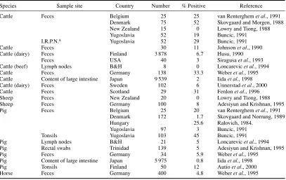
Table 1.2 Listeria monocytogenesin red-meat animals
Species Sample site Country Number % Positive Reference
Cattle Feces Belgium 25 25 van Renterghemet al., 1991
Denmark 75 52 Skovgaard and Morgen, 1988 New Zealand 15 0 Lowry and Tiong, 1988
Yugoslavia 52 19 Buncic, 1991
I.R.P.N.a Yugoslavia 52 29 Buncic, 1991
Cattle Feces 30 11 Johnsonet al., 1990
Cattle (dairy) Feces Finland 3 878 6.7 Husu, 1990
Feces USA 40 3 Siragusaet al., 1993
Cattle (beef) Lymph nodes B&H 8 0 Loncarevicet al., 1994
Cattle Feces Germany 138 33.3 Weberet al., 1995
Cattle Content of large intestine Japan 9 539 2 Iidaet al., 1998
Cattle (dairy) Feces Sweden 102 6 Unnerstadet al., 2000
Cattle Feces Scotland 29 31 Fenlonet al., 1996
Sheep Feces New Zealand 20 0 Lowry and Tiong, 1988
Sheep Feces Germany 100 8 Adesiyun and Krishnan, 1995
Pig Feces Belgium 25 20 van Renterghemet al., 1991
Denmark 172 1.7 Skovgaard and Norrung, 1989
Hungary 25.6 Ralovich, 1984.
Yugoslavia 97 3 Buncic, 1991
Tonsils Yugoslavia 103 45 Buncic, 1991
Pig Lymph nodes B&H 21 5 Loncarevicet al., 1994
Pig Rectal swabs Trinidad 139 5 Adesiyun and Krishnan, 1995
Pig Feces Germany 34 5.9 Weberet al., 1995
Pig Content of large intestine Japan 5 975 0.8 Iidaet al., 1998
Pig Tonsils Finland 50 12 Autioet al., 2000
Horse Feces Germany 400 4.8 Weberet al., 1995
aI.R.P.N., Internal retropharyngeal nodes.
infections and severe and potentially fatal illness in humans. VTEC are the cause of human gastroenteritis that may be complicated by hemorrhagic colitis (HC) or hemolytic-uremic syndrome (HUS).
VTEC strains causing human infections belong to a large, still increasing number of O:H serotypes. A review of the world literature on isolation of non-O157 VTEC (by K.A. Bettelheim) is available on the MicroBioNet website (http://www.sciencenet.com.au). Most outbreaks and sporadic cases of HC and HUS have been attributed to O157:H7 VTEC strains. However, especially in Europe, infections with non-O157 strains, such as O26:H11 or O26:H−, O91:H−, O103:H2, O111:H−, O113:H21, O117:H7, O118:H16, O121:H19, O128:H2 or O128:H−, O145:H−, and O146:H21 are frequently associated with severe illness in humans.
Pathogenicity of VTEC is associated with several virulence factors. The main factor is the ability to form different types of exotoxins (verotoxins). They can be subdivided into a Verotoxin 1 group (Stx1) and a Verotoxin 2 group (Stx2). Characterization of the stx1andstx2 genes revealed the existence of different variants in both Stx groups. At present, threestx1subtypes (stx1, stx1c,andstx1d) and severalstx2gene variants have been described (e.g.stx2, stx2c, stx2d, stx2eandstx2f). Apart from the capability to produce verotoxins, these pathogroups may possess accessory virulence factors such as intimin (eae), VTEC auto-agglutinating adhesin (saa) or enterohemolysin (ehxA). Characterization of
eaegenes revealed the existence of differenteaevariants. At present, 11 genetic variants of theeaegene have been identified and are designated with letters of the Greek alphabet. It is believed that different intimins may be responsible for different host- and tissue cell tropism.
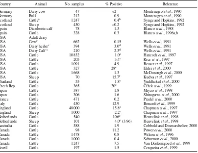
Table 1.3 Escherichia coliO157:H7 in the feces of cattle and sheep
Country Animal No. samples % Positive Reference
Germany Dairy cow 47 <2 Montenegroet al., 1990
Germany Bull 212 0.9 Montenegroet al., 1990
Scotland Cattlea 1 247 0.4b Synge and Hopkins, 1992
Scotland Sheep 450 <0.2 Synge and Hopkins, 1992
Spain Diarrhoeic calf 78 1.3 Blancoet al., 1988
Spain Cattle 328 0.3 Blancoet al., 1996a,b
USA Adult dairy
USA Cowc 662 0.15 Wellset al., 1991
USA Dairy heiferc 394 3.0d Wellset al., 1991
USA Dairy Calfc 210 2.3d Wellset al., 1991
USA Cattle 10 832 1.0e Hancocket al., 1997
USA Cattle 205 3.4e Riceet al., 1997
USA Cattle 1 091 4.9 Besseret al., 1997
USA Cattle 327 28e Elderet al., 2000
USA Cattle 1 668 1.3 McDonoughet al., 2000
USA Sheep 70 15.7f Kudvaet al., 1997
Thailand Cattle 55 1.8e Vuddhakulet al., 2000
Czech Rep Cattle 365 20e ˇC´ıˇzeket al., 1999
Japan Cattle 387 1.8 Miyaoet al., 1998
Japan Cattle 306 1.6 Shinagawaet al., 2000
France Cattle 471 0.2 Pradelet al., 2000
Italy Cattle 450 12.9 Bonardiet al., 1999
England Cattle 48 000 15.8e Chapmanet al., 1997
England Sheep 1 000 2.2e Chapmanet al., 1997
Netherlands Cattle 540 10.6e Heuvelinket al., 1998
Netherlands Sheep 101 4.0e(3.96) Heuvelinket al., 1998
Australia Cattle 588 1.9 Cobbold and Desmarchelier, 2000
Canada Cattle 98 11.2 Poweret al., 2000
Canada Cattle 1 478 0.8 Wilsonet al., 1996
Canada Cattle 1 000 0.4 Schurmanet al., 2000
Canada Cattle 1 247 7.5 Van Donkersgoedet al., 1999
Brazil Cattle 197 1.5 Cerqueiraet al., 1999
B&H, Bosnia and Herzogovina.
aFive calves positive.
bNot stated if H7 or non-motile (NM). cSome herds implicated in human illness.
dOne of 17 from the heifer-calf group wasE. coliO157:NM. eNot stated if H7.
fSeventy sheep where tested over a 16-month period and 11 tested positive at least one time. gHerd implicated in human illness.
E. coliO157:H7 appears to be transient, herd infection may be maintained (Wellset al., 1991; Zhao
et al., 1995; Faithet al., 1996). Drinking water may be a source of dissemination or maintenance of
E. coliO157 on farms (Faithet al., 1996). Growth ofE. coliO157:H7 in rumen fluid is restricted by the pH and volatile fatty acid concentration in well-fed animals, but is not when the animal is fasted for 24–48 h (Rasmussenet al., 1993). The impact of diet on fecal shedding ofE. coliO157:H7 remains unclear (Tkalcicet al., 2000).
Several outbreaks with life-threatening illness resulted in huge efforts to understand VTEC in rumi-nants (SCVPH, 2003).
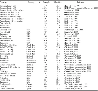
Table 1.4 Type of cattle and incidence ofEscherichia coliO157:H7 in feces
Cattle type Country No. of samples % Positive Reference
Unweaned dairy calf 649 <0.15 Hancocket al., 1994
Unweaned dairy calf 6 894 0.36 NDHEP survey quoted in Zhaoet al., 1995 Unweaned dairy calf<10 days 304 <0.3 Martinet al., 1994
Dairy calf<8 weeks olda 423 1.4 Garberet al., 1995
Dairy calf>8 weeks olda 518 4.8 Garberet al., 1995
Dairy calf 24 h to weaningb 570 1.9 Zhaoet al., 1995
Weaned dairy calf<4 monthsb 395 5.1 Faithet al., 1996
Weaned dairy calf<4 months 560 1.8 Faithet al., 1996
Cow and heiferc 193 4.7 Faithet al., 1996
Bull and steerc 51 3.9 Faithet al., 1996
Weaned calfc 273 2.9 Faithet al., 1996
Cull dairy cow USA 1 668 1.3 McDonoughet al., 2000
Feed-lot cattle USA 327 28 Elderet al., 2000
Dairy cows USA 1 091 4.9 Besseret al., 1997
Cull dairy cow USA 205 3.4d Riceet al., 1997
Weaned heifers USA 3 483 1.7d Hancocket al., 1997
Unweaned calves USA 1 040 1.3d Hancocket al., 1997
Dairy cows USA 4 762 0.4d Hancocket al., 1997
Bull calves 50–100 kg Czech Rep 163 5.5d ˇC´ıˇzeket al., 1999
Bulls 100–200 kg Czech Rep 47 59.6d ˇC´ıˇzeket al., 1999
Bulls 200–400 kg Czech Rep 36 44.4d ˇC´ıˇzeket al., 1999
Bulls 400–600 kg Czech Rep 71 22.5d ˇC´ıˇzeket al., 1999
Bulls slaughtered Czech Rep 48 6.2e ˇC´ıˇzeket al., 1999
Beef cattle England 1 840 13.4e Chapmanet al., 1997
Dairy cattle England 1 661 16.1e Chapmanet al., 1997
Adult cattle Netherlands 540 10.6e Heuvelinket al., 1998
Veal calves Netherlands 397 0.5e Heuvelinket al., 1998
Calves<7 days old Australia 79 1.3e Cobbold and Desmarchelier, 2000
Weanlings 1–14 weeks old Australia 109 5.5e Cobbold and Desmarchelier, 2000
Heifers Australia 106 2.8e Cobbold and Desmarchelier, 2000
Cattle type Australia 588 1.9 Cobbold and Desmarchelier, 2000 Dairy calf<4 month Brazil 64 3.1 Cerqueiraet al., 1999
Beef cattle>2 years Brazil 76 1.3 Cerqueiraet al., 1999
Veal calves Italy 90 0 Bonardiet al., 1999
Feedlot cattle Italy 223 16.6e Bonardiet al., 1999
Dairy cows Italy 137 16.1e Bonardiet al., 1999
Yearling cattle Canada 654 12.4 Van Donkersgoedet al., 1999
Cull cows Canada 593 2 Van Donkersgoedet al., 1999
Calves<4 months Spain 23 4.3 Blancoet al., 1996 a,b
aOne quarter of herds sampled were previously positive in the National Dairy Heifer Evaluation Project (NDHEP); three quarters
of herds sampled were previously negative forE. coliO157. Calves that were older than 8 weeks were up to 4 months old.
bFourteen herds sampled that were previously positive in NDHEP survey; fifty herds sampled that were previously negative for E. coliO157.
cFive herds sampled that were previously positive in Wisconsin survey; seven herds sampled that were previously negative for E. coliO157.
dResults from studies in the USA. eAt % positive=Not stated if H7.
taken at slaughter, higher prevalence of VTEC in cull cows, and highest VTEC O157:H7 prevalence in calves. A European Community report on trends of zoonoses for 2000 (EC, 2002) reported prevalence of VTEC O157 in cattle herds (10% or more), individual bovines (1–5% or more) and beef or minced meat (0–1%).
In Australia, VTEC prevalence among cattle reared specifically for beef production (6.7%) was lower than that in dairy cattle (14.6%). Seasonal variation in shedding results in most cattle being positive in late summer–early autumn (Chapmanet al., 1997; Hancocket al., 1997; De Zutteret al., 1999; Tutenelet al., 2002). Other animals carryingE. coliO157 include sheep, goats, wild deer, pigs, and seagulls (Synge, 1999; Chapman, 2000), feral pigeons (Dell’Omoet al. 1998), and zebu cattle (Kaddu-Mulindwaet al., 2001).
Laegreidet al. (1999) reported the prevalence ofE. coliO157:H7 in beef calves at weaning, prior to arrival at the feed-lot or mixing with cattle from other sources. Thirteen of 15 herds (87%) yielded one, or more than one, isolation ofE. coliO157:H7 in fecal samples. All herds had high prevalence of anti-O157 antibodies (63–100% of individuals within herds seropositive) indicatingE. coliO157:H7 occurrence before weaning and prior to entering feed-lots. Serological evidence suggested that most calves (83%) and all herds (100%) had been exposed toE. coliO157.
The site of colonization of EHEC has been identified as the lymphoid follicle-dense mucosa at the terminal rectum (Nayloret al., 2003).
Elderet al. (2000) estimated the frequency ofE. coliO157:H7 or O157:non-motile (EHEC O157) in feces and on hides within groups of cattle from single sources (lots) at meat processing plants. Of 29 lots sampled, 72% had at least one EHEC O157-positive fecal sample and 38% had positive hide samples. Overall, EHEC O157 prevalence in feces and on hides was 28% (91 of 327) and 11% (38 of 355), respectively.
Thranet al. (2001) suggested that screening fecal samples should not be limited toE. coliO157:H7, and that identification of STEC-positive cattle prior to slaughter should help to reduce the risk of beef contamination.
Other verotoxin-producing serotypes (e.g. O26:H11, O103:H2, O111:NM, O113:H21 and O157:NM) associated with human bloody diarrhea and HUS have also been isolated from sheep, calves, and cattle feces (Dornet al., 1989; Montenegroet al., 1990; Wellset al., 1991).
Cobbold and Desmarchelier (2000) examined 588 cattle fecal samples and 147 farm environmental samples from three dairy farms in southeast Queensland, Australia. STEC were isolated from 16.7% of cattle fecal samples and 4.1% of environmental samples: 10.2% serotyped as O26:H11 and 11.2% as O157:H7, with prevalences in the cattle samples of 1.7% and 1.9%. Prevalences for STEC and EHEC in dairy cattle feces were similar to those derived in surveys within the northern and southern hemispheres. Calves at weaning were identified as the cattle group most likely to be shedding STEC,E. coliO26 or
E. coliO157. Cattle, particularly 1–14-week-old weaning calves, appear to be the primary reservoir for STEC and EHEC on the dairy farm.
Identifying environmental sources ofE. coliO157:H7 in two feed-lots in southern Alberta, to identify management factors associated with the prevalence and transmission, Van Donkersgoedet al. (2001) isolatedE. coliO157:H7 in pre-slaughter pens of cattle from feces (0.8%), feedbunks (1.7%), water troughs (12%) and incoming water supplies (4.5%), but not from fresh total mixed rations. Fresh total mixed rations did not support the growth ofE. coliO157:H7.
Large populations of microorganisms are present on the hide and fleece and are composed of normal resident skin flora (e.g. micrococci, staphylococci and yeasts) and organisms, includingSalmonella
spp. andL. monocytogenes, derived from the environment (soil, pasture and feces). Staph. xylosus
in udders, teat canals, and milk, particularly when animals are mastitic. In colder climates, there is a greater proportion of psychrotrophic flora on the hide and fleece than in warmer climates. Growth on wet hide or fleece can change the numbers of some types of microorganisms. The amount of fecal material on the skin under feed-lot conditions can be large (several kg). Consequently, there is considerable variation in the microflora from animal-to-animal and site-to-site on the hide and fleece.
During transport of sheep and cattle from farm to abattoir, salmonellae, and other organisms shed in the feces (e.g.L. monocytogenesandE. coliO157:H7) will contaminate transport vehicles, markets, and abattoir holding areas. Although thermophilic campylobacters are a relatively fragile organism in the laboratory, they survive well in the environment.
The longer the animals are held before slaughter, the greater the salmonellae contamination of the outside of the animal, and the greater the prevalence of salmonellae in the intestinal tract. Andersonet al.
(1961) found that 0.5% of calves were infected at the market, 0.6% infected when held for only a few hours before slaughter, but 35.6% when the calves were held in lairage for 2–5 days before slaughter. The prevalence of salmonellae in cattle feces can be 10 times that on the farm (Galtonet al., 1954). When sheep awaiting slaughter were held for 7 days, the incidence and numbers of salmonellae on the fleece, in the rumen liquor and in feces increased with time of holding (Grau and Smith, 1974).
In a UK survey (Smallet al., 2002), prevalences ofE. coliO157,Salmonellaspp. andCampylobacter
spp. from swabs taken along the unloading-to-slaughter routes of animal movement in lairages of six commercial abattoirs, three for cattle and three for sheep, were 27.2, 6.1 and 1.1%, respectively, in cattle lairages, and 2.2, 1.1 and 5.6%, respectively, in sheep lairages. On cow hides, prevalences of the three pathogens were 28.8, 17.7 and 0%, respectively, and on sheep pelts 5.5, 7.8 and 0%.
Much of the contamination on the carcass is derived from the hide/fleece contaminated with gut contents. Contamination from the exterior of the animal can be reduced by not accepting for slaughter animals that are visibly dirty. Although this is difficult administratively, there is some evidence that it has helped to improve carcass hygiene, e.g. in the UK.
At the laboratory-scale, sub-atmospheric steam applied to bovine hide pieces inoculated withE. coliO157:H7 in fecal suspensions (McEvoyet al., 2001), effected some reduction in viable numbers, indicating that steam condensing at≤80±2◦C can reduceE. coliO157:H7 when it is present on bovine hide, and suggesting a possible means of reducing cross-contamination to the carcass during slaughter and dressing.
B Pigs
In young piglets, the initial microflora of the intestinal tract is composed principally of high populations ofE. coli, Cl. perfringensand streptococci (Smith, 1961). As the animal grows the numbers of these organisms decline, and non-sporing strict anaerobes become the predominant population in the lower intestine.
Young animals are more susceptible than older animals to infection with salmonellae. Clinical illness was formerly mostly caused by the host-adaptedS.Cholerae-suis, but control measures have significantly reduced the number of outbreaks due to this serotype to<5% of the salmonella isolations reported from pigs in the UK (Hunter and Izsak, 1990), whereasS.Typhimurium andS.Derby accounted for 40–50% of isolations.
tract of 23% of pigs that were fed a mash containing contaminated fish meal, but were found in<2% when the fish meal was not used (Leeet al., 1972). The form in which feed is presented affects the extent of salmonellae excretion. Excretion may be transient only when freshly prepared mash is fed (Lintonet al., 1970). However, the small numbers of salmonellae in the dry meal can grow in the mash during holding of bulk mash and in the residues in pipelines and troughs. This growth increases the rate of infection and the duration of excretion. Pelleting reduces the salmonellae contamination of meal with the extent of reduction dependent on the temperature achieved and the duration of exposure to high temperatures. In pigs raised on pelleted meal, salmonellae were detected in only 1 of 6 047 fecal samples, but were detected in almost all fecal samples after pigs were fed the same meal as a mash for 10 weeks (Edelet al., 1967). Reduced infection rates in pigs have been observed in a number of other studies where pelleted meal was used (Edelet al., 1970; Ghosh, 1972).
Elimination of salmonellae from feeds does not ensure the absence of salmonellae from pig-fattening farms. There are a number of other sources of salmonellae including pigs previously in the pens, birds, rats, and other animals. Breeding sows and boars may be infected. Movement of stock and animal attendants may spread salmonellae. Control of salmonellae contamination on the farm requires a multi-pronged approach (e.g. structural changes to the farm, restriction of movement of stock and personnel, disinfection of pens, feed pelleted at high temperature and by stocking with salmonellae-free animals) (Ghosh, 1972; Linton, 1979). It is very difficult to eradicate salmonellae from the environment of pigs in intensive piggeries (Oosterom and Notermans, 1983; Swanenburget al., 2001).
Nevertheless, in some countries, measures have essentially eradicated salmonellae from pigs and from pork. In five Swedish pig slaughterhouses, each visited six times, with a total of 3 388 samples from pork carcasses and the slaughterhouse environment all cultured negative for salmonellae (Thorberg and Engvall, 2001).
Because salmonellae can cause major economic losses to the swine industry, and the gut is a major reservoir forSalmonella, novel strategies to reduce their concentration in pigs immediately before processing have been explored. Respiratory nitrate reductase activity possessed by salmonellae catalyzes the intracellular reduction of chlorate to chlorite, which is lethal to salmonellae. Weaned pigs orally infected with 8×10⁷cfu of an antibiotic-resistant strain ofS.Typhimurium were treated 8 and 16 h later via oral gavage (10 mL) with 0 or 100 mM sodium chlorate. Chlorate treatment significantly reduced caecal concentrations of salmonellae, the greatest reductions occurring 16 h after receiving the last chlorate treatment, indicating a possible means of reducing numbers of salmonellae before slaughter (Andersonet al., 2001).
Attempts are also being made to identify bacteria that can be used as competitive exclusion cultures to prevent colonization byS.Typhimurium in pigs (Humeet al., 2001a).
Thermophilic Campylobacter spp. are found at a very high frequency (61–100%) in the lower intestinal tract of pigs, often at counts of 103–104cfu/g of feces (Teufel, 1982; Stern and Line, 2000). Presumably, animal-to-animal spread is the major mechanism for this widespread occurrence. The vast majority of strains isolated areC. coli.
Permanent colonization of the gut of neonatal pigs appears to be related to constant exposure of the piglets to feces containing campylobacters and is reduced by early removal of the piglets from the sows and rearing in nurseries isolated from sows (Harveyet al., 2000).Arcobacterspp. can also be isolated from nursing sows and developing pigs (Humeet al., 2001b).
occurring in tongue (14%) and tonsil samples (12%). Six of 50 (12%) carcasses were contaminated with
L. monocytogenes. In the slaughterhouse environment,L. monocytogeneswas detected on two saws, in one drain, on one door and one table. Carcasses were contaminated withL. monocytogenesin two slaughterhouses where mechanical saws used for both brisket and back-splitting also tested positive forL. monocytogenes. Pulsed-field gel electrophoresis typing indicated thatL. monocytogenesfrom the tongue and tonsils can contaminate the slaughtering equipment and in turn spread to carcasses.
Healthy pigs often carry serotypes of Y. enterocoliticathat appear indistinguishable from human pathogenic strains (Table 1.5). The isolation rates from the throat, tonsils and tongue are often higher than those from the cecum or feces (Schiemann, 1989). The carriage rate varies greatly between herds and in different geographic locations. In one survey in England, although non-pathogenic biotypes were frequently encountered, pathogenic strains were rarely found (Table 1.5). In Danish herds, 82% were shown to contain pigs carryingY. enterocolitica, and no association could be found between carriage rate of the organism and different types of herd management (Andersenet al., 1991).
In Norway, IgG antibodies againstY. enterocoliticaO:3 were found in sera from 869 (54.1%) of samples from 1 605 slaughter pigs from 321 different herds. In the positive herds, there were significantly fewer combined herds of piglets and fatteners than fattening herds.
In Denmark, Norway, Sweden, Holland, and Belgium, serotype O:3 is commonly found in pigs. Although serotype O:3 is common in pigs in Eastern Canada, it is rare in Western Canada where O:5,27 occurs in the swine population. In the United States, serotypes O:3, O:5,27, and O:8 have been detected on the tongues of pigs (Table 1.5). The appearance of strains of serovars O:3 and O:9 in Europe, Japan in the 1970s and in North America by the end of the 1980s, is an example of a global pandemic (Tauxe, 2002).
Colonization of pigs appears to be from animal contact rather than from environmental sources. Risk factors included: using a farm-owned-vehicle for transport of slaughter pigs to abattoirs and using straw bedding for slaughter pigs. Epidemiological data suggested that the herd prevalence ofY. enterocolitica
O:3 can be reduced by minimizing contact between infected and non-infected herds (Skjerveet al., 1998). Young pigs become carriers within 1–3 weeks of entering contaminated pens. Within a short time of infection, large numbers (106cfu/g) ofY. enterocolitica are excreted in the feces. This may continue for some weeks before the numbers fall to<100/g (Fukushimaet al., 1983).
Transport of pigs to slaughter appears to result in increased shedding of salmonellae in feces (Williams and Newell, 1967, 1970). Part of the explanation for this may be that the stress of transport increases the flow of material along the intestinal tract. Salmonellae in the cecum and colon can then more readily appear in the feces. However, real differences in salmonellae prevalence in feces have been observed between pigs killed on the farm and at abattoirs (Kampelmacheret al., 1963). Transport vehicles and lairages in which animals are held at abattoirs become contaminated. Cross-contamination of feet, skin and intestinal tract can then take place.
Holding pigs for long times in lairages at abattoirs has long been known to increase the prevalence of salmonellae in the intestinal tract (Tables 1.6 and 1.7). When pigs from one producer were killed at two abattoirs, salmonellae were isolated from 18.5% of pigs killed on the first day, 24.1% on the second day and 47.7% on the third day after leaving the farm (Morganet al., 1987b).
Y. enterocolitica can also be transferred between pigs and appear in low numbers in caecal con-tents when pigs are held for about 20 h in abattoir lairages (Fukushimaet al., 1991). The skin can be contaminated byYersiniaspp. excreted during transport and into the pens.
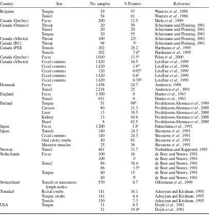
Table 1.5 Yersinia enterocoliticain pigs
Country Site No. samples % Positive Reference
Belgium Tongue 29 97 Wauterset al., 1988
Tonsil 54 61 Wauterset al., 1988
Canada (Quebec) Feces 200 12.5 Mafuet al., 1989
Canada (Ontario) Throat 20 50 Schiemann and Fleming, 1981
Tonsil 20 20 Schiemann and Fleming, 1981
Tongue 20 55 Schiemann and Fleming, 1981
Canada (Alberta) Throat 100 22a Schiemann and Fleming, 1981
Canada (BC) Throat 98 3a Schiemann and Fleming, 1981
Canada (PEI) Tonsils 202 28.2 Hariharanet al., 1995
Tonsils 202 7.4b Hariharanet al., 1995
Canada (Quebec) Feces 1 010 13.5c Pilonet al., 2000
Canada (Alberta) Cecal contents 1 420 16.5 Letellieret al., 1999 Cecal contents 1 420 1.8d Letellieret al., 1999
Cecal contents 120 0.07e Letellieret al., 1999
Cecal contents 1 420 0.6f Letellieret al., 1999
Cecal contents 1 420 0.35g Letellieret al., 1999
Denmark Feces 1 458 24.7 Andersen, 1988
Tonsil 2 218 25 Andersenet al., 1991
England Feces 1 300 0 Hunteret al., 1983
Tonsil 631 0 Hunteret al., 1983
Finland Tongue 51 98h Fredriksson-Ahomaaet al., 1999
Carcass 80 21.3 Fredriksson-Ahomaaet al., 2000
Liver 13 38.5 Fredriksson-Ahomaaet al., 2000
Kidney 13 84.6 Fredriksson-Ahomaaet al., 2000
Heart 8 62.5 Fredriksson-Ahomaaet al., 2000
Japan Feces 1 200 3.8i Fukushimaet al., 1991
Japan Tonsils 140 24.3 Shiozawaet al., 1991
Cecal contents 140 24.3 Shiozawaet al., 1991
Oral cavity swabs 40 85 Shiozawaet al., 1991
Masseter muscles 25 36 Shiozawaet al., 1991
Norway Tonsil 461 31.7 Nesbakken and Kapperud, 1985
Netherlands Feces 100 16 de Boer and Nouws, 1991
100 1j de Boer and Nouws, 1991
Tonsil 86 38.4 de Boer and Nouws, 1991
86 3.5a de Boer and Nouws, 1991
Tongue 40 15 de Boer and Nouws, 1991
40 5j de Boer and Nouws, 1991
Switzerland Tonsils or mesenteric 570 6.7 Offermannet al., 1999 lymph nodes
Trinidad Rectal swabs 141 16.1 Adesiyun and Krishnan, 1995
Tongue swabs 141 6.4 Adesiyun and Krishnan, 1995
Tonsils 150 7.3 Adesiyun and Krishnan, 1995
USA Tongue 31 6.5 Doyleet al., 1981
31 19.4k Doyleet al., 1981
Percent positive is for serotype O:3 (biotype 4), except as indicated by superscripts.
aSerotype O:5,27.
iIn addition, there were 3.6% serotype O:3 (biotype 3) and a single isolate of O:5,27. jSerotype O:9.
Table 1.6 Salmonellae in animals at slaughter
% Samples positive
Country Species Feces MLN HLN Liver Gall bladder Spleen Cecal Rumen Reference
Canada Calf – – 0.7 – – – – – Lammerdinget al., 1988
Swab 3.7% K¨asbohreret al., 2000
Holland Calf 4.9 6 3.1 0.7 6.4 1.2 – – Guineeet al., 1964
Saudi Arabia Sheep/Goats 4.7 14.7 – – – 0.8 – – Nabbut and Al-Nakhli, 1982
Switzerland Swine 0.9e – – – – – – – Offermannet al., 1999
MLN, Mesenteric lymph nodes; HLN, hepatic lymph nodes; PEI, Prince Edward Island.
aCecal samples.
bPigs killed within 3 h=10% positive; pigs killed after 3 days in abattoir holding pens=35%.
cSows killed after 10–14 days in abattoir holding pen=58.2% positive; slaughter hogs killed after 1–3 days=31.3%. dLleocolic lymph node or cecal.
eSamples from tonsils or MLN (Offerman, 1999).
Table 1.7 Prevalence ofSalmonellafecal shedding by animal species and class (various NAHMS national studies)
Year Species and class No. of samples % Positive
1991–1992 Dairy calves 6 862 2.1
1994 Feedlot cattle 4 977 5.5
1995 Swine 6 655 6.0
1996 Dairy cows 4 299 5.4
1997 Beef cow-calf 5 049 1.4
III Primary processing
A Ruminants
Pre-mortem inspection should remove from slaughter excessively dirty and obviously diseased animals. However, inspection cannot prevent slaughter of stock carrying human pathogens in the intestinal tract or on the hide or fleece. During slaughter and dressing, hocks, head, hide, or fleece and viscera are removed. These operations are important. The object is to do this with as little contamination as possible of the exposed sterile carcass tissue and of edible offals. The rumen, lower intestinal tract and the hide and fleece all carry a very large microbial population.
Feces may contain up to 106spores ofCl. perfringens/g (Smith, 1961) as well as salmonellae (Tables 1.6 and 1.7) at levels of up to 108cfu/g, thermophilic campylobacters andL. monocytogenes(Table 1.2). In the feces of healthy bobby calves there can be 106cfuC. jejuni/g. In more adult animals, numbers are fewer (Grau, 1988). Rumen fluid may contain salmonellae andC. jejuniin low numbers. The hide and fleece can carry considerable numbers of salmonellae. Patterson and Gibbs (1978) found up to 4×10⁶cfu salmonellae/g of cattle hair, and 200 cfu salmonellae/cm2have been reported on sheep fleece (Grau and Smith, 1974).L. monocytogenesmay also be on hide and fleece (Lowry and Tiong, 1988). Yeasts (e.g.Candida, CryptococcusandRhodotorulaspp.) usually form only a small percentage of the microflora but can be as high as 12.7% of the microbial load (Dillon and Board, 1991). Hooves usually also carry a large microbial population. Scrapings from cattle hooves have yielded 260 cfu salmonellae/g (Patterson and Gibbs, 1978). Hides and hooves may be heavily contaminated with fecal material, particularly when cattle are intensively raised. Udders may be infected withStaph. aureusand other organisms. A significant percentage ofStaph. aureus strains from mastitic cows, goats, and sheep produced enterotoxin C (Bergdoll, 1989; Gutierrezet al., 1982; Stephanet al., 2001).
The equipment used in the slaughter-dressing operation, and the hands and clothing of personnel can contaminate and spread contamination from animal-to-animal. Unless properly cleaned, saws, steel-mesh gloves, knives, scabbards, and other equipment can carry a high bacterial load and can be sources of salmonellae contamination. Intestinal tract material (rumen and lower intestine) is most likely to be the major source of VTEC (includingE. coliO157:H7), salmonellae,C. jejuni, Cl. perfringens,and other clostridia for carcass and offals. Hide and fleece add most of the mesophilic aerobes (including bacilli) and the psychrotrophs (including psychrotrophic yeasts) to the carcass. The hide and fleece is also a source of staphylococci,L. monocytogenesand clostridia.
The extent and nature of contamination of the carcass and offal meat are reflections of the microbial status of the animal as presented for slaughter, and the care and standards of hygiene and sanitation used. Strict maintenance of food practices of slaughter hygiene in meat production is of central importance, because microbiological hazards are not eliminated in the slaughtering process. Chilling of carcasses and offals prevents growth of mesophilic pathogens and reduces the growth rate of psychrotrophic pathogens and spoilage organisms.
Stunning and bleeding. After animals are stunned, they fall to the floor where the hide can pick up fecal contamination.
The bleeding process should be completed as quickly as possible, although efficiency of bleeding has little effect on microbial growth on meat (Gill, 1991).
The esophagus is cleared from surrounding tissue and tied or clamped close to the rumen (“rodding”) to prevent leakage of rumen fluid, which would contaminate the neck and, during evisceration, the pleural region.
Skinning. Most of the microbial load on the carcass is derived from the skin, hide or fleece during skinning. Bacterial contamination includes the normal skin flora as well as organisms from soil and feces, which are on the skin, and includes yeasts, bacilli, micrococci, staphylococci, corynebacteria, moraxella, acinetobacter, flavobacteria, Enterobacteriaceae,E. coli, salmonellae andListeriaspp. In New Zealand, cattle hide and sheep fleece appear to be the major source ofL. monocytogeneson carcasses (Lowry and Tiong, 1988). The predominant contamination is mesophilic. The percentage of psychrotrophs varies with season and geographic location, being highest in winter and in colder climates. Sometimes animals are washed before slaughter to remove loose dirt. However, this pre-slaughter washing can have a significant effect on microbial contamination of the carcass.
Hocks are removed and incisions through the skin are made along the inside of the legs, along the neck, sternum and abdomen and around the anus. Knives and the operator’s fist are used to separate the skin from the underlying tissue before the rest of the skin is pulled away manually or mechanically. The hands of workmen handling hocks and skin become heavily contaminated, as do their knives, steels, and aprons. Salmonellae can often be found on the hands and equipment of these workers (Stolle, 1981). In one study in Germany, the highest contamination of cattle carcasses with salmonellae was associated with removal of hooves and freeing of the skin around the legs (Stolle, 1981). The incision through the contaminated skin carries microorganisms onto the carcass tissue. The knife blade and handle, and the fist of the operator, as these are used to free the skin, transfer mechanically organisms onto the carcass. Bacterial numbers are highest on regions of the carcass where the initial manual removal of the skin takes place and lowest where the skin is mechanically pulled away (Empey and Scott, 1939; Kelly
et al., 1980). The brisket is a site that is usually considered as a “dirty” site in terms of total bacterial contamination (Robertset al., 1980b). Organisms are also transferred to the carcass when fleece or hide touches exposed tissue, or when exposed tissue is handled by operators.
Cutting the skin around the anus and freeing the anal-sphincter and rectal end of the intestine are major sources of carcass contamination. The hide or fleece around this site and the tail are often contaminated with feces. Care taken during this operation is critical in limiting fecally derived contamination. Samples taken immediately after tissue was exposed during hide removal showed that there was considerably more contamination withE. coliand salmonellae of the perianal and rectal channel than of the hind-leg or brisket (Grau, 1979). The rectal end of the lower intestinal tract of beef animals is often enclosed in a plastic bag to limit contamination of the rectal channel and abdominal cavity. During subsequent carcass trimming, some of the contamination on the fatty tissue around the anal opening is removed. In the operation of releasing the anal-sphincter and rectum of sheep, the operator may handle the anus, and with this hand then handle the exposed tissue of the hind-leg. After the anal-sphincter and rectum are cut free, there can be about a 100-fold increase inE. coliand a significant increase of salmonellae on sheep carcasses without any detectable increase in the total aerobic viable count (Grau, 1986).
During mechanical hide-pulling on cattle, the intestine may be squeezed occasionally through cuts in the abdomen, made from the initial knife incision, and the intestine may rupture contaminating the abdomen and chest regions.
were significantly correlated with carcass contamination (P=0.001), indicating the importance of strict maintenance of good practices of slaughter hygiene in the slaughtering process.
Chapmanet al.(1993a,b) reported prevalence ofE. coliO157:H7 on beef carcasses at abattoir level as 8.0% from rectal swab-negative and 30% from rectal swab-positive cattle. Elsewhere the reported prevalences ofE. coliO157 in beef and veal carcasses were<1% (Daube, 2001).
Byrneet al.(2000) reported that power washing for 3 min significantly reducedE. coliO157:H7 counts on contaminated hides, but did not significantly reduceE. coliO157:H7 counts when transferred to the carcass. Prohibiting access to slaughter facilities of soiled animals is judged by some to be an important preventive measure in the dissemination of food pathogens including VTEC, but the efficacy of prohibiting entry of animals on the basis of selection of visible soiled cattle, to reduce carcass contamination, has been questioned (Van Donkersgoedet al., 1997; Jordanet al., 1999). In Dutch cattle- and calf-slaughtering establishments Heuvelinket al.(2001) obtained a significant reduction of visibly contaminated chilled carcasses (from 22 to 7%) over 4 months by introducing a statutory zero-tolerance policy of visible fecal contamination. Boltonet al.(2001) advocated the application of non-intervention HACCP systems, like the Hygiene Assessment Scheme (HAS) in operation in the UK, as an effective tool for reducing the microbial levels on beef carcasses. After implementation of the HAS system bacterial counts of<2 log10cfu/cm2have been obtained. In addition, knife trimming, water wash, and application of steam vacuum are possible means to reduce or eliminate visible fecal contamination from carcasses (Castilloet al., 1998b). Brown (2000) overviewed the implementation of HACCP in the meat industry.
Barkocy-Gallagheret al. (2001) studied the implied relationships between shedding of VTEC and carcass contamination. Within lots, 68.2% of post-harvest (carcass) isolates matched pre-harvest (ani-mal) isolates. For individual carcasses,>65% of isolates recovered post-evisceration and in the cooler matched those recovered pre-evisceration, suggesting that mostE. coli O157 carcass contamination originates from animals within the same lot and not from cross-contamination between lots.
Castilloet al.(1998c) using a chemical dehairing process under laboratory conditions, found re-ductions of E. coli O157:H7 counts on artificially contaminated bovine skin ranging from 3.4 to >4.8 log10cfu/cm2.
Evisceration. As part of the evisceration process, the brisket is cut, the abdomen is opened and the organs of the thorax and abdomen are removed. Offals are separated from the viscera and inspected. Care is needed to prevent puncture of the rumen during brisket cutting. Similarly, use of the correct style of knife and care by the eviscerator to prevent his knife piercing the rumen or intestine tract is needed. Puncture of the intestine or spillage of its contents can cause massive contamination of the carcass and offals, but this is a rare event. Technological solutions have already been found that allow removal of the rectum without soiling the carcass, e.g. by sealing-off of the rectum with a plastic bag immediately after it has been freed.