The Comparison of GVF Snake Active Contour
Method and Ellipse Fit in Optic Disc Detection for
Glaucoma Diagnosis
Dwi Esti Kusumandari1, Aris Munandar1, and Grace G Redhyka1
1Technical Implementation Unit for Instrumentation Development
Indonesian Institute of Sciences, Bandung, Indonesia
Abstract—Glaucoma nowadays has become the second prevalent cause of blindness in Indonesia following cataract. It induces nerve damage to the optic disc due to the increased pressure in the ocular fluid and result to a decrease in the field of view area, which may later lead to vision loss. One method which is used for diagnosing glaucoma is by observing the shape and size changes of the optic disc and optic cup. This research proposes a method for automatic analysis of the shape and size of the optic disc and optic cup based on fundus image. The proposed method is based on the optic disc and optic cup edge detection, performed by GVF Snake Active Contour. In this paper we compare the system performance of our developed system, GVF Snake Active Contour, with the Ellipse Fit method, which is now used by ophthalmologist as a tool to assist the glaucoma diagnosis. The result shows that the GVF Snake Active Contour system gives better result compare to the Ellipse Fit system. Both of the system give the highest accuracy of performance attained for C/D ratio of area is 84,38% for GVF Snake Active Contour method and 81,25% for Ellipse Fit method.
Keywords-Optic Disc; Optic Cup; Snake Active Contour; Ellipse F it; Glaucoma.
I. INTRODUCTION
Glaucoma is one of the most common causes for blindness worldwide. New statistics reported by WHO have shown that glaucoma is now the second leading cause of blindness globally, and it is estimated to be responsible for 12 percent of global blindness [1]. A research report published by British Journal of Ophthalmology pointed out that the global toll of the disabling eye causing by glaucoma is reaching to 60.5 million by 2010, and rising to almost 80 million by 2020. Especially, women and people living in Asia, Africa, and India will be most badly affected [2].
Glaucoma is characterized by the progressive degeneration of optic nerve fibers and leads to structural changes of the optic nerve head or optic disc. Since, glaucoma is asymptomatic in early stages and the associated vision loss cannot be restored, its early detection and subsequent treatment is essential to prevent visual damaged [3].
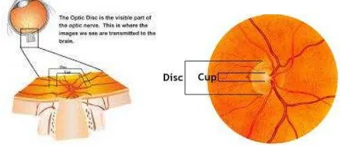
The optic disc can be divided into two distinct zones as shown in Fig.1, a central bright zone called the cup and a peripheral region called the neuroretinal rim where blood vessel and nerve fiber leave the eye. The loss in optic nerve fibers leads to a change in the structural appearance of the optic disc, namely, the enlargement of cup region or thinning of
neuroretinal rim called cupping. The progress of glaucoma and its treatment is assessed by changes in shape of the neuroretinal rim as shown in Fig. 2. Various parameters are estimated and recorded to assess the glaucoma stage. These include the diameter and area of optic disc, diameter and area of optic cup, rim area, mean cup width, etc.
Presently, the shape of the rim is assessed manually, either subjectively by direct inspection or by tracing it from photographs of the optic disc. Both of these methods have been shown to be unreliable since they consume time, require highly-skilled experts, and are highly susceptible to subjective variation and error.
There are some researches done on automated segmentation of the optic nerve head [4-12, 15-26]. The optic disc and cup regions are segmented from monocular retinal images by an automatic optic disc parameterization technique is proposed by Gopal et al. Robustness against variations in an image in optic disc segmentation is achieved by using the local image information in multidimensional feature space [4]. Kavitha et al. proposed two methods to extract the optic disc automatically i.e. using the component analysis method and region of interest based segmentation [5].
A semi-automated method to detect glaucoma using openCV is proposed by Narasimhan et al. The optic disc and cup area are extracted from the region of interest (ROI) by using K-Means clustering technique [6]. Integrating image inpainting and active contour model techniques successfully assisted the correct identification of cup and disc region is presented by Chih-Yin Ho [7]. Automated method for segmented the optic disc using the active contour method with the brightness and edge information is proposed by Chisako [8,9].
Fig. 1. Optic disc and optic cup
2015 International Conference on Automation, Cognitive Science, Optics, Micro Electro-Mechanical System, and Information Technology (ICACOMIT), Bandung, Indonesia, October 29–30, 2015
Fig. 2. Optic nerve head cupping progression
Detection of the optic nerve head in retinal fundus image based on the Hough Transform and active contour models is proposed by Handayani [10]. Later the active contour method is used to obtain accurate optic disc boundary [11]. To provide a better segmentation for images with weak boundaries, the geometric based implicit active contour is employed by Jaspreet et al. [12].
This research concern to the design and implementation of quantitative fundus image analysis software to support glaucoma diagnosis. The proposed method is aimed to serves as an additional diagnostic and assessment tool for ophthalmologist in treating glaucoma disease. We develop image processing techniques and algorithms to identify the optic disc and the optic cup, and then determine diameters of the disc, cup, and rim, in order to calculate the cup-to-disc ratio. We propose GVF Snake Active Contour method to detect optic disc and cup boundary, then evaluated the level of success of this system with a set of performance testing. In this paper we compare the system performance of our developed system, GVF Snake Active Contour, with the Ellipse Fit method, which is now used by ophthalmologist as a tool to assist the glaucoma diagnosis.
II. GVFSNAKE ACTIVE CONTOUR AND ELLIPSE FIT
Active contours, or snakes, are computer-generated curves that move within images to find object boundaries. They are often used in computer vision and image analysis to detect and locate objects, and to describe their shape. Active Contour first introduced in 1987 by Kassel et. al.
The active contour model is defined by an energy function. The energy functional, which is minimized, is a weighted combination of internal and external forces [13]. The internal forces emanate from the shape of the snake, while the external forces come from the image and/or from higher-level image understanding process. The internal forces designed to keep the shape of the curve remains smooth during the deformation process. While the external force is required to move toward the edge model of the object or region of interest in an image.
Snake active contour can be assumed as a parametric curve, where 0≤s≤1, with energy function equation as follow :
���� = ( �′ + �"( ) ) +��(�( )) (1)
The first and second component in the equation 1 shows the internal forces which is determined by the physical properties of the snake . Coefficients α and β determine the extent of the
curve can be deformed, adjust the elasticity and rigidity of the snake. If α and β have a high value, then snake will tend to form a smooth curve. The third component is the external force and can be defined by an edge map. External energy will be minimum when the image gradient have a high value, that is along the edges of the image. This will push snake moves toward the edge.
In this research, we used Gradient Vector Flow (GVF) as an external force. GVF snake begins with the calculation of a field of forces, called the GVF forces, over the image domain. The GVF forces are used to drive the snake, modeled as a physical object having a resistance to both stretching and bending, towards the boundaries of the object. The GVF forces are calculated by applying generalized diffusion equations to both components of the gradient of an image edge map.
Ellipse Fit is a method to create an elliptical contour from a number of points (at least 5 points) with the least square regression method. Ellipse fit used the least square method to estimated the elliptical shape appropriately from a number of points.
Edge detection method of the optic disc and optic cup which now used by some eye hospital in Indonesia was use ellipse fit software. In this research we use MATLAB to simulated the ellipse fit method.
III. DATA ACQUISITION
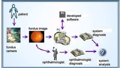
This research proposes a method for automatic analysis of the shape and size of the optic disc and optic cup based on fundus image. The proposed method is based on the optic disc and optic cup edge detection, performed by GVF Snake Active Contour and Ellipse Fit. System design and implementation is conducted on Matlab software. Block diagram system can be seen on Fig. 3
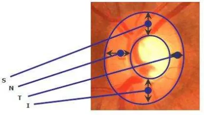
The 64 fundus images eye retina data were collected from Cicendo Eye Hospital Bandung and Bandung Eye Center. The collected data are in RGB 24 bit with size of 1216x1600 pixels, obtained using fundus camera KOWA VK-2. System input is 256x256 pixels, obtained by cropping the digital fundus image of the patient, while system output is the segmentation of the optic disc and optic cup supplemented with information of size criterion which comprises C/D ratio of vertical diameter, C/D ratio of horizontal diameter,
C/D ratio of area, and the thickness of each rim (inferior, superior, nasal and temporal (ISNT)). The illustration of each rim is shown on Fig. 4.
Fig. 4. Rim Inferior (I), Superior (S), Nasal (N), Temporal (T)
The developed system adopts the size criterion threshold value commonly used by ophthalmology to diagnose glaucoma. The normal eye it is found less than 0.5. When the cup-to-disc ratio of diameters are greater than a value of 0.5, the patient is diagnosed as glaucoma. Then we measured cup-to-disc ratio of the areas by comparing the number of pixels inside the cup and the disc regions. For a normal eye, it is found less than 0.3.
With the precise information of cup and disc boundaries, the neuroretinal rim configuration becomes available for the diagnosis of glaucoma. Neuroretinal rim area is calculated by correct verification of ISNT rule and then improve the correct diagnosis of glaucoma at early stages.
To evaluated the level of success of this system, we conducted a set of performance testing. We test the sensitivity, specificity, positive predictive value, negative predictive value and accuracy of the system. We define sensitivity as the probability that the test says a person has the disease when in fact they do have the disease. Whereas specificity is defined as the probability that the test says a person does not have the disease when in fact they are disease free. The positive predictive value of a test is defined as the proportion of people with a positive test result who actually have the disease. And the negative predictive value is defined as the proportion of people with a negative test result who do not have disease [14]. Accuracy of the system is calculated from the overall system’s ability to give the correct diagnosis.
IV. RESULTS AND ANALYSIS
The result of the optic disc and optic cup detection shows that the performance of optic disc and optic cup contour detection by snake active contour is quite satisfying. The entire images of optic disc was evaluated one by one by putting as an input system, processing it, and then compare the
output system, i.e. diameter, area and width of the neuroretinal rim, with the ophthalmologist diagnosis. The result of the edge detection can be seen on Fig. 5.
Fig. 5. The optic disc and optic cup detection
After it determine the size criterion which comprises C/D ratio of vertical diameter, C/D ratio of horizontal diameter, C/D ratio of area, and the thickness (width) of each rim, we can classified it. Classification of diagnosis by ophthalmologist and classification system (GVF Snake Active Contour and Ellipse Fit) based on four feature that used as a parameter in the glaucoma diagnosis.
After the testing of classification system, it conducted performance testing to determine the level of success of the systems as shown in Table 1 and Table 2.
TABLEI. PERFORMANCE OF GVFSNAKE ACTIVE CONTOUR From the results of the performance testing of the systems, it can be seen that the ellipse fit method has the highest accuracy for CD/R area with 81.25%. As we compared with the Snake Active Contour method, the accuracy of the ellipse fit method is lower. From the four criteria that used in glaucoma diagnosis, the accuracy level of CD/R of vertical diameter, CD/R of horizontal diameter and CD/R of area of the ellipse fit system is lower when compared to GVF Snake Active Contour system. Whereas the criteria of width rim, the accuracy of the ellipse fit system is higher.
cup. Quantitative results are goal of this study, so the results of attained for C/D ratio of area is 84,38% for GVF Snake Active Contour method and 81,25% for Ellipse Fit method. The automated determination of cup to disc ratio can be useful for improving the diagnostic efficiency by providing the quantitative data as an additional assessment tool for ophthalmologist in treating glaucoma disease.
ACKNOWLEDGMENT
This research was supported by the thematic program (No. 3425.001.014) through the Bandung Technical Management Unit for Instrumentation Development (Deputy for Scientific Services) and funded by Indonesian Institute of Sciences, Indonesia.
REFERENCES
[1] WHO:http://www.who.int/blindness/causes/priority/en/index7.ht ml,
[2] H. A. Quigley and A. T. Broman, ”The number of people with glaucoma worldwide in 2010 and 2020,” British J. Ophthalmology, vol. 90, pp. 262-267, 2006.
[3] G. Michelson, S. Wrntges, J. Hornegger, and B. Lausen, “The
papilla as screening parameter for early diagnosis of glaucoma,”
Deutsches Aerzteblatt international, vol. 105, pp. 34-35, 2008. [4] D. J. Gopal, S. Jayanthi, and S.R. Krishnadas, “Optic disk and
optic cup Segmentation from monocular colour retinal images
for glaucoma assessment,” IEEE Trans. on Medical Imaging,
vol. 30, no. 6, pp. 1192-12005, March 2011.
[5] S. Kavitha, S. Karthikeyan, and K. Duraiswamy, “Neuroretinal
rim quantification in fundus images to detect glaucoma,” Int. J.
Computer Science and Network Security, vol. 10, no. 6, June 2010.
[6] K. Narasimhan, K. Vijayarekha, K. A. JogiNarayana, P.
SivaPrasad, and V. SatishKumar, “Glaucoma detection from
fundus image using opencv,” Research Journal of Applied
Sciences, Engineering and Technology, Maxwell Scientific Organization, 2012.
[7] Chih-Yin Ho and Tun-Wen Pai, ”An automatic fundus image
analysis system for clinical diagnosis of glaucoma,” Proc. Int. Conf. on Complex, Intelligent, and Software Intensive Systems, pp. 559-564, 2011.
[8] M. Chisako, N. Toshiaki, S. Akira, H. Yuji, Y. Tetsuya, and F. Hiroshi, “Automated determination of cup-to-disc ratio for classification of glaucomatous and normal eyes on stereo retinal
fundus images,” J. Biomedical Optics, vol. 16, no. 9, 096009,
September 2011.
[9] M. Chisako, N. Toshiaki, S. Akira, H. Yuji, H. Takeshi, Y. Tetsuya, and F. Hiroshi, “Automated segmentation of optic disc region on retinal fundus photographs: Comparison of contour
modeling and pixel classification methods,” J. Computer
Methods and Programs in Biomedicine, pp. 23-32, 2011. [10]T. Handayani, W. Ari, and S. Nanik, “Optic nerve head
segmentation using hough transform and active contour,” J.
Telkomnika, vol. 10, no. 3, pp. 531-536, September 2012. [11]M. Madhusudan, K. N. Malaya, and D. Samarendra, “Glaucoma
detection from colour fundus images,” Int. J. Computer and
Communication Technology, vol. 2, issue VI, 2011.
[12]K. Jaspreet and H. P. Sinha, “Automated localization of optic
disc and macula from fundus images,” Int. J. Advanced
Research in Computer Science and Software Engineering, vol. 2, issue 4, pp. 242-249, April 2012.
[13]M. Kass, A. Witkin, and D. Terzopoulos, “Snakes: active contour models,” Int. J. Comput. Vision, vol. 1, pp. 321-331, 1988.
[14]K. A. Anthony, “Understanding diagnostic test 1: sensitivity,
specificity and predictive values,” Journal Compilation, vol. 96,
pp. 338-341, 2006.
[15]M. K. Nath and S. Dandapat, “Techniques of glaucoma detection from color fundus images: a review,” Int J. Image, Graphics and Signal Processing, vol. 9, pp. 44-51, 2012. [16]R. A. Abdel-Ghafar and T. Morris, “Progress towards automated
detection and characterization of the optic disc in glaucoma and diabetic retinopathy,” Medical Informatics and the Internet in Medicine, vol. 32, no 1, pp. 19-25, 2007.
[17]A. Aquino, M. E. Gegundez-Arias, and D. Marin, “Detecting the optic disc boundary in digital fundus images using morphological, edge detection, and feature extraction
techniques,” IEEE Trans. on Medical Imaging, vol. 29, no. 11,
pp. 1860-1869, Nov 2010.
[18] K. Narasimhan and K. Vijayarekha, “An efficient automated
system for glaucoma detection using fundus image,” Journal of
Theoretical and Applied Information Technology, vol. 33, no. 1, pp. 104-110, 2011.
[19]R. Bock, J. Meier, L. G. Nyul, J. Hornegger, and G. Michelson,
“Glucoma risk index: Automated glaucoma detection from color fundus images,” Journal of Medical Image Analysis, vol. 14, pp.
471-481, 2010.
[20]Y. Hatanaka, A. Noudo, C. Muramatsu, A. Sawada, T. Hara, T.
Yamamoto, and H. Fujita, “Vertical cup-to-disc ratio
measurement for diagnosis of glaucoma on fundus images,”
Proc. of SPIE, Medical Imaging : Computer-Aided Diagnosis, vol. 7624, pp. 76243C-1-8, 2010.
[21]M. C. V. S. Mary and B. J. S. Marri, “Automatic optic nerve head segmentation for glaucomatous detection using hough
transform and pyramidal decomposition,” Proceeding of
International Conference on Recent Trends in Computational Methods, Communication and Controls, pp. 33-37, 2012. [22]D. Lamani, Ramegowda and Manjunath, “Fractal dimension as
diagnostic parameter to detect glaucoma,” International Journal
of Innovations in Engineering and Technology, vol. 2, Issue 1, image processing system for automated glaucoma
classification,” Proc. in 52nd
Kolloquium, Technische Universitat Ilmenau, 10-13 September 2007.
[25]N. A. Kumar, M. S. Anuradha, P. SSVD. Vepa and R. Daniel,
“Active contours techniques for automatic detection of
glaucoma,” International Journal of Recent Technology and
Engineering, vol. 1, issue 4, pp. 29-31, 2012.
[26]J. Pruthi and S. Mukherjee, “Computer based early diagnosis of glaucoma in biomedical data using image processing and automated early nerve fiber layer defects detection using feature
extraction in retinal colored stereo fundus images,” International