Pages: 647-652 DOI: 10.13057/biodiv/d170237
Data provision of PIK3CA gene diversity and recombinant plasmids
preparation for control DNA in developing the trastuzumab predictive
response diagnostic kit
DESRIANI1,♥ , BUGI RATNO BUDIARTO1, WIRSMA ARIF HARAHAP2, M. ALI WARISMAN1, AUDREY VANIA CLARISSA OMPUSUNGGU1, DINA ATHARIAH1, FARIDA MIRNAWATI1,
IDA YUSSRIYANI1, FUAD ALAHWANI1, AHMAD RIZQI KURNIAWAN1
1Research Center for Biotechnology, Indonesian Institute of Sciences. Jl. Raya Jakarta-Bogor Km 46, Cibinong, Bogor 16911, West Javat, Indonesia.
Tel.: +62-21-87907604/87907636, Fax.: +62-21-87907612,♥email: [email protected], [email protected]
2Division of Surgical Oncology Medical School of M. Djamil Hospital, Universitas Andalas. Jl. Perintis Kemerdekaan No. 94, Padang 25127, West
Sumatra, Indonesia
Manuscript received: 30 May 2016. Revision accepted: 6 August 2016.
Abstract. Desriani, Budiarto BR, Harahap WA, Warisman MA, Ompusunggu AVC, Athariah D, Mirnawati F, Sriyani IY, Alahwani F, Kurniawan AR. 2016. Data provision of PIK3CA gene diversity and recombinant plasmids preparation for control DNA in developing the trastuzumab predictive response diagnostic kit. Biodiversitas 17: 647-652. HER-2 overexpression is well known as a poor prognostic factor for breast cancer patients. Targeted therapy can be carried out using monoclonal antibody named trastuzumab. Some reports have higlighted the core problem of HER2 positive-breast cancer resitance on trastuzumab due to incorrect in selecting HER2 status patient who will reciept the drug and the emergence of PIK3CA mutations especially in exon 9 and 20 which is the downstream of HER-2 pathway. In this study, data provision of PIK3CA gene and preparation of plasmid to support developing the trastuzumab predictive response diagnostic kits will be reported. Based on direct DNA sequencing result, two samples of 68 breast cancer patients exhibited mutation at exon 20 H1047R, while another three samples showed silent mutation (T1025T) at the same exon. On the other hand, careful strategy should be considered for exon 9 analysis, since we found that almost 68 samples sequenced none of them were exon 9 positive (pesudogene). Two prepared plasmids, pGEMT-easy PIK3CA exon 9 and 20 will be applied as control PIK3CA gene for qPCR SYBR green I-based PIK3CA genotyping, while pGEMT easy HER-2 will be applied as a reference gene for scoring HER-2 status. The standard curve equation of plasmid-cloned HER2 gene amplification was Y=-3,0472x + 46,465, R2= 0,99 with qPCR efficiency was 115%, respectively. Inconclusion, data provision and control DNA preparation of predicted factors for breast cancer patients who positively respond to trastuzumab are very fundamental important aspects for the development of trastuzumab response diagnostic kit which is based on Indonesian population genetics profile.
Keywords: Breast cancer, DNA, PIK3CA, HER-2, resistance trastuzumab, mutation
INTRODUCTION
Cancer is one of the leading causes of death worldwide. In 2012, it was approximately 8.2 million deaths caused by cancer. Lung cancer, liver, stomach, colorectal, and breast cancer are the biggest cause of cancer deaths each year. Cancer is regulated by many genes, known as oncogenes which express oncoprotein. The emergence of oncogenes could be as results of mutation, amplification and so forth. Some of cancer diagnostic test have been developed not only in the genomic level but also in the proteomic level as well. Oncotype DX test is an example of detection techniques in genomic level. Oncotype DX test has been manufactured and commercialized. With this test, as many as 21 cancer biomarker genes can be detected at the genomic level. These targeted genes were determined based on American Society of Clinical Oncology (ASCO) and National Comprehensive Cancer Network (NCCN)’s guideline and it has widely been applied by Oncologist in therapying and treating their cancer patient. At the protein level, oncoprotein detection in serum can be analyzed using two-dimensional gel electrophoresis (2-DE). Examination
of information obtained can be used for the same purposes as the Oncotype test (Wang et al. 2003)
for the targeted drug in term of HER2 status. Some reports have shown the high resistance incidence due to trastuzumab treatment for non-target ones. False interpretation of immunohistochemistry (IHC) results in scoring of HER-2 status as the main factor for that incident. Another factor is phophatidylinositol 3-kinases/ PIK3CA gene mutation. In breast epithelial cells, this gene acts as a regulator of the cellular growth, cell migration, survival, apoptosis and proliferation. This gene located on HER-2 signaling pathway, encodes the p110α catalytic subunit of the PI3K enzyme. These mutations cause the lipid kinase activity increased two times higher, producing an increase of phosphorylated AKT protein and hence inducing oncogenic transformation. These mutations are generally clustered in exon 9 and 20 of PIK3CA gene. Mutations in PIK3CA are also found in other exons but in very rare frequency. Patients harbor simultaneously mutations at E545K and H1047R position have been reported to be more resistant to therapy than other mutant types based on in vitro study. ESMO at 2014 has officially issued that detection of PIK3CA mutation in cancer patients is required to predict their response to trastuzumab. Furthermore, PIK3CA mutations have also been reported as a potential biomarker for predicting prognostic status in breast cancer patients. Indeed, PIK3CA mutations is associated with increased tumor aggressiveness (Kurebayashi et al. 2001; Gallia et al. 2006; Kato et al. 2007; Hale et al. 2008; Zhao et al. 2008). So far, targeted therapy using trastuzumab given to breast cancer patient is solely based on IHC result. The high percentage of resistance to trastuzumab in single used reached over 60-80%, indicating that HER-2 test which is only refer to IHC alone is not enough. Moreover, IHC test is currently known to be subjective due to the factors of operator skill and the type of antibody used which potentially lead to misreading. Based on this finding, the quantifying of HER-2 status using other molecular methods with complement to existed methods is significant to be developed (Clifford and Hudis 2007; Siddig et al. 2008; Breyer 2009; Cremoux et al. 2012; Alaoui-Jamali et al. 2015). In a diagnostic kit for cancer detection, the standard reagent preparation was one of the important things to be well prepared.
Here, the development of trastuzumab predictive response diagnostic kits through PIK3CA gene data provision and plasmids preparation to support the diagnostic kit study will be reported. We provided diversity data of PIK3CA gene exon 9 and 20 detected with Sanger DNA sequencing. We have also prepared three recombinant plasmids as part of manufacture the predictive factor detection kit at genomic level for patient responsiveness against trastuzumab where two of them were applied for PIK3CA genotyping in exon 9 and exon 20 while the other for HER-2 status scoring. The diversity data in term of PIK3CA mutation obtained in this study and the PIK3CA gene-contained recombinant plasmids can be as a basis for creating and validating new cancer detection kit which is unique only for PIK3CA genotypes originated from Indonesian population. Furthermore, HER2-contained recombinant plasmid can be applied as part of standard kit preparation in qPCR to overcome the subjectivity problem
of IHC methods, avoiding false interpretation that may occurs. The prepared control plasmids could significantly contribute to the quality controls and quality assurance program of cancer detection kit for predictive factors to trastuzumab treatment.
MATERIALS AND METHODS Genomic DNA isolation from Breast Cancer Tissue
The research was conducted using ethical clearance issued by Indonesia Ministry of Health. Breast cancer tissues were provided from several hospitals in West Sumatra province, Indonesia. The fresh tissue samples were stored at -80°C. Extraction was done using PureLink® Lysate-Mini Kit from Invitrogen. The Genomic DNA obtained then was confirmed using electrophoresis with 1.5 % agarose, visualized with UV-Transilluminator.
The PIK3CA gene amplification and sequencing For HER-2 primer were forward Primer (5'-TGA TCT GCC CAC AGA CTC-3 ') and reverse Primer (5'-TCT CAT CGT CCG CTT GTA CC-3'), for PIK3CAexon 9 5’ AGT AAC AGA CTA GCT AGA GAC AAT 3’, reverse primer 5’CTG TGA CTC CAT AGA AAA TCT 3’, Primer for
PIK3CA gene exon 20 were (5’-TTT TTT CCT TCT CCA
TCA TTT CTA-3’, reverse primer (3’-GTT TCA GGA GAT GTG TTA CAA-5’). PCR composition used are 12.5 µL of DreamTaq Green PCR Polymerase, 0.25 µL of forward primer, 0.25 µL of reverse primer, 5.5 µL of MilliQ nuclease-free water, and 0.25 µL of genomic DNA. The PCR condition used as follows: pre-heat at 95°C for 5 minutes then followed by 35 cycle of denaturation at 95°C for 30 seconds, annealing at 50.9°C for 30 seconds, extension at 72°C for 30 seconds. After the PCR process completed, the gene was confirmed using DNA electrophoresis with 1.5% agarose, visualized with UV-Transilluminator. Gene sequencing was done using Applied Biosystem® 3100 Genetic Analyzer based on Sanger method.
Purification, ligation, transformation
PCR product was purified using Wizard SV® Gel from Promega, then was inserted to pGEM®-T Easy following manufacture instruction. Transformation to E. coli DH5α competent cells were done with heat shock method and were spread in LB media containing with 100 ppm of ampicillin, 50mM of X-Gal, and 1 mM of IPTG for selecting targeted and non-targeted E. coli. Positive bacteria colonies (containing recombinant plasmids) will be white while the negative bacteria will be blue.
PCR colony for screening targeted E. coli
methods, except for HER-2 confirmation we used 5’ -CCAGCCCTCTGACGTCCAT-3’ for forward primer and 5’-CGTGTACTTCCGGATCTTCTGCTG-’3 for reverse primer which recognized inside HER-2 producing 116 bp PCR product. Positive clone than were cultured for overnight in an incubator shaker at the speed of 150 rpm and temperature 37°C.
Plasmid extraction
Plasmid extraction was done using High-Speed Plasmid Mini Kit from Geneaid. The plasmid result then visualized with UV-Transilluminator.
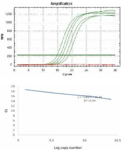
Control curve standardization for HER-2 amplification pGEMT-easy HER-2 serial dilutions were started from 12.5 ng diluted by two times for each in five spots. Dilutions were used as plasmid copy number standards to generate a standard curve and to quantify her-2 chromosomal DNA copies. General formula used: (6.02 x 10²³copies/mol) x (concentration in g/μ L) / (MW in g/mol)
= copies/μ L(Mendoza et al. 2013).
qPCR experimental conditions
For amplification and data collection we used CFX-96 Real-Time PCR from Biorad. Reactions were carried out in triplicates, SYBR-Green from KappaBiosains®, 1µM of
each primer. Cycling conditions were 95˚C for 5 min, 35 cycles; at 95˚C for 30 sec; at 60˚C for 10 sec and at 72˚C for 30 sec.
RESULTS AND DISCUSSION PIK3CA data provision
There are many theories which trastuzumab resistance problems arise. Some of report showed that mutation in PIK3CA gene especially in the hotspot area such as in exon 9 and 20 contributed to the therapy implication. Patients with mutation in this hotspots area have been shown unresponsive toward trastuzumab therapy.
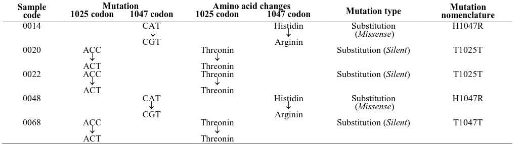
Based on sequencing method, two samples among 68 breast cancer samples obtained from West Sumatra province of Indonesia showed mutation in exon 20 H1047R (2.94%) and three samples showed T1025T silent mutation
(4.41%). While for exon 9 study careful strategy should be considered, since we found almost 68 samples showed mutation at A1634C (E545A) position and a base deletion at nucleotide 1659 which referred as pseduogene (Samuel et al. 2004). Mutation in exon 20 H1047R was reported to have oncogenic capability and it was responsible for trastuzumab resistance problems, while T1025T position has no reports implicate to cancer development. Mutation in PIK3CA gene quite varies around 8%-40% and the contribution of this mutation to prognostic implication is still controversial (Levine et al. 2005; Mangone et al. 2012; Arsenic et al. 2014). According to Li et al. (2006), mutation in exon 20 of PIK3CA gene were predominated in breast cancer. To confirm the data obtained in this study, the large number for PIK3CA study is needed for future works.
Although the percentage of mutations were low in Indonesian breast cancer especially in West Sumatra province, the detection system is important to be prepared since H1047R has oncogenic capabilities. According to Meyer et al. 2013, PIK3CA H1047R was able to induce mammary tumor growth compared to its wild type and also this mutant showed more oncogenic pontency compared with PIK3CA E545K in the transgenic mouse models. Furthermore, PIK3CA mutations was reported associated with lower tumor stages which mean it could be as a biomarker for early tumor detection (Rudd et al. 2011; Dumont et al. 2012). Prepared recombinant plasmids and the results of PIK3CA genotyping could be used for developing and validating a new method for predictive factor therapy with trastuzumab in genomic level in future.
Control DNA preparation for pGEM-T easy HER-2 and PIK3CA gene exons 9 and 20
HER2 gene as a predictive factor of treatment with trastuzumab using was successfully cloned into pGEM-T easy plasmid as a vector. This prepared plasmid was required as a control to support targeted detection kit development. Furthermore, the performance of the kit such as sensitivity, specificity, reproducibility and suitability are prerequisite to be performed before the kit commercially produced. The property of the kit and all the associated reagents should fulfill the sensitive, specificity, fast and easy to interpret the result (Yang and Rothman 2004).
Table 1. Mutations profile in exon 20 of PIK3CA gene
Mutation Amino acid changes
Sample
code 1025 codon 1047 codon 1025 codon 1047 codon Mutation type nomenclatureMutation
Figure 1. Breast cancer genomic DNA (arrow)
Figure 2. PCR product for HER-2 (480 bp), PIK3CA exon 9 (124 bp) and exon 20 (619 bp). DNA ladder marker: 100 bp DNA ladder (left) and 1Kb plus DNA ladder (right)
Figure 3. PCR colony of E.coli DH5α for HER-2 (116 bp), PIK3CA exon 9 (124 bp) and exon 20 (619 bp). DNA ladder marker: 1 Kb plus DNA ladder.
Genomic DNA was used as template for PCR amplification of exon 9 and 20 of PIK3CA as well as HER-2 gene. The genomic extraction result is shown in the Figure 1. The amplification, insertion and transformation of targeted genes for each were shown in Figure 2.
The results of the preparation of plasmid were sequenced to confirm the targeted gene (data not shown). Three recombinant plasmids prepared then were preserved in E. coli DH5α bacterial strain as glycerol stocks for future usage.
pGEM-T easy PIK3Ca exon 9 and 20
Mostly cancer biomarker detection method was based on PCR methods, which were massive, high-trough put and rapid (Kristensen and Hanse 2009). Prepared recombinant plasmids could be used as reference gene in conducting breast cancer genotyping in Indonesia based on PCR methods. The commercial products currently developed and commonly used are ARMS and probe technique. Both of the technique provides a negative and positive control reaction in the detection kit. By using a prepared plasmid not only could be applied as control part of the kit, but also could be used to test and to develop new techniques easier and cheaper avoiding limited sample usage. Here below the plasmid map of pGEMT easy PIK3CA for exon 9 and 20.
pGEM-T easy HER-2 for standard curve calculation in scoring HER-2
post-qPCR data processing can seriously affect the interpretation of the results. If there is no reference gene it is needed the wisdom of researchers in executing the data processing. The standard curve in real time PCR approach may have advantages. The standard curve method simplifies calculations and avoids practical and theoretical problems. (Mendoza et al. 2013). pGEM-T easy HER-2 was successfully tested for a standard curve preparation for HER-2 scoring application.
Figure 4. Map pGEM-T easy PIK3Ca exon 9 (left) and exon 20 (right)
619 bp 124 bp
61
9
bp
12
4
bp
11
6
bp
Figure 5. Standard curve of HER-2 amplification calculation
In the figure showed above, the curve is used as reference to calculate the copy number and to score the over expression of HER-2 by comparing between breast cancer and normal patients. The standard curve equation was Y=-3.0472x + 46.465, R2= 0.99, the efficiency was 115%. This equation meets the requirements in qPCR theory. Quantifying HER-2 satus using qPCR was expected to minimize the subjectivity in selecting HER2 positive candidate among patients tested. HER-2 quantification detection kit is already commercialized, such as by Roche. The kit provides specific primers, hybridization probes, positive and negative control reaction (Beyser et al. 2001). Since the probes frequently used for the DNA labeling process hence it directly causes the kits price becoming slightly expensive. In our future detection kit development, the application of such probe is avoidable in order to minimize the price so that the kit will be more affordable especially for Indonesia market.
PIK3CA data provision and three recombinant plasmids preparation in our study are important aspect as a part of supporting in development of diagnostic kit of predicted factor for patients which is responsive to trastuzumab at genomic level based on Indonesian population. The provided kit could be applied for determination of HER2 amplification status and PIK3CA genotyping that those are becoming the major cause for resistance towards trastuzumab. Furthermore, the availability of these kits is expected helping the oncologist from inappropriate treatment of trastuzumab administrated-patients.
ACKNOWLEDGEMENTS
We thank the research funding from LIPI grant numbers SP DIPA-079.01.2.450083/2015.3403.002 (Principal investigator: Dr. Eng. Desriani).
REFFERENCES
Alaoui-Jamali MA, Morand GB, da Silva SD. 2015. ErbB polymorphisms: in sights and implications for response to targeted cancer therapeutics. Front Genet 6: 1-9.
Arsenic R, Lehmann A, Budczies J, Koch I, Prinzler J, Tebbe AK, Schewe C, Loibl S, Dietel M, Denkert C. 2014. Analysis of PIK3CA mutations in breast cancer subtypes. Appl Immunohistochem Mol Morphol 22 (1): 50-56.
Breyer JP. 2009. Heritable variation of ERBB2 and breast cancer risk. Cancer Epidemiol Biomark Prevent 18: 1252-1258.
Beyser K, Reiser A, Gross C, Moller C, Tabiti K, Ruschoff. 2001. Real time quantification of HER-2/neu gene amplification by light cycler polymerase chain reaction (PCR)-a new research tool. Biochemica 2: 15-18.
Clifford A, Hudis MD. 2007. Trastuzumab-mechanism of action and use in clinical practice. New England J Med 357: 39-51.
Cremoux P, Spyratos F, Bieche I. 2012. Outcome Impact of PIK3CA mutations in HER2-positive breast cancer patients treated with trastuzumab. Br J Cancer 108: 1807-1809.
Dumont Ag, Dumont SN, Trent JC. 2012. The favorable impact of PIK3CA mutation on survival: an analysis of 2587 patient with breast cancer. Chinese J Cancers 31 (7): 327-334.
Gallia GL, Rand V, Siu I, Eberhart CG, James CD, Marie SKN, Oba-Shinjo SM, Carlotti CG, Caballero OL, Simpson AJG, Brrock MV, Massion PP, Carson BS, Riggins G.J. 2006. PIK3CA gene mutation in pediatric and adult glioblastoma multiforme. Mol Cancer Res 4: 709-14. breast cancer. Cancer Res 68 (15): 6084-6091.
Hynes NE, Dey JH. 2009. PIK3 inhibition overcomes trastuzumab resistance: blockade of ErbB2/ErbB3 is not always enough. Cancer Cells 15: 353-355.
Kato S, Iida S, Higuchi T, Ishikawa T, Takagi Y, Yasuno M, Enomoto M, Uetake H, Sugihara K. 2007. PIK3CA mutation is predictive of poor survival in patients with colorectal cancer. Intl J Cancer 121: 1771-1778.
Kristensen LS, Hansen LL 2009. PCR-based methods for detecting single-locus DNA methylation biomarkers in cancer diagnostics, prognostics, and response to treatment. Clin Chem 55: 1471-1483. Kurebayashi J. 2001. Biological and clinical significance of HER2
overexpression in breast cancer. Breast Cancer 8: 45-51.
Li SY, Rong M, Grieu F, Laccopeta B. 2006. PIK3CA mutations in breast cancer are associated with poor outcome. Breast Cancer Res Treat 96: 91-5.
Levine DA, Bogomolniy F, Yee CJ, Lash A, Barakat RR. Borgen PI, Boyd J. 2005. Frequent mutation of the PIK3CA gene in ovarian and breast cancer. Clin Cancer Res 11 (8): 2875-2878.
Mangone FR, Bobrovnitchaia IG, Salaorni S, Manuli E, Nagai MA. 2012 .
PIK3CA Exon 20 mutations are associated with poor prognosis in
breast cancer patients. Clinics 67: 1285-290.
Mendoza, G., Portillo A, Olmos-Soto J. 2013. Accurate breast cancer diagnosis through real-time PCR her-2 gene quantification using immunohistochemically-identified biopsies. Oncology Lett 5: 295-298.
Meyer DS, Koren S, Leroy C, Brinkhaus H, Muller U, Klebba I, Muller M, Cardiff RD, Alj MB. 2013. Expression of PIK3CA mutant E545K in the mammary gland induces heterogeneous tumors but is less potent than mutant H1047R. Oncogenesis 2: 1-6.
Paplomata E, O’Regan R. 2014. The PI3K/AKT/mTOR pathway in breast
cancer: targets, trials and biomarkers. Ther Advanc Med Oncol 6 (4): 154-166.
Rudd ML, Price JC, Fogoros S, Godwin AK, Sgroi DC, Merino MJ, Bell DB. 2011. A unique spectrum of somatic PIK3CA (p110α ) mutations
within primary endometrial carcinomas. Clinicall cancer research 17: 1331-1340.
Siddig A, Mohamed AO, Kamal H, Awad S, Hassan AH, Zilahi E, Al-Haj M, Bernsen R, Adem A. 2008. HER‐2/neu Ile655Val polymorphism
and the risk of breast cancer. Ann NY Acad Sci 1138: 84-94. Wang L, Zhang Q, Zhang J, Sun S, Guo H, Jia W, Wang B,.Shao Z, Wang
Z, Hu X. 2011. PI3K pathway activation results in low efficacy of both trastuzumab and lapatinib. BMC Cancer 11 (248): 1-10. Wang W, Sun J, Nimtz M, Decker WD, Zeng AP. 2003. Protein
identification from two-dimensional gel electrophoresis analysis of
Klebsiella pneumoniae by combined use of mass spectrometry data
and raw genome sequences. Proteome Sci 1: 1-9.
Yang S, Rothman RE. 2004. PCR-based diagnostics for infectious diseases: uses, limitations, and future applications in acute-care settings. Lancet Infect Dis 4: 337-348.