Coating of L-Arginine Modified Silica on Magnetite
through Two Different Sol-Gel Routes
Amaria
1,2, Suyanta
2, and Nuryono
2,* 1Department of Chemistry, Faculty of Mathematics and Natural Sciences, Universitas Negeri Surabaya, Jl. Ketintang, Surabaya 60231, Indonesia
2
Department of Chemistry, Faculty of Mathematics and Natural Sciences, Universitas Gadjah Mada, Sekip Utara, PO BOX BLS 21, Yogyakarta 55281, Indonesia
Received February 27, 2017; Accepted May 2, 2017
ABSTRACT
In this research, magnetite coated with L-arginine modified silica (Fe3O4/SiO2-GPTMS-Arg) has been
synthesized through a sol-gel process at a room temperature in two Routes. In Route 1, a precursor of sodium silicate solution (source of SiO2), 3-glycidoxypropyltrimethoxysilane (GPTMS) as a coupling agent and L-arginine
(Arg) as the source of functional groups were added sequentially to magnetite nanoparticles (Fe3O4). Gelling was
carried out by adding HCl solution dropwise to the mixture to reach pH of 7.0. The product was washed with water and ethanol and then dried at 65 °C for 1 day. In Route 2, sodium silicate solution was added to a mixture of GPTMS and L-arginine, and then the sol obtained was added into magnetite nanoparticles. The results were characterized with FTIR spectroscopy, X-ray diffraction, atomic absorption spectroscopy and volumetric method to identify functional groups, crystal size, iron ions released and amino groups content, respectively. The results showed that Fe3O4/SiO2-GPTMS-Arg has been successfully synthesized through both two routes. Route 1, however, gave
product of Fe3O4/SiO2-GPTMS-Arg more stable and more content of amino groups than Route 2. The presence of
amino groups leads to the application of the product for metal ion removal from aqueous solution.
Keywords:L-arginine; magnetite; silica; sol-gel; coating
ABSTRAK
Pada penelitian ini, magnetit terlapis dengan silika termodifikasi L-arginin (Fe3O4/SiO2-GPTMS-Arg) telah
disintesis melalui proses sol-gel pada suhu kamar dalam dua rute. Pada Rute 1, prekursor larutan natrium silikat (sumber SiO2), 3-glisidoksipropiltrimetoksisilan (GPTMS) sebagai senyawa penghubung dan L-arginin sebagai
gugus fungsional permukaan, ditambahkan secara berurutan ke dalam nanopartikel magnetit (Fe3O4). Pembentukan
gel dilakukan dengan penambahan larutan HCl tetes demi tetes untuk mencapai pH 7. Produk dicuci dengan air dan etanol, kemudian dikeringkan pada 65 °C selama 1 hari. Pada Rute 2, larutan natrium silikat ditambahkan ke dalam campuran GPTMS dan L-arginin, kemudian sol yang terbentuk dimasukkan ke dalam magnetit. Hasil-hasil yang didapat dikarakterisasi dengan spektroskopi FTIR, diffraksi sinar-X, spektroskopi serapan atom, dan metode volumetri, masing-masing untuk mengidentifikasi gugus fungsional, ukuran kristal, kadar ion besi yang terlarut, dan kadar gugus amino. Hasil penelitian menunjukkan bahwa Fe3O4/SiO2-GPTMS-Arg telah berhasil disintesis melalui
kedua rute, namun Rute 1 menghasilkan Fe3O4/SiO2-GPTMS-Arg lebih stabil dan kadar gugus aminonya lebih besar
daripada Rute 2. Adanya gugus amino memberikan aplikasi produk untuk penghilangan ion logam dari larutan.
Kata Kunci:L-arginin; magnetit; silika; sol-gel; pelapisan
INTRODUCTION
In the last few years, the magnetic material has been very interesting, because of the nature and its potential, which is very profitable. Magnetite (Fe3O4),
one of magnetic iron oxides, has favorable magnetic characteristics. It is one of the most studied materials, due to the highly potential application in many fields, not only in industrial fields (ceramics, catalyst, energy
dissolved in acidic medium. Therefore, before being used in various fields it is very important to stabilize the magnetite nanoparticles.
The stabilization of magnetite with an inorganic material, such as silica [6-9] may produce magnetite nanoparticle surface coated with silica and enhance the performance of magnetite. Methods for preparation of silica coated magnetite have been reported by various methods, such as direct silanation [10], sol-gel [11-12] dense-liquid and two-step coating of silica [13]. However, aminopropyltriethoxysilane film silanized on maghemite with direct silanation method shows less stable after leaching test with acid solution [10]. The sol-gel process is a method commonly applied to coat mild particles using a precursor, such as tetraethoxy-orthosilicate (TEOS) in an organic solvent, to produce porous material. The presence of Si-OEt terminal groups of TEOS is reported to be responsible for the porous nature of the coated film [13]. Synthesis of mesoporous silica using a mixture of sodium silicate (Na2SiO3)
solution and TEOS reported by Liu et al. [14] shows that addition of TEOS can induce larger pore diameter. Higher the content of TEOS more porous particles and the higher the content of sodium silicate gets smaller pore diameter.
The presence of silanol groups on the silica surface can be modified with various functional groups. The addition of basic organic compounds such as L-arginine stabilized magnetite nanoparticles [15-16]. To attach organic functional groups on the magnetite coated silica, a coupling agent is needed. A coupling agent of 3-glycidoxypropyltrimethoxysilane (GPTMS) reported by Davis et al. [17] forms a network of silica and organic epoxy. Methoxy groups of GPTMS are hydrolyzed to produce silanol, which then condenses to form the silica network [18]. The opening of the epoxy ring may occur at room temperature and an organic network may be formed using amine containing compounds [17-19]. Each active hydrogen in the amino group can open and bind to one epoxy group.
In this work, we synthesized L-arginine modified silica coated on magnetite nanoparticle surface using linker of GPTMS through a sol-gel process. Since the synthesis involved some precursors namely magnetite, sodium silicate solution, L-arginine, and GPTMS, the sequence of mixing precursors (mixing Route) may affect the characters of the product. Therefore, this paper also reports the synthesis of magnetite coated with L-arginine modified silica conducted through two different Routes. On Route 1, an interaction of precursors (magnetite, sodium silicate, GPTMS, L-arginine) was carried out sequentially. On Route 2, mixing of precursors was conducted in two stages. GPTMS and L-arginine were mixed firstly and then
suspension of magnetite. We demonstrate the differences of both types of Route in term of the crystal size, chemical stability, and the elements composition.
EXPERIMENTAL SECTION
Materials
Magnetite (Iron (II)(III)) oxide powder, GPTMS (≥ 98%) were purchased from Sigma-Aldrich. Sodium silicate solution (25.5-28.5% SiO2), L-arginine
powder, ethanol absolute (99.5%), hydrochloric acid (HCl 37%), sodium hydroxide (NaOH) were purchased from Merck and used as received.
Instrumentation
FTIR spectrophotometer (Shimadzu FTIR 8400S) was used to identify functional groups of materials, diffractometer X-ray (XRD Bruker D8 Advance 206276) was used for determination of material phase and the crystal size. The concentration of iron ion in solution was determined by absorption atomic spectro-photometer (AAS, Analyst 100 Perkin Elmer). The element composition of materials was measured with energy dispersive X-ray (EDX Carl Zeiss 9 EVO MA 10 series 1454). The measurement of pH solution was conducted with a pH meter (pH/ion 510 Eutech Oacton). A set of equipment for acid-base titration was used to analyze the content of amino groups. The magnetic value of coated magnetite samples was measured with vibrating sample magnetometer (VSM) type OXFORD YSMI.2H
Procedure
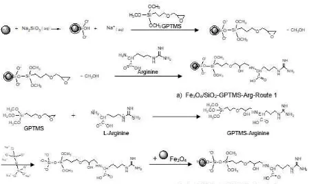
Coating of L-Arginine modified silica on magnetite through Route 1 (Fe3O4/SiO2-GPTMS-Arg Route-1)
Fig 1.The reaction model of coating L-arginine modified silica on magnetite through a) Route1 and b) Route-2
Coating of L-Arginine modified silica on magnetite through Route-2 (Fe3O4/SiO2-GPTMS-Arg Route-2)
Route 2 consisted of two mixtures, namely mixture of GPTMS-arginine-silicate sol and magnetite suspension using the same amount of composition as Route 1. The first mixture was prepared by mixing GPTMS, L-arginine and stirring for 15 min and then was added with sodium silicate solution and stirred for 2 h. The second mixture was prepared by suspending magnetite in 5 mL of distilled water and sonicated for 5 min. The GPTMS-Arginine-silicate sol then was incorporated into the magnetite suspension, sonicated for 15 min and stirred mechanically. Lastly, the similar treatment was carried out to Route 1. The difference of reaction steps of both Routes was modeled in Fig. 1.
Characterization
Samples of Fe3O4/SiO2-GPTMS-Arg obtained were
characterized by identification of crystal structure and size with X-ray diffraction. X-ray diffractograms were recorded at a 2θ range of 5-80° in scan speed 5°/min from X-ray diffractometer. The energy source used Cu-Kα radiation (λ 0.154 nm) at voltage 40 kV and current of 30 mA. Identification of functional group was conducted with FTIR spectrophotometer Shimadzu 8400S at a wave number 400-4000 by KBr pellet technique. The element composition of the coated magnetic synthesized was analyzed with Energy Dispersive X-ray Spectroscopy (EDX).
Estimation of amino group content. The content of arginine in the product was estimated based on the number of amino groups analyzed with volumetric analysis [21]. Twenty-five milliliters of 0.05 M HCl solution was added to the 0.05 g of the coated magnetite sample and shaken for 15 h. HCl concentration found in the residue was measured by titration with 0.05 M NaOH solution using phenolphthalein as the indicator. The number of HCl mmol g-1) interacting with amino groups was calculated using Equation 1.
M1 M2
25Concentration of amino groups = mmol g 0.05
(1)
where M1 and M2 are the initial and final concentration
of HCl, respectively.
The characteristics of L-arginine modified-silica coated magnetite identified with methods described previously are presented and evaluated in the following sections.
Functional Groups of Fe3O4/SiO2-GPTMS-Arg
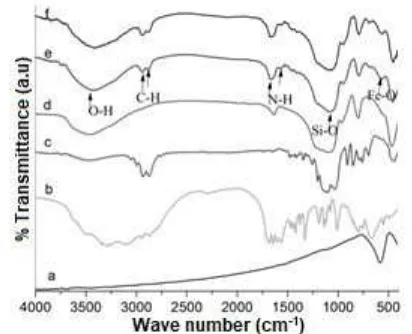
The success in coating magnetite with L-arginine modified silica is showed by comparing the FTIR spectra of precursor and the products. FTIR spectra of magnetite, L-arginine, GPTMS and silica precursors are presented in Fig. 2a-d and FTIR spectra of Fe3O4/SiO2
-GPTMS-Arg Route1 and 2 are presented in Fig. 2e and 2f. In Fig. 2a, 2e, and 2f are observed absorption peaks corresponding to the Fe-O vibration from magnetite phase at 585, 569, and 563 cm-1, respectively [24]. It indicates that the magnetite is coated with arginine-silica. The phenomenon can be explained by the formation of Fe-O-Si bond. Hydrogen atoms of Fe-O-H on the surface have been replaced with Si atom to form Fe-O-Si. Si atom is larger than H, so the bond is longer and weaker, therefore the absorption bands shift to lower wave numbers [25].
In Fig. 2d is observed absorption at 3474, 1092, 800, 469 cm-1, corresponding to stretching vibration of O-H (Si-OH), asymmetric and symmetric stretching vibration and bending vibration of Si-O(Si-O-Si), respectively. Spectra Fig. 2e from Fe3O4/SiO2
-GPTMS-Arg Route 1 shows that magnetite has been coated by silica, based on specific absorption at 3441, 1086, 795, and 459 cm-1. Moreover, FTIR spectra of Fe3O4/SiO2
-GPTMS-Arg Route 2 (Fig. 2f) shows specific bands of silica at 3402, 1090, 795, and 451 cm-1, assigned to –OH (Si-OH silanol) groups stretching vibration, asymmetric and symmetric stretching and bending vibration of siloxane Si-O(Si-O-Si), respectively [20,26].
The absorption peak associated with GPTMS and L-arginine can be seen at 2940 and 2839 cm-1 corresponding to asymmetric and symmetric stretching vibration of C-H, respectively (Fig. 2b and 2c) [26], and also appears in Fig. 2e with wave number at 2943, 2881 cm-1and in Fig. 2f at 2937 and 2878 cm-1. Characteristic absorption bands related to guanidine functional groups of arginine are seen at 1560 and 1684 cm-1 (Fig. 2b) assigning to bending vibration of N-H and stretching vibration of C=N [26-28]. In this study, FTIR spectra of Fe3O4/SiO2-GPTMS-Arg Route 1 and 2 (Fig. 2e and 2f)
show the bands at 1565, 1664 and 1574, 1668 cm-1, respectively. In addition, the bands at 1256 and 908 cm-1 are ascribed to symmetric and asymmetric stretching vibration of the C-O-C the epoxy ring of GPTMS [29], which appears in Fig. 2c at 1196 and 910 cm-1. FTIR spectra of Fe3O4/SiO2-GPTMS-Arg Route 1 (Fig. 2e)
Fig 2. FTIR spectra: a) magnetite, b) L-arginine, c) GPTMS, d) silica e) Fe3O4/SiO2–GPTMS-Arg-Route
1 and f) Fe3O4/SiO2-GPTMS-Arg-Route 2
shows the peak completely disappears. It indicates that epoxy ring has been opened and covalently bound to N atom of L-arginine. The FTIR spectra of Fe3O4/SiO2
-GPTMS-Arg Route 2 shows the peaks are almost completely disappeared. It appears at 1200 cm-1 in a weak intensity, nevertheless formed -OH functional group of the C-OH is seen at wave numbers 1063 cm-1 [26]. The bands at 1668, and 1574 cm-1 related to stretching vibration of C=N and bending vibration of N-H of guanidine group from L-arginine [28]. Other supports of the presence of the amino group from L-arginine on the coated magnetite were confirmed by analysis elemental composition determined from EDX and volumetric data.
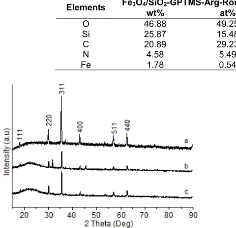
Structure and Crystal Size
X-ray diffraction method was performed to analyze crystal structure of coated and uncoated magnetite,and the XRD pattern is presented in Fig. 3. The structure of magnetite does not change with presence of silica and L-arginine. From XRD patterns of L-arginine-modified silica-coated magnetite through Route 1 and 2 can be seen a decrease of peak intensity on the index field (220), (311), (400), (511), (440) and (533) (JCPDS card no. 19-0629) in comparison to uncoated magnetite. In addition, it is observed amorphous phase of SiO2 in 2θ 20-26
degrees (Fig. 3b and c) confirmed with JCPDS No.46-1045, for SiO2. In Fig. 3b, coated magnetite Route 1
can be seen a phase change, namely maghemite (Fe2O3) [30]. This may be attributed to the effect of
Table 1.Structural properties of L-arginine modified silica-coated magnetite
Sample 2θ(°) d311(Å) D(nm)
Fe3O4 uncoated 35.346 2.54 36.00
Fe3O4/SiO2-GPTMS-Arg route 1 35.498 2.53 37.16
Fe3O4/SiO2-GPTMS-Arg route 2 35.506 2.53 45.32
Table 2.Chemical stability of coated magnetite presented in dissolved Fe
Sample Fedissolved(mol g-1) Condition Ref
Fe3O4/SiO2-SH (50/50) 1.88 HCl 1 M 23
Fe3O4-D 2678.60 HCl pH 2.5 33
Magnetic-APTES 607.00 HCl 0.01 M 10
Fe3O4 10.95 x 10⁻⁵ HCl 1 M This work
Fe3O4/SiO2-GPTMS-Arg-route 1 0.86 x 10⁻⁵ HCl 1 M This work
Fe3O4/SiO2-GPTMS-Arg-route 2 3.01 x 10⁻⁵ HCl 1 M This work
Table 3. Elemental composition of L-arginine modified silica-coated magnetite nanoparticles Route 1 and 2 based on Energy Dispersive X-ray Spectroscopy (EDX) data
Fe3O4/SiO2-GPTMS-Arg-Route 1 Fe3O4/SiO2-GPTMS-Arg -Route 2
Elements
wt% at% wt% at%
O 46.88 49.25 47.03 51.25
Si 25.87 15.48 28.50 17.69
C 20.89 29.23 17.48 25.38
N 4.58 5.49 3.76 4.68
Fe 1.78 0.54 3.23 1.01
Fig 3. XRD patterns of a) magnetite, b) Fe3O4/SiO2
-GPTMS-Arg Route 1 and c) Fe3O4/SiO2-GPTMS-Arg
Route 2
Analysis of the index plane peaks of the samples Route 1 and 2 based on XRD pattern, d311spacing and
the crystal size are obtained and presented in Table 1. In general, coating increases crystal size and slightly decreases d311spacing. From Table 1 can be seen that
crystals size of L-arginine modified silica-coated magnetite is greater than that of uncoated magnetite. The crystal size is calculated using Scherer equation (2).
K D
cos
(2)
K is a constant of 0.9, λ is the wavelength of light diffraction (1.54 Å), β is a diffraction peak full-width half maximum (FWHM) in radian and cos θ is the angle of diffraction peaks [30-31]. The crystals size of
Fe3O4/SiO2-GPTMS-Arg Route 2 (45.32 nm) is greater
than that of Route 1 (37.16 nm). This can be explained, that in Route 2 the epoxy group of GPTMS react with nitrogen atom of L-arginine to form covalent bond of C-N and to produce crystals [32] greater size than in Route 1.
The value of spacing basal in Table 1 shows a slight decrease in magnetite nanoparticles coated with L-arginine-modified silica (2.53 Å) compared to uncoated magnetite (2.54 Å). It is possible that the presence of arginine-modified silica in the magnetite surface leads to result in the distance between the molecules of magnetite decrease.
Chemical Stability
The chemical stability of Fe3O4/SiO2-GPTMS-Arg
Route 1 and 2 was examined in acid solution (hydrochloric acid 1 M for 1 day). The chemical stability of Fe3O4/SiO2-GPTMS-Arg sample was based on the
quantity of iron dissolved. The quantity of iron dissolved from Fe3O4/SiO2-GPTMS-Arg was measured by atomic
absorption spectroscopy and results were expressed in Table 2. The quantity of iron dissolved for Fe3O4/SiO2
-GPTMS-Arg Route 1 (0.85910-5
mol g-1) is smaller than that of Route 2 (3.0110-5
mol g-1). In hydrochloric acid solution, Fe3O4 are dissolved to produce Fe
2+
and Fe3+ions, according to Equation (3)
3 4(s) (aq) 3(aq) (aq) 2 (I)
Fe O 8HCl 2FeCl FeCl 4H O (3)
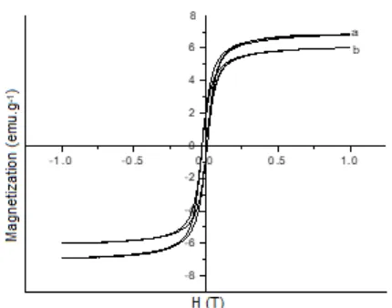
Fig 4. Magnetization curve of Fe3O4/SiO2/GPTMS-Arg
(a) Route 1 and (b) Route 2
acid medium than Route 2. When magnetite is added to sodium silicate solution, reaction between silicate monomer with magnetite surface to form complex of ≡Fe-OSi(OH)2OX or (≡FeO)2-Si(OH)OX [34]. While in
Route 2, magnetite is added latest after GPTMS, L-arginine and sodium silicate were mixed, it may produce arginine-modified silica-coated magnetite film incompletely.
Elemental Composition and Estimation of Amino Groups Content
Elemental composition analysis was performed with EDX and the result is expressed in Table 3. It is observed that the percentage of Fe is smaller than that of Si due to silica derived from sodium silicate and GPTMS. Based on these data, we performed the semi-quantitative surface analysis by calculating theoretically the weight ratio of silicon to iron (Si/Fe) in the Route 1 and 2 of 14.5 and 8.8, respectively. It is evidence that a number of silica coated on magnetite through Route 1 is higher than that of Route 2. Based on the theoretical calculation of the weight ratio Si/Fe is 12.47. The difference is predicted because there is a small portion of Fe released when the product was washed with water. Carbon content in the product resulted in Route 1 and 2 is 20.89 and 17.48%, and nitrogen content in Route 1 and 2 is 4.58 and 3.76%, respectively. The mole ratio of carbon to nitrogen in Route 1 and 2 is 5.8:1 and 5.44:1, respectively. The observation data is greater than based on the theoretical calculation (C:N = 3.75:1). This is probable due to the polymerization of carbon chains.
These data support the results of stability tests on the acidic medium, in which the quantity of iron dissolved in Fe3O4/SiO2-GPTMS-Arg-Route 1 (0.8610
-5
mol g-1) is smaller than that in Route 2 (3.0110-5
mol g-1). In addition, mass percentage of nitrogen in Fe3O4/SiO2
-to Fe3O4/SiO2-GPTMS-Arg-Route 2 (3.76%). Also, the
quantity of nitrogen confirmed by the estimation of amino group content of magnetite coated with L-arginine modified silica (Fe3O4/SiO2-GPTMS-Arg) was
found to be 0.364 and 0.266 mmol g-1for Route 1 and 2, respectively The presence of amino groups leads to the application of the product Fe3O4/SiO2-GPTMS-Arg
for metal ion removal from aqueous solution.
Magnetic Properties
Magnetic property of Fe3O4/SiO2-GPTMS-Arg was
examined with a vibrating sample magnetometer (VSM) and the results are shown in Fig. 4. The magnetization of Fe3O4/SiO2-GPTMS-Arg Route 1 and
2 are 6.82 and 6.02 emu.g-1, respectively. The results are agreed with the previous researchers reporting that the coating decreases a value of the magnetization. Nuryono et al. [23] reported that the magnetite coated with thiol-silica decreases the saturation magnetization value from 70.39 to 14.05 emu.g-1. Although the saturation magnetization of the magnetite coated with L-arginine modified silica is smaller than the native magnetite (55-78 emu.g-1) [24], the attractive forces to the external magnetic field is still be proceed. Therefore, the purpose to separate the magnetic product using a magnet can be achieved
CONCLUSION
In this study, L-arginine modified silica-coated magnetite through two different sol-gel routes using GPTMS to link silica and L-arginine has been synthesized successfully. The L-arginine modified silica-coated magnetite through Route 1 (mixing sequentially) is more stable against acidic medium and contains more amino group than that through Route 2 (mixing in two steps). The presence of amino groups in L-arginine-silica coated on magnetite leads to the application of the product for metal ion removal from aqueous solution.
ACKNOWLEDGEMENT
The authors thank to the Ministry of Research, Technology and Higher Education of the Republic of Indonesia for financial support through BPPDN Scholarship Program.
REFERENCES
pathogens,J. Hazard. Mater., 192 (3),1539–1547. [2] Yantasee, W., Warner, C.L., Sangvanich, T.,
Addleman, R.S., Carter, T.G., Wiacek, R.J., Fryxell, G.E., Timchalk, C., and Warner, M.G., 2007, Removal of heavy metals from aqueous systems with thiol functionalized superparamagnetic nanoparticles, Environ. Sci. Technol., 41 (14), 5114–5119.
[3] Afkhami, A., Saber-Tehrani, M., and Bagheri, H., 2010, Modified maghemite nanoparticles as an efficient adsorbent for removing some cationic dyes from aqueous solution, Desalination, 263 (1-3), 240–248.
[4] Pan, D., Tan, L., Qian, K., Zhou, L., Fan, Y., Yu, C., and Bao, X., 2010, Synthesis of highly ordered and hydrothermally stable mesoporous materials using sodium silicate as a precursor,Mater. Lett., 64 (13), 1543–1545.
[5] Gupta, A.K., and Gupta, M., 2005, Synthesis and surface engineering of iron oxide nanoparticles for biomedical applications, Biomaterials, 26 (18), 3995–4021.
[6] Yang, D., Hu, J., and Fu, S., 2009, Controlled synthesis of magnetite-silica nanocomposites via a seeded sol-gel approach, J. Phys. Chem. C, 113 (18), 7646–7651.
[7] Feng, G., Hu, D., Yang, L., Cui, Y., Cui, X.A., and Li, H., 2010, Immobilized-metal affinity chromatography adsorbent with paramagnetism and its application in purification of histidine-tagged proteins,Sep. Purif. Technol., 74 (2), 253–260. [8] Hong, R.Y., Li, J.H., Qu, J.M., Chen, L.L., and Li,
H.Z., 2009, Preparation and characterization of magnetite/dextran nanocomposite used as a precursor of magnetic fluid, Chem. Eng. J., 150 (2-3), 572–580.
[9] Ahangaran, F., Hassanzadeh, A., and Nouri, S., 2013, Surface modification of Fe3O4@SiO2
microsphere by silane coupling agent, Int. Nano Lett., 3, 3–7.
[10] Xu, Z., Liu, Q., and Finch, J.A., 1997, Silanation and stability of 3-aminopropyl triethoxy silane on nanosized superparamagnetic particles: I. Direct silanation,Appl. Surf. Sci., 120 (3-4), 269–278. [11] Deng, Y.H., Wang, C.C., Hu, J.H., Yang, W.L., and
Fu, S.K., 2005, Investigation of formation of silica-coated magnetite nanoparticles via sol-gel approach,Colloids Surf., A., 262 (1-3), 87–93. [12] Popovici, M., Gich, M., and Savii, C., 2006,
Ultra-light sol-gel derived magnetic nanostructured materials,Rom Rep. Phys., 58 (3), 369–378.
[13] Liu, Q., Xu, Z., Finch, J.A., and Egerton, R.A., 1998, Novel two-step silica-coating process for engineering magnetic nanocomposites, Chem. Mater.,10 (8), 3936–3940.
[14] Liu, J., Yang, Q., Zhao, X.S., and Zhang, L., 2007, Pore size control of mesoporous silicas from mixtures of sodium silicate and TEOS,
Microporous Mesoporous Mater., 106 (1-3),
62–67.
[15] Wang, Z., Zhu, H., Wang, X., Yang, F., and Yang, X., 2009, One-pot green synthesis of biocompatible arginine-stabilized magnetic nanoparticles,Nanotechnology, 20 (46), 465606. [16] Park, J.Y., Choi E.S., Baek, M.J., and Lee, G.H.,
2009, Colloidal stability of amino acid coated magnetite nanoparticles in physiological fluid,
Mater. Lett., 63 (3-4), 379–381.
[17] Davis, S.R., Brough, A.R., and Atkinson, A., 2003, Formation of silica/epoxy hybrid network polymers,
J. Non-Cryst. Solids, 315 (1-2), 197–205.
[18] Gizdavic-Nikolaidis, M.R., Zujovic, Z.D., Edmonds, N.R., Bolt, C.J., and Easteal, A.J., 2007, Spectroscopic characterization of GPTMS/DETA and GPTMS/EDA hybrid polymers, J. Non-Cryst. Solids,353 (16-17), 1598–1605.
[19] Innocenzi, P., Kidchob, T., and Yoko, T., 2005, Hybrid organic-inorganic sol-gel materials based on epoxy-amine systems,J. Sol-Gel Sci. Technol., 35 (3), 225–235.
[20] Hastuti, S., Nuryono, and Kuncaka, A., 2015, L-arginine-modified silica for adsorption of gold (III),
Indones. J. Chem., 15 (2), 108–115.
[21] Donia, A.M., Yousif, A.M., Atia, A.A., and Elsamalehy, M.F., 2014, Efficient adsorption of Ag(І) and Au(ІІІ) on modified magnetic chitosan with amine functionalities, Desalin Water Treat., 52 (13-15), 2537–2547.
[22] Wang, J., Zheng, S., Shao, Y., Liu, J., Xu, Z., and Zhu, D., 2010, Amino-functionalized Fe3O4@SiO2
core–shell magnetic nanomaterial as a novel adsorbent for aqueous heavy metals removal, J. Colloid Interface Sci., 349 (1), 293–299.
[23] Nuryono, N., Rosiati, N., Rusdiarso, B., Sakti, S.C., and Tanaka, S., 2014, Coating of magnetite with mercapto modified rice hull ash silica in a one-pot process,Springerplus, 3 (1), 515.
[24] Casillas, P.E.G., Gonzalez, C.A.R., and Pérez, C.A.M., 2012, “Infrared Spectroscopy of Functionalized Magnetic Nanoparticles” inInfrared Spectroscopy-Materials Science, Engineering and Technology, Theophile Theophanides ed., Rijeka, Croatia: Intech, 405–420.
[25] Yadav, L.D.S., 2005, Organic Spectroscopy, Springer-Science & Business Media, B.V., Dordrecht, 52–64.
H., Ohto, K., Inoue, K., and Alam, S., 2013, N-aminoguanidine modified persimmon tannin: a new sustainable material for selective adsorption, preconcentration and recovery of precious metals from acidic chloride solution, Bioresour Technol., 129, 108–117.
[28] Kumar, S., and Rai, S.B., 2010. Spectroscopic studies of L-arginine molecule,Indian J. Pure Appl. Phys., 48, 251–255.
[29] Sales, J.A.A., Prado, A.G., and Airoldi, C., 2002, The incorporation of propane-1,3-diamine into silylant epoxide group through homogeneous and heterogeneous routes, Polyhedron, 21 (25-26), 2647–2651.
[30] Mascolo, M.C., Pei, Y., and Ring, T.A., 2013, Room temperature co-precipitation synthesis of magnetite nanoparticles in a large pH window with different bases,Materials, 6 (12), 5549–5567.
Synthesis of magnetic nanoparticles coated with covalently bonded carboxymethyl cellulose,Int. J. ChemTech. Res., 9 (5), 1–6.
[32] Xu, H., Xu, Z., Yang, L., and Wang, Q., 2011, “One-pot” preparation of basic amino acid-silica hybrid monolithic column for capillary electrochromatography, J. Sep. Sci., 34 (16-17), 2314–2322.
[33] Matei, E., Predescu, C., Berbecaru, A., Predescu, A., and TruşCǍa, R., 2011, Leaching tests for synthesized magnetite nanoparticles used as adsorbent for metal ions from liquid solutions,Dig. J. Nanomater. Bios., 6 (4), 1701–1708.