Activated platelets in patients with severe hypertriglyceridemia:
effects of triglyceride-lowering therapy
Frits H. de Man
a,*, Rienk Nieuwland
b, Arnoud van der Laarse
a, Fred Romijn
b,
Augustinus H.M. Smelt
c, Jan A. Gevers Leuven
c,d, Augueste Sturk
baDepartment of Cardiology,C5-P,Leiden Uni6ersity Medical Centre,P.O.Box9600,2300RC Leiden,The Netherlands bDepartment of Clinical Chemistry,Leiden Uni6ersity Medical Centre,Leiden,The Netherlands
cDepartment of Internal Medicine,Leiden Uni6ersity Medical Centre,Leiden,The Netherlands dTNO-Pre6ention and Health,Gaubius Laboratory,Leiden, The Netherlands
Received 30 April 1999; received in revised form 28 October 1999; accepted 6 December 1999
Abstract
Hypertriglyceridemia, a risk factor for cardiovascular disease, has been associated with hypercoagulability, but whether platelet activation is implicated is unknown. This study was designed to compare the in vivo platelet activation status between patients with severe hypertriglyceridemia and age- and sex-matched control subjects, and to evaluate the effects of triglyceride-lowering therapy. Sixteen patients with primary hypertriglyceridemia were included in a double-blind, placebo-controlled cross-over trial with 400 mg bezafibrate once daily. Platelet activation was analysed by double label flow cytometry, using monoclonal antibodies against GP53, P-selectin, and platelet-bound fibrinogen. Surface expression of the lysosomal membrane protein GP53 was significantly higher in the hypertriglyceridemic patients at baseline as compared to the group of age- and sex-matched controls (16.394.8% vs. 8.993.4%, respectively, PB0.001). No differences in the expression of P-selectin and fibrinogen binding were observed. In response to bezafibrate therapy, the expression of GP53 in the patient group decreased from 16.394.8% to 13.194.1% (P=0.018). The expression of P-selectin and fibrinogen binding was not affected by bezafibrate therapy. In conclusion, patients with hypertriglyceridemia have an increased in vivo platelet activation status, which can be improved by bezafibrate therapy. © 2000 Elsevier Science Ireland Ltd. All rights reserved.
Keywords:Triglycerides; Flow cytometry; Bezafibrate; Platelet activation
www.elsevier.com/locate/atherosclerosis
1. Introduction
Platelets play an important role in the pathogenesis of atherosclerosis and acute coronary syndromes [1,2]. The beneficial effects of anti-platelet agents in sec-ondary prevention trials support this hypothesis [3]. Activation of platelets has been demonstrated in pa-tients with unstable angina pectoris and acute myocar-dial infarction [4,5]. Also the classical cardiovascular risk factors such as diabetes, smoking and hyperlipo-proteinemia have been associated with increased platelet reactivity or platelet activation [6 – 8].
Hypertriglyceridemia (HTG) is a risk factor for
car-diovascular disease in both men and women [9]. In addition to the characteristic lipoprotein profile, which includes low levels of high density lipoprotein (HDL) cholesterol and the presence of atherogenic small, dense low density lipoprotein (LDL) particles, hypertriglyce-ridemia has been associated with various derangements of the hemostatic system. Plasma fibrinogen levels are often elevated in hypertriglyceridemic patients [10,11]. Also, activated factor VII and plasminogen activator inhibitor-1 levels are elevated in these patients and correlate with total plasma triglyceride levels [12,13]. These results indicate that hypertriglyceridemia is asso-ciated with a hypercoagulable state and impaired fibrinolysis. The effects of triglycerides on platelet func-tion are less uniformly described [7,14]. Previous studies used platelet aggregometry to indirectly assess platelet reactivity ex vivo. This technique is difficult to perform * Corresponding author. Tel.: +31-71-5262020; fax: +
31-71-5266809.
E-mail address:a.van –der–[email protected] (F.H. de Man).
in turbid plasma samples. The development of whole blood flow cytometry [15] provides a tool to address the platelet activation status directly and specifically, by measuring the presence of activation antigens on the platelet surface.
Activation of platelets is initiated by the binding of agonists to specific receptors [16]. As a result, the glycoprotein (GP) IIb – IIIa complex changes its confor-mation, thereby revealing the binding site for fibrino-gen. In addition, the membranes of the a-granules, dense granules and lysosomes fuse with the platelet cell-membrane to release the granular contents. As a consequence, novel activation-dependent membrane glycoproteins become exposed, which in the resting platelet were present only in the granule membranes. In the present study, three of these activation-dependent antigens were determined: P-selectin (CD62P), an a -granule-derived transmembrane protein; GP53 (CD63) a lysosomal membrane protein; and platelet-bound fibrinogen.
The present study was designed to directly assess the platelet activation status in patients with hypertriglyce-ridemia as compared to healthy age- and sex-matched control subjects. In addition, the effects of triglyceride-lowering therapy were studied in a double-blind, placebo-controlled trial with 400 mg bezafibrate once daily.
2. Materials and methods
2.1. Patients and control subjects
The study group consisted of 16 unrelated patients (14 males and two females) with primary hypertriglyce-ridemia who were recruited from the outpatient lipid clinic of the Leiden University Medical Centre. The diagnosis primary hypertriglyceridemia was based on the means of two fasting blood samples obtained after a dietary period of at least 8 weeks. The diagnostic criteria for endogenous hypertriglyceridemia were: total serum triglyceride (TG)\4.0 mmol/l; very low density lipoprotein (VLDL) cholesterol\1.0 mmol/l; and low density lipoprotein cholesterol (LDL-C)B4.5 mmol/l. Exclusion criteria were the apoE2E2 phenotype, sec-ondary hyperlipidemia (renal, liver or thyroid disease, fasting glucose\7.0 mmol/l, and alcohol consumption of more than 40 g/day) and the use of anti-platelet or anti-coagulant drugs. Patients with a medical history of cardiovascular disease were not included in the study. Lipid lowering drugs, if used, were stopped 6 weeks before entering the study. Patients were urged to adhere to the dietary advice during the study. Seventeen nor-molipidemic, age- and sex-matched control subjects (16 males and one female) were recruited in response to a newspaper advertisement.
2.2. Study design and blood sampling
The patients were randomised to receive in a double-blind cross-over fashion a fixed dose of bezafibrate, 400 mg once daily, and placebo for 6 weeks. The two periods in which medication was taken were separated by a wash-out period of 6 weeks. Before and after each treatment period of 6 weeks, fasting venous blood samples were obtained from the participants for lipid measurements. The platelet activation status was as-sessed at the end of the placebo and treatment periods. From the control subjects, fasting blood samples were obtained at baseline under identical conditions. In-formed consent was obtained from each patient and the protocol was approved by the institutional Medical Ethics Committee (protocol number P183/96).
2.3. Lipids and lipoproteins
Venous blood was collected after an overnight fast. Serum was obtained after centrifugation at 1500×gfor 15 min at room temperature. Three millilitres of fresh serum was ultracentrifuged for 15 h at 232 000×g at 15°C in a TL-100 tabletop Ultracentrifuge, using a TLA-100.3 fixed angle rotor (Beckman, Palo Alto, CA, USA). The ultracentrifugate was carefully divided in a density (d)B1.006 and d=1.006 – 1.25 g/ml fraction, designated as the VLDL and LDL – HDL fraction, respectively. The triglyceride and cholesterol concentra-tions were measured enzymatically using test kits (Boehringer, Mannheim, Germany). HDL cholesterol was measured in the LDL – HDL fraction after precipi-tation of apoB-containing particles with phospho-tungstic acid and MgCl2. ApoE phenotyping was performed by isoelectric focusing according to Havekes et al. [17]. ApoB48 was measured in the dB1.006 g/ml fraction with SDS-PAGE. The mean apoB48 concen-tration was 493% (range 0 – 10%) of apoB100 concentration.
2.4. Preparation of whole blood samples for flow cytometry
mg/ml FITC-anti-P-selectin, 16 mg/ml FITC-anti-fibrinogen (platelet-bound FITC-anti-fibrinogen) and 5 mg/ml FITC-labelled control antibody (IgG1 isotype). After careful mixing and 15 min incubation at room tempera-ture in the dark, 5ml of ten-fold diluted phycoerythrin-conjugated streptavidin was added. Incubation continued for another 15 min at room temperature. Finally, 2.5 ml of buffer A was added, containing 0.2% formaldehyde (final concentration).
2.5. Antibodies
The following murine MoAbs were used. Anti-GPIb (CLB-MB45), directed against the a-chain of GPIb, was obtained from the Central Laboratory of the Netherlands Red Cross Blood Transfusion Service (Amsterdam, The Netherlands). Anti-GPIb was bi-otinylated as described by Hnatowitch et al. [18]. FITC-labelled MoAbs against P-selectin (CD62P) and GP53 were obtained from Immunotech (Marseille, France). FITC-labelled chicken anti-human fibrinogen poly-clonal antibodies were obtained from Biopool AB (Umea˚, Sweden) and FITC-labelled IgG1 control anti-bodies were purchased from Becton & Dickinson (San Jose, CA, USA). Phycoerythrin-conjugated streptavidin was obtained from Dakopatts (Glostrup, Denmark).
2.6. Flow cytometric analysis
Double label flow cytometry was performed as de-scribed previously [19,20]. After collection, blood sam-ples were analysed in a FACScan flow cytometer with CellQuist software (Becton Dickinson, San Jose, CA, USA). Forward light scatter and sideward light scatter were set at logarithmic gain. Platelets were identified by analysing the phycoerythrin-GPIb content of particles at 585 nm. Regions were identified, corresponding to platelet-derived microparticles (R1), platelets (R2) and
platelet – platelet, platelet – leukocyte, platelet-derived-microparticle(s) – leukocyte or platelet – erythrocyte complexes (R3). The surface expression of activation markers was determined in a population of 5000 platelets (R2) by analysing the fluorescence intensity of the FITC-labelled antibodies at 515 nm. The threshold for platelet activation was set at 2% fluorescence-posi-tive platelet activation with an FITC-labelled IgG1 control antibody. Platelet count and volume were mea-sured on a Sysmex™ SE-9000 analyser (TOA Medical Electronics, Kobe, Japan).
To verify our method of platelet activation analysis by expression of the activation markers, whole blood obtained from eight healthy volunteers, and coagulated with 0.10 mmol/l sodium citrate (final concentration) was stimulated with 10 mmol/l adenosine diphosphate in vitro. The mean fluorescence intensity (MFI) for FITC-labelled anti-P-selectin (CD62P) increased from 3.690.5 to 10.894.3 and MFI for FITC-labelled anti-GP53 (CD63) increased from 5.690.8 to 9.791.5 (mean9S.D.). These data indicate that the procedure used to label and fix the platelets can detect platelet activation by increased antigen expression of activation markers.
2.7. Statistical analyses
Results are presented as the mean9S.D. Mean dif-ferences between the control and patient groups were calculated with the Mann – WhitneyU-test. Only differ-ences in sex distribution between the patient group and control group were calculated with the Fisher’s exact test. The results at the end of the placebo and bez-afibrate period were analysed pairwise using the Wilcoxon paired signed-ranks test. Findings were re-garded to be statistically significant when the probabil-ity of these data under the null hypothesis was less than 0.05. Statistical analyses were performed with SPSS/
PC+™ software (SPSS Inc., Chicago, IL, USA).
3. Results
3.1. Patient characteristics
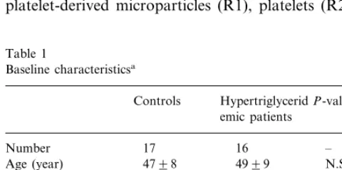
As shown in Table 1, the patient and control groups were comparable with regard to age and sex. Serum triglyceride levels were significantly higher in the HTG patients as compared to the control subjects. Although both LDL-C and high density lipoprotein cholesterol (HDL-C) levels were lower in the patient group, very low density lipoprotein cholesterol (VLDL-C) concen-trations were markedly elevated and accounted for the elevation in total serum cholesterol concentrations. Table 1
TTG (mmol/l) 10.2194.44 B0.001 TC (mmol/l) 5.0390.99 7.7991.84 B0.001
0.2990.18
VLDL-C (mmol/l) 4.4792.23 B0.001 0.002
aValues are presented as the mean9S.D. TC, total cholesterol;
TTG, total triglycerides.
bNumeric variables were analysed with the Mann–WhitneyU-test.
Table 2
Effects of treatment with placebo and bezafibrate on serum lipids and lipoproteinsa
Before placebo During placebo Before bezafibrate During bezafibrate
16
Number 16 16 16
12.2098.58 11.4197.77 3.8391.51* TTG (mmol/l) 10.4895.31
7.6592.46 8.0892.75
aValues are presented as the mean9S.D. TC, total cholesterol; TTG, total triglycerides; All values are mean9S.D. based on triplicate
measurements.
*PB0.001 as compared to the corresponding value before bezafibrate therapy using the Wilcoxon paired signed-ranks test. **PB0.01 as compared to the corresponding value before bezafibrate therapy using the Wilcoxon paired signed-ranks test. ***PB0.05 as compared to the corresponding value before bezafibrate therapy using the Wilcoxon paired signed-ranks test.
3.2. Effect of therapy on serum lipid and lipoprotein le6els
All subjects concluded the study without any side-ef-fects. The effects of bezafibrate and placebo are shown in Table 2. Placebo therapy did not influence serum lipid and lipoprotein levels. Bezafibrate therapy resulted in a significant reduction in serum TG (−66%, PB 0.001), serum cholesterol levels (−27%,PB0.001) and VLDL-C levels (−68%,PB0.001). LDL-C and HDL-C levels increased by 29% (P=0.003) and 12% (P=
0.011), respectively.
3.3. Platelet analyses
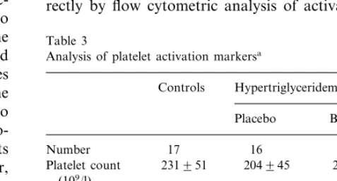
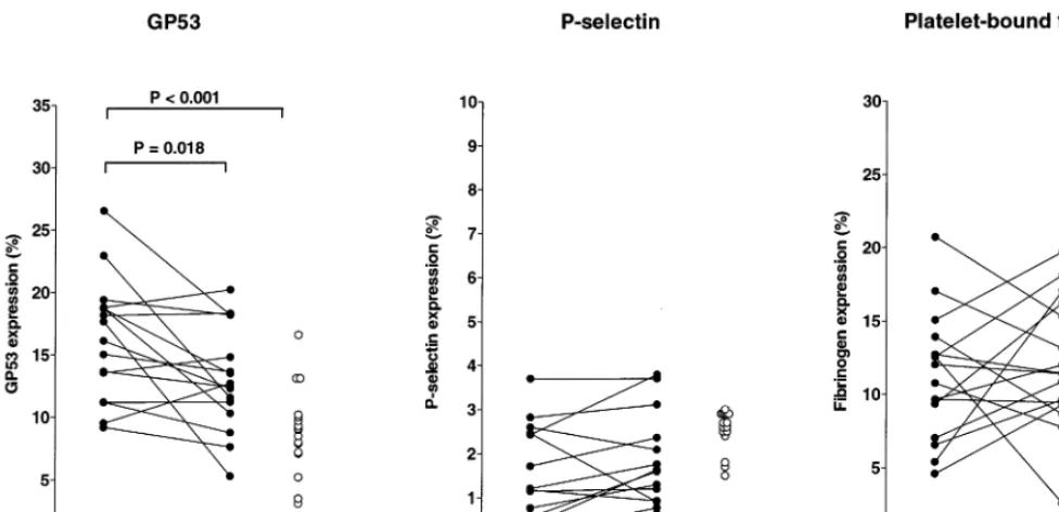
The first objective was to compare the expression of GP53, P-selectin and platelet fibrinogen binding be-tween control subjects and HTG patients. Since placebo therapy did not influence serum lipid levels in the patient group, the platelet activation data obtained during placebo therapy were used as ‘baseline’ values and compared with the platelet activation data of the control group. As shown in Table 3 and Fig. 1, no differences in the expression of P-selectin and fibrino-gen binding were found between the control subjects and HTG patients (during placebo therapy). However, the expression of GP53 was significantly higher in the HTG patients (16.394.8%) as compared to the group of control subjects (8.993.4%, PB0.001; range 3.1 – 16.6% and median 9.1%). Platelet count and mean platelet volume did not differ between the HTG pa-tients and control subjects (Table 3).
Bezafibrate therapy resulted in an improvement of the lipoprotein profile in the patient group (Table 2). The mean effects of this therapy on the expression of platelet activation markers are shown in Table 3. The expression of P-selectin and binding of fibrinogen did not change in response to bezafibrate therapy. During bezafibrate therapy, the high expression of GP53 in HTG patients decreased from 16.394.8% to 13.19
4.1% (P=0.018), but remained higher than the levels in the control group (PB0.05). Although mean platelet volume was not affected by both therapy modalities, the platelet number increased in response to bezafibrate therapy (+8%, P=0.011; Table 3). The individual changes in GP53, P-selectin and platelet-bound fibrino-gen expression upon bezafibrate therapy are presented in Fig. 1. The P-selectin expression did not change substantially within the patients and was similar to the values obtained in the healthy controls. The changes in platelet-bound fibrinogen were not significant.
4. Discussion
In this study we investigated the in vivo platelet activation status in hypertriglyceridemic patients di-rectly by flow cytometric analysis of
activation-depen-Table 3
Analysis of platelet activation markersa
Hypertriglyceridemic patients
Platelet count 204945 (109/l)
10.0391.20 9.9891.17 Mean platelet 9.9190.50
volume (fl)
aValues are presented as the mean9S.D. bPresented as percentage activated platelets.
*PB0.05 as compared to the corresponding value on placebo therapy using the Wilcoxon paired signed-ranks test.
**PB0.001 as compared to the corresponding value in the hyper-triglyceridemic group on respectively placebo therapy and bezafibrate therapy, using the Mann–WhitneyU-test.
Fig. 1. Expression of platelet activation markers. Expression of GP53 (left panel), P-selectin (middle panel) and platelet-bound fibrinogen (right panel) on platelets obtained from 16 hypertriglyceridemic patients (solid circles) during placebo and bezafibrate therapy compared with 17 age-and sex-matched control subjects (open circles). Analysis of the platelet activation markers was performed by double label flow cytometry, using antibodies against GP53, P-selectin and platelet-bound fibrinogen. The surface expression of the lysosomal membrane protein GP53 was significantly higher in the hypertriglyceridemic patients as compared to the group of controls. No differences in the expression of P-selectin and fibrinogen binding were observed. In response to bezafibrate therapy, the expression of GP53 in the patient group decreased, whereas the expression of P-selectin and fibrinogen binding was not affected.
dent platelet surface antigens. In previous studies, platelet reactivity was assessed in vitro by platelet ag-gregometry [7,14]. A major problem of this technique is that light transmission measurements are influenced by turbidity of plasma, a common phenomenon in this patient group. Other investigators have tried to avoid this problem by defining an upper limit of the serum triglyceride level [21] or using an impedance aggregome-ter technique [14]. A second problem of platelet aggre-gometry is the fact that the in vivo platelet activation status is not assessed directly but is merely implied from platelet reactivity in vitro. In contrast, the flow cyto-metric analysis of activation-dependent platelet surface antigens is not influenced by turbidity of blood samples and directly reflects the platelet activation status in vivo. To our knowledge, this is the first report describ-ing the in vivo platelet activation status of hypertriglyc-eridemic patients.
Our first objective was to compare the platelet activa-tion status of HTG patients with age- and sex-matched control subjects. The surface expression of GP53 was higher in the patient group as compared to the group of healthy volunteers. The platelet-activation markers P-selectin and platelet-bound fibrinogen were comparable in both groups. Thus, only one out of three platelet activation markers showed a higher expression in the HTG group. GP53 is a member of the lysosome-associ-ated membrane proteins (LAMP) which become
rapid loss of P-selectin in vivo. This decrease was paralleled by an increase in soluble P-selectin. Thus, P-selectin is exposed transiently at the platelet surface after activation and dissociates from the platelet, imply-ing that the expression of P-selectin may not be a good marker of platelet activation in vivo. Also in contrast to GP53, no differences were found in the amount of fibrinogen bound to the surface of patient versus con-trol platelets. Upon platelet activation, fibrinogen binds to the activated GPIIb – IIIa complex. Resting platelets do not bind fibrinogen, but when platelets are activated the GPIIb – IIIa complex changes its conformation and thus becomes eligible to bind fibrinogen. This change in the fibrinogen receptor conformation is highly re-versible [26]. Therefore, the extent of surface binding of fibrinogen may be a transient phenomenon, which makes it less suitable as a marker for the platelet activation status. The finding that different platelet activation markers provide seemingly discrepant results is not uncommon in clinical studies. A number of studies have reported increased expression of GP53 only, or of one of the other markers, in a variety of diseases [20,27 – 29]. We conclude that an increased platelet activation status, as measured by surface GP53 exposure, is present in hypertriglyceridemic patients.
Several mechanisms may explain the increased platelet activation status in patients with hypertriglyce-ridemia. First, platelet activity in hyperlipidemias may be related to changes in the lipid composition of platelet membranes. Increased plasma cholesterol levels have been shown to decrease the platelet membrane fluidity [30]. These cholesterol-enriched rigid platelet membranes show an enhanced platelet responsiveness by increasing the number and affinity of platelet thrombin receptors [31]. Malle et al. [32] studied platelet membrane fluidity in other types of dyslipi-demia. Interestingly, platelets from HTG patients demonstrated increased membrane fluidity as compared to healthy control subjects. So, platelet membrane rigidity does not seem to be a plausible explanation for the enhanced platelet activation status in the HTG group.
A second mechanism that may be involved is oxida-tive stress. Several studies have demonstrated that oxi-dative stimuli may activate platelets [33,34]. A characteristic lipoprotein pattern that can be observed in hypertriglyceridemia includes relatively low concen-trations of LDL and HDL cholesterol, which are caused by exchange of lipids between the VLDL pool on one hand and the LDL – HDL pool on the other. In this process, the LDL particles become triglyceride-en-riched and relatively cholesterol ester-depleted. As the LDL particles are progressively lipolysed in the circula-tion, the triglycerides are degraded and a small, dense particle remains. These small, dense LDL particles are regarded as potentially atherogenic since they show a
low resistance to oxidative modification [35]. However, earlier we reported that small dense LDL of HTG patients is not associated with low resistance to oxida-tion [36]. At present it is not clear whether small, dense LDL particles in these patients are associated with an increased tendency to oxidative modification [35,37] and, consequently, with an increased platelet activation [38].
A third explanation may be the platelet activating potential of VLDL, the lipoprotein fraction that pri-marily accumulates in hypertriglyceridemia. Several studies have demonstrated that VLDL stimulates platelet aggregation [39,40]. Van Willigen et al. [41] studied the effect of LDL on fibrinogen binding to the GPIIb – IIIa complex. Interestingly, incubation of platelets with LDL induced a rapid and dose-dependent increase in fibrinogen binding to platelets. No increase was observed after modification of the lysine residues of LDL, and therefore the effect was suggested to be receptor-mediated. It was speculated that LDL changes the exposure of the GPIIb – IIIa complex into a more active configuration. The principal candidate ligand of LDL is apolipoprotein (apo) B-100, which mediates the clearance of LDL from the circulation, as ligand for the LDL-receptor. Interestingly, apoB-100 contains a sub-stantial number of lysine residues that play an impor-tant role in the receptor-mediated clearance [42]. In hypertriglyceridemia, LDL concentrations are low and the principal lipoprotein fraction that accumulates is VLDL. Since VLDL, like LDL, contain apoB-100, VLDL may influence the platelet activation status as well by changing the conformation of the GPIIb – IIIa complex via apoB-100. Another study, however, found that LDL augments platelet reactivity via a receptor-in-dependent mechanism, by inhibiting the Na+/H+ an-tiport in the platelet membrane [43]. Whether this ion exchanger is influenced by VLDL as well remains to be determined. Further studies are needed to elucidate the mechanisms that could be involved in the interaction between VLDL and platelets.
In conclusion, our data indicate that hypertriglyce-ridemia is associated with an increased expression of the lysosomal membrane protein GP53, which is indica-tive of an increased in vivo platelet activation status. Triglyceride-lowering therapy by bezafibrate results in a significant reduction of the serum lipid levels and a modest reduction in GP53 expression.
Acknowledgements
References
[1] Moliterno DJ, Granger CB. Differences between unstable angina and acute myocardial infarction. The pathological and clinical spectrum. In: Topol EJ, editor. Acute Coronary Syndromes. New York: Marcel Dekker, 1998:67 – 103.
[2] Fuster V, Badimon L, Badimon JJ, Chesebro JH. The pathogen-esis of coronary artery disease and the acute coronary syn-dromes. New Engl J Med 1992;326:310 – 8.
[3] Antiplatelet Trialists’ Collaboration. Secondary prevention of vascular disease by prolonged antiplatelet treatment. Br Med J 1988;296:320 – 31.
[4] Becker RC, Tracy RP, Bovill EG, Mann KG, Ault K. The clinical use of flow cytometry for assessing platelet activation in acute coronary syndromes. TIMI-III Thrombosis and Anticoag-ulation Group. Cor Art Dis 1994;5:339 – 45.
[5] Schultheiss HP, Tschoepe D, Esser J, Schwippert B, Roesen P, Nieuwenhuis HK, Schmidt SC, Strauer B. Large platelets con-tinue to circulate in an activated state after myocardial infarc-tion. Eur J Clin Invest 1994;24:243 – 7.
[6] Hawkins RI. Smoking, platelets and thrombosis. Nature 1972;236:450 – 2.
[7] Carvalho AC, Colman RW, Lees RS. Platelet function in hyper-lipoproteinemia. New Engl J Med 1974;290:434 – 8.
[8] Strano A, Davi G, Patrono C. In vivo platelet activation in diabetes mellitus. Semin Thromb Hemost 1991;17:422 – 5. [9] Hokanson JE, Austin MA. Plasma triglyceride level is a risk
factor for cardiovascular disease independent of high-density lipoprotein cholesterol level: a meta-analysis of population-based prospective studies. J Cardiovasc Risk 1996;3:213 – 9.
[10] Simpson HC, Meade TW, Stirling Y, Mann JI, Chakrabarti R, Woolf L. Hypertriglyceridaemia and hypercoagulability. Lancet 1983;i:786 – 90.
[11] Avellone G, Di Garbo V, Cordova R, Raneli G, De Simone R, Bompiani GD. Fibrinolysis in hypertriglyceridaemic subjects in response to venous occlusion. Blood Coagul Fibrinolysis 1993;4:429 – 33.
[12] Zitoun D, Bara L, Basdevant A, Samama MM. Levels of factor VIIc associated with decreased tissue factor pathway inhibitor and increased plasminogen activator inhibitor-1 in dyslipidemias. Arterioscler Thromb Vasc Biol 1996;16:77 – 81.
[13] Green D, Chamberlain MA, Ruth KJ, Folsom AR, Liu K. Factor VII, cholesterol, and triglycerides. The CARDIA Study. Coronary Artery Risk Development in Young Adults Study. Arterioscler Thromb Vasc Biol 1997;17:51 – 5.
[14] Riess H, Merk W, Falkner C, Hiller E. Increased in vitro platelet aggregation in hypertriglyceridemias. Thromb Res 1986;41:281 – 9.
[15] Shattil SJ, Cunningham M, Hoxie JA. Detection of activated platelets in whole blood using activation-dependent monoclonal antibodies and flow cytometry. Blood 1987;70:307 – 15. [16] Kroll MH, Schafer AI. Biochemical mechanisms of platelet
activation. Blood 1989;74:1181 – 95.
[17] Havekes LM, de Knijff P, Beisiegel U, Havinga JR, Smit M, Klasen E. A rapid micromethod for apolipoprotein E phenotyp-ing directly in serum. J Lipid Res 1987;28:455 – 63.
[18] Hnatowitch J, Verzin F, Ruscowski M. Investigations of avidin and biotin for imaging applications. J Nucl Med 1987;28:1294. [19] Abrams CS, Ellison N, Budzynski AZ, Shattil SJ. Direct
detec-tion of activated platelets and platelet-derived microparticles in humans. Blood 1990;75:128 – 38.
[20] Konijnenberg A, Stokkers EW, van der Post JA, Schaap MC, Boer K, Bleker OP, Sturk A. Extensive platelet activation in preeclampsia compared with normal pregnancy: enhanced ex-pression of cell adhesion molecules. Am J Obstet Gynecol 1997;176:461 – 9.
[21] Pazzucconi F, Mannucci L, Mussoni L, Gianfranceschi G, Maderna P, Werba P, Franceschini G, Sirtori CR, Tremoli E. Bezafibrate lowers plasma lipids, fibrinogen and platelet aggre-gability in hypertriglyceridaemia. Eur J Clin Pharmacol 1992;43:219 – 23.
[22] Nieuwenhuis HK, van Oosterhout JJ, Rozemuller E, van Iwaar-den F, Sixma JJ. Studies with a monoclonal antibody against activated platelets: evidence that a secreted 53 000-molecular weight lysosome-like granule protein is exposed on the surface of activated platelets in the circulation. Blood 1987;70:838 – 45. [23] Metzelaar MJ, Wijngaard PL, Peters PJ, Sixma JJ, Nieuwenhuis
HK, Clevers HC. CD63 antigen. A novel lysosomal membrane glycoprotein, cloned by a screening procedure for intracellular antigens in eukaryotic cells. J Biol Chem 1991;266:3239 – 45. [24] Silverstein RL, Febbraio M. Identification of
lysosome-associ-ated membrane protein-2 as an activation-dependent platelet surface glycoprotein. Blood 1992;80:1470 – 5.
[25] Michelson AD, Barnard MR, Hechtman HB, MacGregor H, Connolly RJ, Loscalzo J, Valeri CR. In vivo tracking of platelets: circulating degranulated platelets rapidly lose surface P-selectin but continue to circulate and function. Proc Natl Acad Sci USA 1996;93:11877 – 82.
[26] van Willigen G, Akkerman JW. Protein kinase C and cyclic AMP regulate reversible exposure of binding sites for fibrinogen on the glycoprotein IIB – IIIA complex of human platelets. Biochem J 1991;273:115 – 20.
[27] Tschoepe D, Schultheiss HP, Kolarov P, Schwippert B, Dannehl K, Nieuwenhuis HK, Kehrel B, Strauer B, Gries FA. Platelet membrane activation markers are predictive for increased risk of acute ischemic events after PTCA. Circulation 1993;88:37 – 42. [28] Murakami T, Komiyama Y, Masuda M, Kido H, Nomura S,
Fukuhara S, Karakawa M, Iwasaka T, Takahashi H. Flow cytometric analysis of platelet activation markers CD62P and CD63 in patients with coronary artery disease. Eur J Clin Invest 1996;26:996 – 1003.
[29] Broijersen A, Karpe F, Hamsten A, Goodall AH, Hjemdahl P. Alimentary lipemia enhances the membrane expression of platelet P-selectin without affecting other markers of platelet activation. Atherosclerosis 1998;137:107 – 13.
[30] Shattil SJ, Cooper RA. Membrane microviscosity and human platelet function. Biochemistry 1976;15:4832 – 7.
[31] Tandon N, Harmon JT, Rodbard D, Jamieson GA. Thrombin receptors define responsiveness of cholesterol-modified platelets. J Biol Chem 1983;258:11840 – 5.
[32] Malle E, Sattler W, Prenner E, Leis HJ, Karadi I, Knipping G, Romics L, Kostner GM. Platelet membrane fluidity in type IIA, type IIB and type IV hyperlipoproteinemia. Atherosclerosis 1991;87:159 – 67.
[33] Salvemini D, de NG, Sneddon JM, Vane JR. Superoxide anions enhance platelet adhesion and aggregation. Br J Pharmacol 1989;97:1145 – 50.
[34] Iuliano L, Pedersen JZ, Pratico D, Rotilio G, Violi F. Role of hydroxyl radicals in the activation of human platelets. Eur J Biochem 1994;221:695 – 704.
[35] de Graaf JC, Hendriks JC, Demacker PN, Stalenhoef AF. Identification of multiple dense LDL subfractions with enhanced susceptibility to in vitro oxidation among hypertriglyceridemic subjects. Normalization after clofibrate treatment. Arterioscler Thromb 1993;13:712 – 9.
[36] De Man FH, Princen HM, van Duyvenvoorden W, Hollaar L, Smelt AH, Gevers Leuven JA, van der Laarse A. Hypertriglyce-ridemia is associated with an enhanced resistance to oxidative stress. J Am Coll Cardiol 1999;33(suppl A):319A.
[38] Ardlie NG, Selley ML, Simons LA. Platelet activation by oxida-tively modified low density lipoproteins. Atherosclerosis 1989;76:117 – 24.
[39] Aviram M, Brook JG. Characterization of the effect of plasma lipoproteins on platelet function in vitro. Haemostasis 1983;13:344 – 50.
[40] Mochizuki M, Takada Y, Urano T, Nagai N, Nakano T, Nakajima K, Takada A. The in vitro effects of chylomicron remnant and very low density lipoprotein remnant on platelet aggregation in blood obtained from healthy persons. Thromb Res 1996;81:583 – 93.
[41] van Willigen G, Gorter G, Akkerman JW. LDLs increase the exposure of fibrinogen binding sites on platelets and secretion of dense granules. Arterioscler Thromb 1994;14:41 – 6.
[42] Mahley RW, Innerarity TL, Weisgraber KB, Oh SY. Altered metabolism (in vivo and in vitro) of plasma lipoproteins after selective chemical modification of lysine residues of the apo-proteins. J Clin Invest 1979;64:743 – 50.
[43] Nofer JR, Tepel M, Kehrel B, Wierwille S, Walter M, Seedorf U, Zidek W, Assmann G. Low-density lipoproteins inhibit the Na+/H+ antiport in human platelets. A novel mechanism en-hancing platelet activity in hypercholesterolemia. Circulation 1997;95:1370 – 7.